DOI:
10.1039/C5RA17851G
(Paper)
RSC Adv., 2015,
5, 96158-96164
Detection of nonfluorescent cyhalothrin in honey by a spheral SiO2-based particle coating with thin fluorescent molecularly imprinted polymers film†
Received
2nd September 2015
, Accepted 27th October 2015
First published on 27th October 2015
Abstract
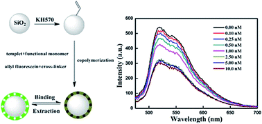
In this study, we report a general protocol for making core–shell SiO2@KH570-MIP based on the surface modification of SiO2 beads for the selective detection of ultra trace cyhalothrin. We first prepared the fluorescent surface molecularly imprinted polymer (SMIP) spheres via copolymerization of acrylamide with allyl fluorescein in the presence of cyhalothrin to form recognition sites. The experimental results showed that the fluorescence quenching of SiO2@KH570-MIP for cyhalothrin was much higher than that of the structural analogue composite, which illustrated good recognition capacity and selectivity of SiO2@KH570-MIP for the template. In addition, a linear relationship could be obtained at a lower concentration range of 0–2.5 nM with a correlation coefficient of 0.99698 described by the Stern–Volmer equation. The results of practical detection suggest that the developed method satisfactorily determines cyhalothrin in honey samples. This study therefore demonstrated the potential of SiO2-based SMIP spheres for the recognition and detection of cyhalothrin in food.
1. Introduction
Pyrethroid insecticides are widely used for pest-control in agriculture areas because of relatively low mammalian toxicity and high activity against a broad spectrum of insect pests. Nevertheless, for their widespread usages and less solubility, pyrethroid insecticides are usually removed by solid sorbent, resulting in low residue concentration in the environment and enrichment of agricultural products. Some of the pyrethroid insecticides are not only known to cause disturbed consciousness and seizures by affecting the central nervous system of humans, but they are also suspected to have endocrine-disrupting effects, which can adversely affect reproduction and sexual development, as well as the immune system.1–5 Thus, sensitive analytical methods that involve a small amount of solvent and are sensitive to trace levels of pesticide residues in food must be employed in monitoring pyrethroid insecticides. Usually, pyrethroid insecticides are determined by gas chromatography coupled with electron-capture detection (GC-ECD), mass spectrometry (GC-MS), or liquid chromatography-electrospray ionization mass spectroscopy (LC-MS).6–8 The MS instruments are very expensive but exhibit high selectivity and sensitivity.9 These methods are also complicated to operate, time-consuming and require a tedious sample pretreatment. The method in this study not only has the advantages of high sensitivity, simplicity, efficiency, speed, and convenience but also could deal with complex samples. The interference of coexisting substances in the specimen material can be eliminated by this coupling technology, and the selectivity of MIPs can be combined with the high sensitivity of fluorescence detection.
An optosensing system, especially applying fluorescence technology, is a useful method for detecting environmental pollutants10 because it has the advantages of high sensitivity, low detection limit, and low cost.11 In view of these properties, fluorescence technology would be appropriate for detecting pyrethroid pesticides at low concentrations. However, not all the analytes can be directly detected by fluorescence technology. Phosphors with strong fluorescence could be used to recognize the targets with no fluorescence through chemical reactions. It is well known that fluorescein and its derivatives are well known detector as a fluorescent probe in food testing, which are generally used to detect heavy metal ions and analyze environmental pollution based on the variations of fluorescence,12,13 because this type of compound has a high extinction coefficient and high fluorescent quantum yield in an aqueous or organic solution.14 In addition, the Stoke's shift15 is big enough to avoid self-absorption, which improves the validity of fluorescence detection. However, fluorescein and its derivatives are not very selective in recognizing target molecules and are not able to distinguish between the proper target and analogues.16 Recently, considerable effort has been devoted to develop functional receptors with synthetic counterparts as recognition elements for the simple, rapid, selective, and sensitive detection of pesticides.
Molecular imprinting technique (MIT) has been known as an attractive method to develop artificial receptors, which are used to prepare molecularly imprinted polymers (MIPs).17,18 MIPs are frequently used to separate certain molecules as a type of established and powerful particle with specific recognition sites. After extraction of the template, the spatial cavity that is complimentary in shape for the desired analyte is formed in addition to a combination of hydrogen bonding, hydrophobic and/or electronic interactions. MIPs looking like a recognition system by nature are characterized by their capability of recognizing and binding target molecules with high affinity and selectivity.19 By virtue of the super-cross-linked nature, MIPs can tolerate extreme physical and chemical treatment such as high temperatures, pressures, extreme pH values, organic solvents, acids, and bases.20 To date, the simplest method that can be used for the production of MIP in the laboratory involves conventional free-radical solution polymerization. A highly cross-linked monolithic polymer is obtained upon copolymerization of functional monomer(s) with an excess of cross-linking agent in the presence of a template. When the polymer particles are discrete, grinding and sieving of the monolith is necessary. Unfortunately, the extraction of template molecules from the thick polymer network is considerably difficult due to the high cross linking nature of MIPs, which results in low binding kinetics and small binding capacity.21 By coating the MIPs film onto a solid support, the surface-imprinting technique provides an alternative way to improve mass transfer and reduce permanent entrapment of the template. To date, SiO2 spheres, which possess a vast surface area, physical robustness and thermal stability, have been widely used in the surface imprinting process.22,23
In this study, we proposed a novel fluorescent SiO2@KH570-MIP by the surface-imprinting technique for the optical detection of ultra trace cyhalothrin. Fluorescent SiO2@KH570-MIP was obtained using cyhalothrin as template, acrylamide (AM) as functional monomer, allyl fluorescein as fluorophore, ethylene glycol dimethacrylate (EGDMA) as crosslinker, azobisisobutyronitrile (AIBN) as initiator and acetonitrile as porogen. The characterization of the fluorescent SiO2@KH570-MIP spheres was performed through Fourier transform infrared spectroscopy (FT-IR), scanning electron microscopy (SEM) and transmission electron microscopy (TEM). Fluorescence spectra were obtained on a fluorescence spectrophotometer. The selectivity of the fluorescent SiO2@KH570-MIP particles and detection of target molecules were elucidated by the different fluorescent intensities of the cyhalothrin and the similar pyrethroid pesticides. Desired pyrethroid pesticide fluorescent SiO2@KH570-MIP particles were obtained for the quantitative analysis of ultra trace pesticide residue.
2. Experimental section
2.1 Materials
Cyhalothrin, cyfluthrin, fenvalerate and bifenthrin were purchased from Yingtianyi standard sample company (Beijing, China). N,N-Dimethylformamide (DMF, 99.5%), acrylamide (AM), ethylene glycol dimethacrylate (EGDMA), 2,2′-azobis (2-methylpropionitrile) (AIBN), tetraethyl orthosilicate (TEOS) and γ-methacryloxypropyltrimethoxysilane (KH570) were obtained from Aladdin-reagent. Double distilled water was prepared in-house and used for cleaning processes. All other chemicals used were of analytical grade and obtained commercially.
2.2 Fluorescence measurement
The samples for the spectral measurements were prepared by dispersing 100.0 mg of fluorescent-MIP particles in 100.0 mL alcohol solution. 5.0 mL of the solutions were then poured into various amounts of cyhalothrin (0–10 nM) in 5.0 mL of ethanol. The mixture was stirred thoroughly before measurement. Fluorescence spectra were obtained on a Cary Eclipse fluorescence spectrophotometer under a 450 nm excitation light source.
2.3 Characterization
Infrared spectra (4000–400 cm−1) were obtained on a Nicolet NEXUS-470 FT-IR apparatus (USA) using KBr disks. Scanning electron microscopy (SEM) images were recorded by a JSM-7001-F. The energy dispersive spectrometer was an Inca Energy-350 X-ray photoelectron spectrometer (Oxford, Britain). Fluorescence intensity was measured by Cary Eclipse fluorescence spectrophotometer (Varian, USA). Transmission electron microscopy (TEM) images were recorded by a JEM-2100 (HR) electron microscope.
2.4 Synthesis
2.4.1 Preparation of allyl fluorescein. Allyl fluorescein was prepared according to a procedure from the literature.24 In a typical synthetic procedure (Scheme S1†), a mixture of fluorescein (2.00 g, 6.0 mmol), allyl bromide (2.42 g, 20.0 mmol), K2CO3 (4.97 g, 36.0 mmol), hydroquinone and iodine (trace), and dry DMF (60 mL) was heated and stirred at 71 °C in the dark under N2 for 25 h. The solvent was removed under reduced pressure. The crude product was recrystallized from carbon tetrachloride, and the resulting product was separated by column chromatography on silica.
2.4.2 Preparation of SiO2@KH570 beads. First, SiO2 beads were prepared according to the Stöver process. Typically, 2 mL of ammonium hydroxide was dissolved in 25 mL ethanol and 25 mL double distilled water by sonication for 15 min in a 100 mL round-bottomed flask. Then, 2.0 mL of TEOS was added into the flask. The mixture was reacted for 24 h at room temperature with continuous stirring. Second, 1 mL of KH570 was added into the flask, and the mixture was reacted for another 24 h. The resulting product was separated from the solvent by centrifugation, and washed five times with ethanol and double distilled water. Finally, the SiO2@KH570 beads obtained were dried under vacuum overnight at 40 °C.
2.4.3 Preparation of fluorescent SiO2@KH570-MIP particles. 150 mg SiO2@KH570 beads were added to a 100 mL round-bottomed flask and dispersed in 60 mL acetonitrile by sonication for 30 min. Then, cyhalothrin, AM, allyl fluorescein and EGDMA were dissolved in the round-bottomed flask. The solution was degassed in an ultrasonic bath for 5 min then sparged with oxygen-free nitrogen for 10 minutes. The flask was then submerged in a thermostatically controlled oil bath and then the mixture was stirred. The temperature was increased from room temperature to 60 °C over 2 h. Then, free-radical initiator AIBN was added to the flask, and the flask was maintained at 60 °C for further 24 h. Accurately measured amounts of the reactants were mixed in the correct order, as shown in Table S1.† At the end of the reaction, the particles were collected from the reaction medium by centrifugation, and were then cleaned successively with methanol/acetic acid (100 mL, 90/10 v/v) to remove the templates by Soxhlet extraction. Finally, the product was dried in vacuo overnight at 40 °C. Fluorescent SiO2@KH570-NIP were prepared under nominally duplicate conditions to SiO2@KH570-MIP in the absence of the cyhalothrin template. By gravimetric analysis, the yields of fluorescent-MIP and fluorescent-NIP were found to be 62% and 58%, respectively.
2.5 The adsorption experiments of fluorescent SiO2@KH570-MIP particles
The samples for the spectral measurements were prepared by dispersing 100.0 mg of fluorescent SiO2@KH570-MIP particles in 100.0 mL alcohol solution. 5.0 mL of the solutions were then poured into various concentration of the cyhalothrin (0–10.0 nM) in 5.0 mL of ethanol. The mixture was stirred thoroughly before measurement. The fluorescence intensity was detected with the fluorescence spectrophotometer. The fluorescence quenching efficiency of the fluorescent SiO2@KH570-MIP particles with cyhalothrin was calculated using the Stern–Volmer equation (eqn (1)). A linear relationship could be described by plotting [(I0/I) − 1] as a function of concentration (nM). The same procedure was performed for the fluorescent SiO2@KH570-NIP.I0 is the initial fluorescence intensity in the absence of analyte, I is the fluorescence intensity in the presence of related concentrations of cyhalothrin, KSV is the Stern–Volmer quenching constant, and C (nM) is the concentration of cyhalothrin.
2.6 Selectivity and interference experiments
To further evaluate the selectivity of the fluorescent SiO2@KH570-MIP beads, the potential interference of several structurally related compounds upon the determination of cyhalothrin were studied and compared. The fluorescent SiO2@KH570-MIP beads were added to 10 mL of methanol solutions containing 1.0 nM L−1 of cyfluthrin, fenvalerate or/and bifenthrin. The mixture was agitated for 4 h at room temperature. Then, the fluorescence intensity was detected with the fluorescence spectrophotometer and the [(I0/I) − 1] values were calculated from eqn (1) with the fluorescence data. The fluorescent SiO2@KH570-NIP was also evaluated with the same procedure.
3. Results and discussion
3.1 Characterization of fluorescent SiO2@KH570-MIP
FTIR spectra. The FTIR spectra of SiO2@KH570 and fluorescent SiO2@KH570-MIP are shown in Fig. 1. As shown in Fig. 1a, the observed feature around 1094 cm−1 is assigned to the asymmetric stretching vibration of Si–O–Si bonds. The peaks at 953 and 800 cm−1 are attributed to the stretching vibrations of Si–O–H and Si–O, respectively. The peak at 1634 cm−1 proves that SiO2 beads had been modified successfully by KH570. In Fig. 1b, the characteristic peaks of EGDMA at 1731 cm−1 are attributed to the C
O stretch. The typical peaks at 3439 cm−1 represent the stretching vibration of secondary amine groups of AM. At the same time, the band appearing at 1683 cm−1 is attributed to the C
O stretching of carbonyl group of AM. Furthermore, the characteristic peak at 1387 cm−1 is attributed to the stretch of C–N of AM. The characteristic peaks verified the successful polymerization of the functional monomer (AM) with the crosslinker (EGDMA). These FT-IR spectra suggest that the fluorescent SiO2@KH570-MIP particles had been successfully prepared.
 |
| Fig. 1 FT-IR spectra of (a) SiO2@KH570 and (b) SiO2@KH570-MIP particles. | |
SEM and TEM images. The morphological structure and particle size distribution of SiO2@KH570 and fluorescent SiO2@KH570-MIP are provided by SEM and TEM. As shown in Fig. S1,† the SEM image of the SiO2@KH570 beads reveals a relatively uniform size distribution with a mean diameter of about 260 nm. A thin imprinting layer and much smaller core–shell particles can be obtained on this basis. The SiO2@KH570-MIPs particles have a regular spherical morphology with a decrease in the amount of monomer and crosslinker, as shown in Fig. S2.† It is also obvious that the particles are uniformly dispersed. Fig. S3† shows the TEM image of SiO2@KH570-MIP with the decrease in the amount of monomer and crosslinker. It is clearly observed that the particles have a regular spherical morphology under the optimal conditions. A 10 nm imprinting layer on the surface of SiO2@KH570 core can also be seen. Thus, the SiO2@KH570-MIP was successfully prepared. The morphology of fluorescent SiO2@KH570-MIP was evaluated by SEM and TEM from the resulting patterns shown in Fig. S2 and S3.† They clearly show that the thickness of the shell film of core–shell structure is about 10 nm under the optimal conditions. Thus, it is confirmed that the fluorescent SiO2@KH570-MIP particles with a core–shell structure have been successfully prepared. The size of the fluorescent SiO2@KH570-MIP was about 270 nm. Thus, fluorescent SiO2@KH570-MIP has a large surface to selectively recognize the target molecule.
3.2 Fluorescence properties and analytical applications of the fluorescent SiO2@KH570-MIPs
The adsorption experiments were carried out using fluorescent SiO2@KH570-MIP or fluorescent SiO2@KH570-NIP in varying concentrations of cyhalothrin in methanol at room temperature. The fluorescence spectrophotometer was used to monitor the fluorescence intensity of values at an excitation wavelength of 450 nm. Different fluorescent spectra could be obtained by varying the fluorescence intensity of the SiO2@KH570-MIP particles with the concentration of cyhalothrin. To further study the fluorescence property of the particles, the fluorescence emission spectra of allyl fluorescein and fluorescent SiO2@KH570-MIP particles were recorded (Fig. 2). It was observed that the peak position of fluorescence emission for fluorescent SiO2@KH570-MIP particles was about 517 nm, which is a blue shift of 35 nm as compared to the free allyl fluorescein (552 nm) in ethanol. This blue shift in the fluorescence spectra might be responsible for the difference of the allyl fluorescein environments in ethanol and in the polymer matrix as a result of the change in the dielectric environment of allyl fluorescein molecules upon loading into the polymer matrix. Quantitative analysis of cyhalothrin was performed by monitoring the fluorescence emission intensity at 517 nm of the ethanol dispersion.
 |
| Fig. 2 Fluorescence spectra of allyl fluorescein and SiO2@KH570-MIPs. | |
To investigate the fluorescence quenching mechanism of SiO2@KH570-MIP particles with cyhalothrin, the quenching efficiency of SiO2@KH570-MIP particles was evaluated by the Stern–Volmer equation, as follows:
where
I0 is the initial fluorescence intensity in the absence of analyte,
I is the fluorescence intensity in the presence of
C, and
KSV is quenching constant with cyhalothrin.
As shown in Fig. 3, the fluorescence intensity decreased with increasing cyhalothrin concentration. In Fig. 3, it is noticed that the fluorescence intensity of fluorescent SiO2@KH570-MIP (Fig. 3a) is quenching with the increasing cyhalothrin concentration, and the reduced degree of fluorescent SiO2@KH570-MIP was notably higher than that of the fluorescent SiO2@KH570-NIP (Fig. 3b). It is illustrated that the spatial adsorption sites could be incorporated into the SiO2@KH570-MIP matrix, but none in SiO2@KH570-NIP. Therefore, it is also confirmed that the SiO2@KH570-MIP particles had been successfully prepared.
 |
| Fig. 3 Response of (a) SiO2@KH570-MIP and (b) SiO2@KH570-NIP to cyhalothrin in the concentration range from 0 to 10.0 nM. | |
As shown in the inset of Fig. 4a, the good linearity of the method was investigated by the relationship of the fluorescence intensity against the concentration of cyhalothrin in the range from 0 to 2.5 nM. The linear equation of SiO2@KH570-MIP particles was (I0/I) − 1 = 0.26795Cc + 0.01604 (where C is the concentration of cyhalothrin in nM, and (I0/I) − 1 is the relative fluorescence intensity), and the corresponding correlation coefficients (R2) were R2 = 0.99698. The limit of detection was evaluated using 3s/S, and is found to be 0.004 nM, where s is the standard deviation of the blank signal, and S is the slope of the linear calibration plot. The linear Stern–Volmer relationship suggested that the quenching arises from either a static mechanism by the quenching of a bound complex or a dynamic mechanism by the quenching of a bimolecular collision of the excited states. However, as shown in the inset of Fig. 4b, the relationship of the fluorescence intensity against the concentration of cyhalothrin in the range from 0 to 0.5 nM was worse with R2 = 0.99382. In the case of SiO2@KH570-MIP particles, a specific binding effect of the recognition site to cyhalothrin results in the better affinity of cyhalothrin. The obtained high quenching efficiency of the SiO2@KH570-MIP resulted from specific recognition sites for a template molecule that was created during the course of imprinting.
 |
| Fig. 4 Dependence of fluorescence quenching efficiency on cyhalothrin concentration. Insets show the linear equations of (a) SiO2@KH570-MIP and (b) SiO2@KH570-NIP. | |
3.3 Selective recognition of the fluorescent SiO2@KH570-MIP
To further study the selective recognition properties of the SiO2@KH570-MIP particles, cyfluthrin, fenvalerate and bifenthrin were selected to act as the competitors; their structures are similar to that of cyhalothrin. The experiment was performed by adding SiO2@KH570-MIP ethanol dispersion into each solution (1.0 nM), and the fluorescence intensity was analyzed.
Fig. 5a shows that the fluorescence quenching efficiency of SiO2@KH570-MIP for cyhalothrin was considerably higher than that of the structural analogues. The results showed that none of the competitors being evaluated lead to any significant fluorescence quenching. To further investigate the competitive quenching amount of cyfluthrin, fenvalerate and bifenthrin, three competitive pesticides were added into cyfluthrin solution in turn to form blend solutions, and the concentrations of both cyfluthrin and the competitive pesticides were 1.0 nM. As can be seen from Fig. 5b, the reduction in the fluorescence intensity of SiO2@KH570-MIP particles was not obvious with the three competitive pesticides, and the competitors being evaluated did not give any significant interference. It can be proven that the SiO2@KH570-MIP particles provided high selectivity to cyhalothrin, which shows that the higher selectivity for cyhalothrin results from its specific binding affinity of cyhalothrin due to an efficient imprinting effect. The fluorescence quenching of SiO2@KH570-NIP particles was invisible because of non-specific recognition sites in the SiO2@KH570-NIP particles.
 |
| Fig. 5 Quenching amount of SiO2@KH570-MIP and SiO2@KH570-NIP by different types of 1.0 nM pyrethroids (a); test for the interference of different pyrethroids on the fluorescence response (b). | |
3.4 Application to honey sample analysis and reuse
To assess the applicability of SiO2@KH570-MIP particles to a practical food treatment, 5.0 g honey sample dissolved into cyhalothrin solution (0–10.0 nM) was analyzed by the equation (I0/I) − 1 = 0.26795Cc + 0.01604, and the corresponding results are listed in Table 1. The results clearly demonstrate that the SiO2@KH570-MIP particles can produce good recovery in the range from 0 to 2.5 nM and can be effectively applied in the detection of cyhalothrin from honey samples.
Table 1 Recovery of cyhalothrin in honey samples with cyhalothrin solution at different concentration levels
|
Concentration taken (nM) |
Found (nM) |
Recovery (%) |
Cyhalothrin |
0.00 |
0.001 |
— |
Cyhalothrin |
0.10 |
0.094 |
94 |
Cyhalothrin |
0.25 |
0.261 |
104 |
Cyhalothrin |
0.50 |
0.572 |
114 |
Cyhalothrin |
1.00 |
1.021 |
102 |
Cyhalothrin |
2.50 |
2.496 |
100 |
Cyhalothrin |
5.00 |
2.687 |
54 |
Cyhalothrin |
10.00 |
2.853 |
29 |
To appraise the regeneration for SiO2@KH570-MIP particles, 1.0 nM cyhalothrin solutions were first prepared. 100 mg of SiO2@KH570-MIP particles were added into 100 mL of cyhalothrin solution and incubated for 2.0 h before measurement. After the test, the SiO2@KH570-MIP particles containing cyhalothrin were washed with methanol–acetic acid solution (100 mL, 90/10 v/v) for 24 hours using a Soxhlet extractor, collected by centrifugation, and rinsed 3 times with ethanol. As shown in Fig. S4,† the SiO2@KH570-MIP particles certify that they can be reused for not less than five times without a remarkable loss in signal intensity.
4. Conclusions
A core–shell nanostructure fluorescent SiO2@KH570-MIP based on the surface of SiO2 beads was synthesized and investigated for selective detection of cyhalothrin. The adsorption of fluorescent SiO2@KH570-MIP was detected with a fluorescence spectrophotometer. The fluorescence intensity was analyzed using the Stern–Volmer equation. The experimental results showed that the fluorescence quenching of fluorescent SiO2@KH570-MIP for cyhalothrin was considerably higher than that of the structurally analogue composite and the fluorescent SiO2@KH570-NIP, which illustrates good adsorption capacity and selectivity of fluorescent SiO2@KH570-MIP for the template. To sum up, it is noteworthy that the fluorescent SiO2@KH570-MIP not only has a fluorescence property, but also has rapid detection and high selective recognition for the target molecules. Thus, molecular imprinting with fluorescence spectrum analysis was established to determine cyhalothrin.
Acknowledgements
This study was financially supported by the National Natural Science Foundation of China (No. 21107037, No. 21407057, No. 21407064, No. 21176107 and No. 21277063), Natural Science Foundation of Jiangsu Province (No. BK2011461, No. BK2011514), National Postdoctoral Science Foundation (No. 2013M530240), Postdoctoral Science Foundation funded Project of Jiangsu Province (No. 1202002B) and Programs of Senior Talent Foundation of Jiangsu University (No. 12JDG090), Ph.D. Innovation Programs Foundation of Jiangsu Province (No. CXZZ13_0681).
References
- M. Fernandez-Alvareza, M. Llomparta, J. P. Lamasa, M. Loresa, C. García-Jares, R. Cela and T. Dagnac, Development of a matrix solid-phase dispersion method for the simultaneous determination of pyrethroid and organochlorinated pesticides in cattle feed, J. Chromatogr. A, 2009, 1216, 2832–2842 CrossRef PubMed.
- J. You, D. L. Wang and M. J. Lydy, Determination of pyrethroid insecticides in sediment by gas chromatography-ion trap tandem mass spectrometry, Talanta, 2010, 81, 136–141 CrossRef CAS PubMed.
- L. N. Vandenberg, T. Colborn, T. B. Hayes, J. J. Heindel, D. R. Jacobs Jr, D. H. Lee, J. P. Myers, T. Shioda, A. M. Soto, F. S. vom Saal, W. V. Welshons and R. T. Zoeller, Regulatory decisions on endocrine disrupting chemicals should be based on the principles of endocrinology, Reprod. Toxicol., 2013, 38, 1–15 CrossRef CAS PubMed.
- M. D. Pine, J. K. Hiney, B. Lee and W. L. Dees, The pyrethroid pesticide esfenvalerate suppresses the afternoon rise of luteinizing hormone and delays puberty in female rats, Environ. Health Perspect., 2008, 116, 1243–1247 CrossRef CAS PubMed.
- J. M. Pan, L. Z. Li, H. Hang, R. R. Wu, X. H. Dai, W. D. Shi and Y. S. Yan, Fabrication and evaluation of magnetic/hollow double-shelled imprinted sorbents formed by Pickering emulsion polymerization, Langmuir, 2013, 29, 8170–8178 CrossRef CAS PubMed.
- X. Z. Shi, J. H. Liu, A. L. Sun, D. X. Li and J. Chen, Group-selective enrichment and determination of pyrethroid insecticides in aquaculture seawater via molecularly imprinted solid phase extraction coupled with gas chromatography-electron capture detection, J. Chromatogr. A, 2012, 1227, 60–66 CrossRef CAS PubMed.
- I. San Román, M. L. Alonso, L. Bartolomé and R. M. Alonso, Hollow fibre-based liquid-phase microextraction technique combined with gas chromatography-mass spectrometry for the determination of pyrethroid insecticides in water samples, Talanta, 2012, 100, 246–253 CrossRef PubMed.
- R. le Grand, S. Dulaurent, J. M. Gaulier, F. Saint-Marcoux, C. Moesch and G. Lachâtre, Simultaneous determination of five synthetic pyrethroid metabolites in urine by liquid chromatography-tandem mass spectrometry: application to 39 persons without known exposure to pyrethroids, Toxicol. Lett., 2012, 210, 248–253 CrossRef CAS PubMed.
- D. L. Wang, D. P. Weston and M. J. Lydy, Method development for the analysis of organophosphate and pyrethroid insecticides at low parts per trillion levels in water, Talanta, 2009, 78, 1345–1351 CrossRef CAS PubMed.
- C. B. Liu, Z. L. Song, J. M. Pan, X. Wei, L. Gao, Y. S. Yan, L. Z. Li, J. Wang, R. Chen, J. D. Dai and P. Yu, Molecular imprinting in fluorescent particle stabilized Pickering emulsion for selective and sensitive optosensing of λ-cyhalothrin, J. Phys. Chem. C, 2013, 117, 10445–10453 CAS.
- Y. Li, C. K. Dong, J. Chu, J. Y. Qia and X. Li, Surface molecular imprinting onto fluorescein-coated magnetic nanoparticles via reversible addition fragmentation chain transfer polymerization: a facile three-in-one system for recognition and separation of endocrine disrupting chemicals, Nanoscale, 2011, 3, 280–287 RSC.
- O. Y. F. Henry, D. C. Cullen and S. A. Piletsky, Optical integration of molecularly imprinted polymers and development of MIP sensors: a review, Anal. Bioanal. Chem., 2005, 382, 947–956 CrossRef CAS PubMed.
- C. C. Mi, J. P. Zhang, H. Y. Gao, X. L. Wu, M. Wang, Y. F. Wu, Y. Q. Di, Z. R. Xu, C. B. Mao and S. K. Xu, Multifunctional nanocomposites of superparamagnetic (Fe3O4) and NIR-responsive rare earth-doped up-conversion fluorescent (NaYF4:Yb, Er) nanoparticles and their applications in biolabeling and fluorescent imaging of cancer cells, Nanoscale, 2010, 2, 1141–1148 RSC.
- G. R. Sun, M. Y. Berezin, J. D. Fan, H. Lee, J. Ma, K. Zhang, K. L. Wooley and S. Achilefu, Bright fluorescent nanoparticles for developing potential optical imaging contrast agents, Nanoscale, 2010, 2, 548–558 RSC.
- M. Nakamura and K. Ishimura, One-pot synthesis and characterization of three kinds of thiol-organosilica nanoparticles, Langmuir, 2008, 24, 5099–5108 CrossRef CAS PubMed.
- J. Jose and K. Burgess, Syntheses and properties of water-soluble Nile Red derivatives, J. Org. Chem., 2006, 71, 7835–7839 CrossRef CAS PubMed.
- X. T. Shen and L. Ye, Molecular imprinting in Pickering emulsions: a new insight into molecular recognition in water, Chem. Commun., 2011, 47, 10359–10361 RSC.
- E. Verheyen, J. P. Schillemans, M. V. Wijk, M. A. Demeniex, W. E. Hennink and C. F. van Nostrum, Challenges for the effective molecular imprinting of proteins, Biomaterials, 2011, 32, 3008–3020 CrossRef CAS PubMed.
- J. O. Mahony, K. Nolan, M. R. Smyth and B. Mizaikoff, Molecularly imprinted polymers-potential and challenges in analytical chemistry, Anal. Chim. Acta, 2005, 534, 31–39 CrossRef CAS.
- P. A. G. Cormack and A. Z. Elorza, Molecularly imprinted polymers: synthesis and characterization, J. Chromatogr. B: Anal. Technol. Biomed. Life Sci., 2004, 804, 173–182 CrossRef CAS PubMed.
- Q. Li, H. J. Su and T. W. Tan, Synthesis of ion-imprinted chitosan-TiO2 adsorbent and its multi-functional performances, Biochem. Eng. J., 2008, 38, 212–218 CrossRef CAS.
- W. J. Cheng, Z. J. Liu and Y. Wang, Preparation and application of surface molecularly imprinted silica gel for selective extraction of melamine from milk samples, Talanta, 2013, 116, 396–402 CrossRef CAS PubMed.
- J. Li, X. B. Zhang, Y. X. Liu, H. W. Tong, Y. P. Xu and S. M. Liu, Preparation of a hollow porous molecularly imprinted polymer using tetrabromobisphenol A as a dummy template and its application as SPE sorbent for determination of bisphenol A in tap water, Talanta, 2013, 117, 281–287 CrossRef CAS PubMed.
- Q. H. Liu, J. Liu, J. C. Guo, X. L. Yan, D. H. Wang, L. Chen, F. Y. Yan and L. G. Chen, Preparation of polystyrene fluorescent microspheres based on some fluorescent labels, J. Mater. Chem., 2009, 19, 2018–2025 RSC.
Footnote |
† Electronic supplementary information (ESI) available. See DOI: 10.1039/c5ra17851g |
|
This journal is © The Royal Society of Chemistry 2015 |
Click here to see how this site uses Cookies. View our privacy policy here.