
Antibacterial properties and cytocompatibility of bio-based nanostructured carbon aerogels derived from silver nanoparticles deposited onto bacterial cellulose†
Ning Yan‡
a,
Yabin Zhou‡b,
Yudong Zheng*a,
Shuang Qiaoa,
Qun Yua,
Zhongzheng Lia and
Haiyang Lua
aSchool of Materials Science and Engineering, University of Science and Technology Beijing, Beijing 100083, PR China. E-mail: 383064167@qq.com; 18813120584@163.com; hanqianyu@163.com; 1805246283@qq.com; ustblhy@163.com; zhengyudong@mater.ustb.edu.cn; Tel: +86-010-62330802
bSchool of Chemistry and Biological Engineering, University of Science and Technology Beijing, Beijing 100083, PR China. E-mail: zhouyabin_zyb@163.com
First published on 29th October 2015
Abstract
An AgNP (silver nanoparticle)-deposited bio-based nanostructured carbon aerogel (denoted as p-BC/AgNP) with a three-dimensional (3D) porous network structure was fabricated by carbonizing AgNP bacterial cellulose (denoted as BC/AgNP). The unique 3D nano-network structure gave the hydrophobic p-BC/AgNP good mechanical properties and water reabsorption capacity. AgNPs uniformly dispersed onto the fibers, combining more firmly with fibers after the carbonization process, and no rapid release of Ag+ occurred in an in vitro release test. As an antibacterial material, p-BC/AgNP has an excellent antibacterial effect on Escherichia coli (E. coli) and Staphylococcus aureus (S. aureus), for which the inhibition rate was over 99%. Moreover, p-BC/AgNP was good for cell attachment and normal proliferation, which was revealed by fluorescence microscopy, field-emission scanning electron microscopy and an MTT assay in Bel-7402 cells. Because of all the above, p-BC/AgNP might be a desirable antibacterial material that could be used as a new kind of wound dressing, implant, and drug release carrier, and in other biomedical fields.
1. Introduction
With high porosity, large specific surface area and high electrical conductivity, carbon aerogels have been widely recognized as promising materials for capacitors, catalyst supports, adsorbents and gas sensors.1–4 Traditionally carbon aerogels are obtained from resorcinol formaldehyde organic aerogel pyrolysis under an inert atmosphere, thus forming a highly crosslinked carbon network structure.5 Recently, with unique physical properties such as low density, high porosity, high flexibility, high specific surface area and good electrical conductivity, bio-based nanostructured carbon aerogels with a three-dimensional network structure have attracted the attention of several researchers.6–8Bacterial cellulose (BC) is a polysaccharide synthesized from Acetobacter xylinus. Due to its high elastic modulus, biocompatibility, good water holding capacity, ultrafine three-dimensional network structure, low cost and easy accessibility, BC is a promising biopolymer widely used in the paper and food industries, sewage purification, acoustics, optics and medical fields.9–11 To improve the antibacterial properties of BC in medical fields, BC is combined with different materials, such as Ag,12 chitosan,13 ZnO,14 TiO2,15 montmorillonite (MMT)16 and other antibacterial materials,17 of which AgNPs are one of the most widely used antimicrobial materials. However, the aggregation tendency of AgNPs (Ag nanoparticles) in synthesis, the weak bonding between the fibers and the AgNPs, as well as the rapid release behavior of AgNPs in a short time, may all cause cell toxicity and even more serious tissue reactions. Meanwhile, the water absorption capacity of BC is difficult to restore after dehydration, so the release capacity of AgNPs is limited and the antibacterial properties will be reduced. Some researchers have proposed several methods to solve these problems, as Table 1 shows. But they always tend to solve one of the problems by changing the synthetic methods or raw materials.
| Raw materials | Synthetic methods | Source |
|---|---|---|
| Vegetable cellulose, silver nanoparticles | UV radiation | 18 |
| Bacterial cellulose, silver nanoparticles | NaBH4 reduction | 19 |
| Bacterial cellulose, silver nanoparticles | Hydrothermal reduction | 12 |
| Ti–Cu | Closed field unbalanced magnetron sputtering (CFUBMS) | 20 |
| Au, polycarbonate | Gamma radiation assisted diffusion | 21 |
| Magnetic graphene oxide–TiO2 (MGO–TiO2) | Ultrasonic mixing | 22 |
| ZnO films | Spray pyrolysis | 23 |
| Ag nanoparticles, mesoporous sintered activated carbon | Glucose reduction | 24 |
One of the recently reported bio-based nanostructured carbon aerogels, called pyrolysis BC (p-BC) aerogel, has also caused great repercussions.25 Its unique nanostructure and pore size distribution properties make it a suitable candidate for flexible solid-state energy storage devices, oil/water separation, catalyst supports, sensors and so forth.26–28 In addition, researchers have used different materials in combination with p-BC to improve its structure and electrical properties, thus expanding its application in the energy, acoustics and environmental fields.29–34 Moreover, we think this “green” bio-based carbon material with a 3D nano-network structure should have better biological compatibility and match with tissues. But until now we found no literature reporting on its biomedical applications.
Herein, we report a new, convenient, low-cost method to fabricate an AgNP-deposited bio-based nanostructured carbon aerogel with excellent antimicrobial properties, strong reabsorption of water, and good mechanical properties. Then, it is carbonized in a nitrogen gas atmosphere. The synthesis process and structure of BC/AgNP were investigated by XRD (X-ray diffractometry), SEM (scanning electron microscopy), contact angle measurements as well as EDS (energy dispersive spectrometry) tests. The in vitro Ag release properties of p-BC/AgNP, water holding capacities and rehydration ability were measured in PBS buffer solution. Resilience was measured in air and PBS buffer solution respectively. The antibacterial activities of p-BC/AgNP against E. coli and S. aureus were demonstrated by disk diffusion and shake flask test methods. The effects of p-BC/AgNP on cell growth were investigated through cultivation of Bel-7402 cells.
2. Materials and methods
2.1 Chemicals and materials
Bacterial cellulose membrane was provided by Hainan Yida Food Co. Ltd. (China). Silver nitrate (AgNO3) and sodium hydroxide (NaOH) were purchased from Sinopharm Chemical Reagent Beijing Co. Ltd. Diamine tetraacetic acid (EDTA), ethylene tetrazolium bromide (3-(4,5-dimethyl-2-thiazolyl)-2,5-diphenyl-2H, MTT) and trypsin were provided by Merck. Dulbecco’s modified eagle medium (DMEM) was purchased from Gibco BRL. Other reagents and solvents were purchased from domestic suppliers and used as received.2.2 Preparation of BC/AgNP and p-BC/AgNP
The BC/AgNP membranes were synthesized using an in situ hydrothermal method according to our recent report.35 The bacterial cellulose membranes were washed with deionized water and then immersed in 0.1 M NaOH solution at 95 °C for 1 h to remove impurities. Then silver ammonia solution (Tollens reagent, 0.05 M) was prepared. The purified BC membranes were soaked in silver ammonia solution for 24 h and then thoroughly washed to remove residual chemicals on the surface. Subsequently, BC membranes were kept in a warm water bath at 95 °C for 30 min. BC/AgNP hybrid gel-membranes were obtained.The membranes of BC and BC/AgNP were cut into a rectangular or cubic shape with a sharp blade, frozen by liquid nitrogen (−196 °C), and dried in a freeze dryer (Labconco Corporation, USA). After sublimation of ice, BC and BC/AgNP aerogels were generated. The obtained BC and BC/AgNP aerogels were transferred into a tubular furnace for carbonization under a nitrogen flow. The BC and BC/AgNP precursors were first heated at a heating rate of 2 °C min−1 to 500 °C, at which the temperature was kept for 1 h, then heated at 5 °C min−1 to 800 °C, at which the temperature was held for 2 h to allow complete pyrolysis, and thirdly cooled at 5 °C min−1 to 500 °C and finally naturally cooled to room temperature to yield black and ultralight aerogels. p-BC and p-BC/AgNP respectively represents BC and BC/AgNP after carbonization.
2.3 Physical characterization
The microstructures of BC, BC/AgNP, p-BC and p-BC/AgNP were observed by SEM (Apollo 300, and 10 kV). All of the samples were freeze-dried and then coated with a thin layer of gold through a sputter coater in advance. Elemental maps of the AgNPs in p-BC/AgNP were obtained using EDS (Oxford).CA (contact angles) of p-BC and p-BC/AgNP were measured with an optical contact angle meter (OCA20, Dataphysics Inc) at ambient temperature. Water drop volumes were 3 μL PBS.
The phase identification and crystalline structure of BC, BC/AgNP, p-BC and p-BC/AgNP were studied by a Rigaku D/max-RB X-ray diffractometer (Rigaku Corporation, Japan). The samples were scanned from 10° to 70° at a speed of 10° min−1. The data were processed using MDI/JADE6.0 software.
The compressive tests of p-BC and p-BC/AgNP in air and PBS were performed by using a TA.XTplus Texture Analyzer, equipped with two flat-surface compression stages and 500 N load cells at 25 °C.
2.4 Water holding capacity and reabsorption ability
The water holding capacities of the BC, BC/AgNP, p-BC and p-BC/AgNP samples were determined gravimetrically after immersing these samples into PBS at 25 °C. The samples were removed from water after 0.5 h, 1 h, 2 h, 4 h, 8 h, 12 h and 24 h, then weighed after removing the surface water. The water absorption capacity was calculated as| WA = (Mt − M0)/M0 | (1) |
For the measurement of the reabsorption ability, the absorbed samples BC, BC/AgNP, p-BC and p-BC/AgNP were compressed, with excess water absorbed by filter paper, and then put into an incubator (60 °C). After 24 h, the dried samples were weighed (noted as M1) and immersed into PBS again at 25 °C. The samples’ weight after absorbing water again after 24 h was noted as Mr. The reabsorption rate was calculated as
| WR = (Mr − M1)/M1 | (2) |
At least three samples were performed for each measurement.
2.5 Release of silver in vitro
The kinetics of silver release from the prepared BC/AgNP and p-BC/AgNP samples was studied. BC/AgNP and p-BC/AgNP were cut into squares with a side length of 15 mm. These samples were then immersed in 50 mL phosphate buffered saline (PBS, pH = 7.4) at 37 °C. 5 mL of solution was taken at regular time intervals (0.5 h, 1 h, 2 h, 3 h, 6 h, 12 h, 24 h, 36 h, 48 h and 72 h) from the remaining liquid, and 5 mL of concentrated nitric acid was added to the solution to turn silver into silver ions completely, and the amount of Ag released was analyzed using flame atomic absorption spectrophotometry (FAAS).2.6 Antibacterial tests
Antibacterial activity was assessed using Gram-negative Escherichia coli (E. coli, ATCC 25922) and Gram-positive Staphylococcus aureus (S. aureus, ATCC 25923). S. aureus and E. coli were cultured on Luria–Bertani (LB) agar plates. Two methods, the zone of inhibition and dynamic shake flask method, were adopted to evaluate the antibacterial activity according to our previous work.12 The test specimens pure BC and BC/AgNP were cut into a disk shape with a 6 mm diameter. p-BC and p-BC/AgNP were evaluated by disk diffusion assay, whereby a 6 mm disk saturated with a p-BC or p-BC/AgNP composite patch (with the same amount of BC and BC/AgNP) was placed onto an agar plate.36 All samples were required to be sterilized by autoclaving for 20 min at 121 °C.For the zone of inhibition, before culturing in an incubator at 37 °C for 24 h, samples were pressed into intimate contact with an agar culture medium inoculated with E. coli or S. aureus. Subsequently, the zone of bacterial inhibition was monitored.
In the shake flask test method, 60 μL of suspension containing the bacteria was dropped onto the samples. After culturing for 24 h at 37 °C, the samples were then washed with the suspension and put into a sterilized centrifugal tube with 1 mL LB culture solution. The tube was vigorously shaken for 30 s to detach the bacteria from the sample surface. Subsequently, the detached bacteria suspension was diluted 10 times. 2 mL of the diluted bacteria suspension was introduced onto the LB plates. Negative control samples were blank LB plates in which 5 mL suspension of the bacteria inoculum and 5 mL of 0.9% physiological saline were uniformly distributed. After culturing for 24 h at 37 °C, the active bacteria were counted according to the National Standard of China GB/T 4789.2 protocol. The bacteriostatic rate was determined using the following formula:
 | (3) |
2.7 Cell culture
Bel-7402 cells were purchased from Shanghai Institutes for Biological Sciences (Shanghai, China). The cell lines were cultured in DMEM medium (Hyclone, Thermo Scientific, San Jose, CA) supplemented with 10% fetal bovine serum (FBS; Hangzhou Sijiqing Biological Engineering Materials Co., Ltd., Hangzhou, China) solution. The cell lines were cultured in 10 cm plates, subcultured three times a week, and incubated in a humidity-controlled CO2 incubator at 37 °C with 5% CO2.Bel-7402 cells were used to investigate the morphology after carbonization with BC and BC/AgNP. The cell line Bel-7402 used in the experiments was a kind of malignant HCC cell with a high growth rate and great ability to excrete AFP,37 and it has been frequently used as a model to study the cytotoxicity of samples,38,39 and to evaluate cell adhesion, spreading and proliferation.40 In the in vitro experiment, Bel-7402 cells were cultured in 96-well plates for 12 h. Subsequently, suspensions of BC and BC/AgNP, p-BC and p-BC/AgNP powders at concentrations of 0.05, 0.1 and 0.2 mg mL−1 were placed in 96-well plates containing the cells. After 24 h, the cell morphology was observed using an optical microscope (Life Technologies, Eugene, OR, USA). Meanwhile, after 1, 3 and 7 days, the cells were washed with PBS and fixed in 2% paraformaldehyde for 30 min, and then permeabilized with 0.1% Triton X-100 in PBS for 30 min. Nuclei were stained by incubating the cells with DAPI (1 mg mL−1) and examined under a fluorescence microscope (Life Technologies, Eugene, OR, USA).
Cell viability was tested by MTT assays with different concentrations of samples after 24 h, 48 h and 72 h. Briefly, 100 μL of MTT (5 mg mL−1) was added into each well followed by incubation for different times at 37 °C in a 5% CO2 incubator. Subsequently, the medium was removed carefully and 150 μL of dimethyl sulfoxide (DMSO) was added to dissolve the formazan formed previously. Absorbance was recorded at 570 nm with a microtiter plate reader (Tecan Sunrise, Tecan Group AG, Zürich, Switzerland). Cells cultured with the medium alone were used as a control and their cell viability was considered as 100%.
After one day’s incubation of Bel-7402 cells with samples on the cover glass, the cells were washed with PBS, fixed with 2.5% glutaraldehyde at 4 °C for 30 min, and washed again with PBS. Next, cells were soaked in osmium tetroxide and washed with PBS. Then the cells were dehydrated by using an ethanol gradient (50%, 75%, 90%, 95% and 100%) and dried in a vacuum drying oven. Finally, the cells were coated with gold and observed by SEM.
2.8 Statistical analyses
Statistical analysis of data was performed by one-way analysis of variance (ANOVA), assuming a confidence level of 95% (p < 0.05) for statistical significance. All the data were expressed as means ± standard deviation (SD).3. Results and discussion
3.1 Structure of p-BC/AgNP
The general strategy for fabricating AgNP deposited nanostructured carbon aerogels by in situ reduction and carbonization is illustrated in Fig. 1A. Ag nanoparticles can be easily formed in situ in the interspace of nanoscale BC fibers, and various groups on the fibers were decomposed in the pyrolysis process and turned into more “green” fibers (no harmful functional groups). Therefore, it is very easy and meaningful to produce AgNP-deposited carbon aerogels. To prevent the collapse of the network of BC, a freeze-drying technique was used to remove water from BC and BC/AgNP to form a porous cellulose nanofiber aerogel (Fig. 1B left). The dried aerogel was then pyrolyzed under flowing nitrogen gas at 800 °C to generate a black, ultralight and flexible aerogel (Fig. 1B right).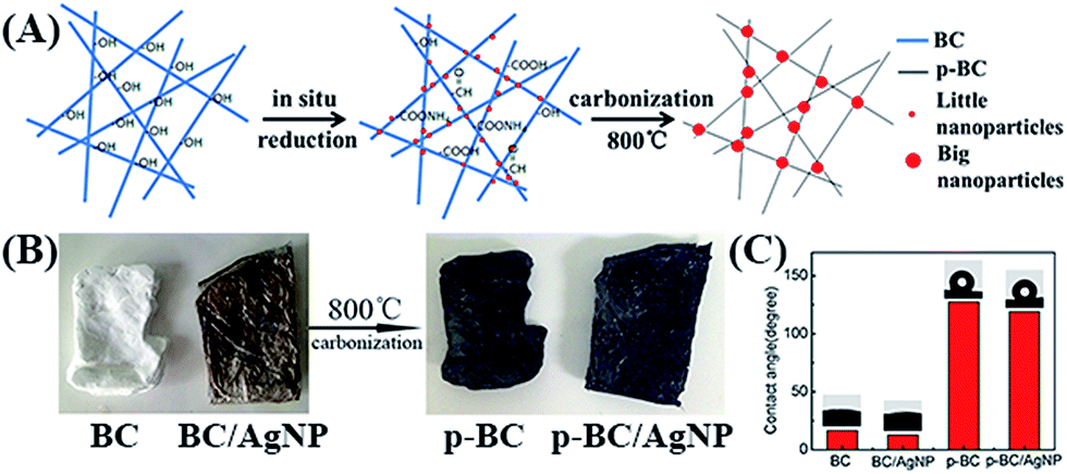 | ||
| Fig. 1 (A) Schematic representation of the fabrication process of AgNP deposited carbon aerogels by in situ reduction and carbonization; (B) the macrophotographs of BC, BC/AgNP, p-BC and p-BC/AgNP; (C) contact angles (CA) of BC, BC/AgNP, p-BC and p-BC/AgNP. | ||
The SEM images in Fig. 2 show the morphologies of the BC, BC/AgNP, p-BC and p-BC/AgNP. BC aerogels have a porous, interconnected, well-organized 3D network structure, which was formed through self-assembly in the bacterial culture process (Fig. 2A). The high-magnification SEM image indicates that the nanofibers with a diameter of 20–80 nm are highly interconnected with large numbers of junctions (Fig. 2B). After the carbonization treatment, the porous 3D structure of BC aerogels remained intact, but the diameters of the nanofibers decreased to 10–50 nm and some nanofibers were broken (Fig. 2E and F), due to the carbonization of the BC and evaporation of volatile species such as CO, CO2, methanol, and acetic acid during pyrolysis.43 Compared to pure nanofibers, BC loaded AgNPs possessed a 3D network with uniform silver nanoparticles evenly distributed. The granularity analysis (by Image-Pro Plus 6.0) results show that the particle diameters of more than 80% of the silver nanoparticles generated on the BC nanofibers were in the range of 20–40 nm (the inset image in Fig. 2C). However, most particle sizes in p-BC/AgNP were doubled, in which the average diameter of particles was about 80 nm (the inset image in Fig. 2G). This is mainly due to the heating process; the high energy meant that the nanoparticles aggregated in order to reduce the free enthalpy. However, due to the obstruction of the BC network, most nanoparticles are not fully aggregated. In addition, silver particles were not lost or shed from the nanofibers after pyrolysis. On the contrary, they were evenly distributed and anchored on the fibers, and the nanofibers still retained the original excellent 3D network structure (the image in Fig. 2G). EDS analysis of p-BC/AgNP further confirmed the existence of silver in BC (Fig. 2I). Although there was O element in the EDS spectrum, silver oxides did not occur in XRD.
 | ||
| Fig. 2 SEM images of (A and B) pure BC, (C and D) BC/AgNP, (E and F) p-BC, (G and H) p-BC/AgNP, (I) EDS analysis of p-BC/AgNP and (J) XRD of BC, BC/AgNP, p-BC and p-BC/AgNP. | ||
The XRD patterns of pure BC, BC/AgNP, p-BC and p-BC/AgNP are shown in Fig. 2J. The peaks at 2θ = 14.7°, 16.8° and 23.2° of the BC sample could be attributed to the characteristic diffraction peaks of the (110), (1![[1 with combining macron]](https://www.rsc.org/images/entities/char_0031_0304.gif) 0) and (200) lattice planes of the BC structure, respectively.44,45 The peak at 2θ = 26.2° of the p-BC sample could be attributed to the characteristic diffraction peak of the (002) lattice plane of graphite. This diffraction peak belongs to the typical characteristic peaks of carbon materials and reflects the degree of graphitization.25 In the patterns of BC/AgNP and p-BC/AgNP, there are some strong diffraction peaks located at 2θ = 38.1°, 44.3°, 64.4°, 77.5° and 81.5° corresponding to the (111), (200), (220), (311) and (222) planes of the Ag crystal, respectively.46,47 No characteristic diffraction peaks of Ag2O, AgO, AgCl or other silver oxides appear in the patterns. On the basis of the results of XRD characterization, it is indicated that BC/AgNP and p-BC/AgNP were successfully prepared by the previous methods. The average size of Ag particles in BC/AgNP and p-BC/AgNP is 26.24 and 74.76 nm, respectively, calculated from the (111) plane diffraction peak of the Ag crystal by the Scherrer equation,48 identifying with the SEM results.
0) and (200) lattice planes of the BC structure, respectively.44,45 The peak at 2θ = 26.2° of the p-BC sample could be attributed to the characteristic diffraction peak of the (002) lattice plane of graphite. This diffraction peak belongs to the typical characteristic peaks of carbon materials and reflects the degree of graphitization.25 In the patterns of BC/AgNP and p-BC/AgNP, there are some strong diffraction peaks located at 2θ = 38.1°, 44.3°, 64.4°, 77.5° and 81.5° corresponding to the (111), (200), (220), (311) and (222) planes of the Ag crystal, respectively.46,47 No characteristic diffraction peaks of Ag2O, AgO, AgCl or other silver oxides appear in the patterns. On the basis of the results of XRD characterization, it is indicated that BC/AgNP and p-BC/AgNP were successfully prepared by the previous methods. The average size of Ag particles in BC/AgNP and p-BC/AgNP is 26.24 and 74.76 nm, respectively, calculated from the (111) plane diffraction peak of the Ag crystal by the Scherrer equation,48 identifying with the SEM results.
It has been reported that many novel carbon aerogels had higher compressive properties than the traditional aerogels.27 Herein, the compressive stress–strain diagrams of p-BC and p-BC/AgNP are presented in Fig. 3A–D.
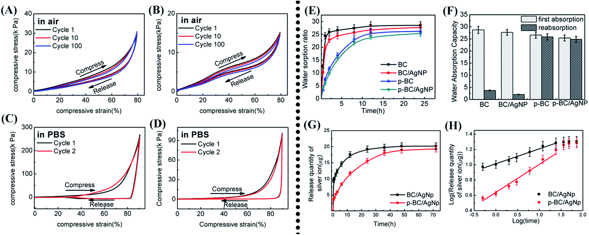 | ||
| Fig. 3 The compressive stress to reach 90% strain of p-BC (A) and p-BC/AgNP (B) at an engineering strain rate of 1 mm s−1 is plotted as a function of cycle number for 100 cycles in air; mechanical property measurements as PBS is pushed out and sucked in during 90% compressive strain for p-BC (C) and p-BC/AgNP (D); (E) water sorption ratio of BC, BC/AgNP, p-BC and p-BC/AgNP after 0.5 h, 1 h, 2 h, 4 h, 8 h, 12 h and 24 h of immersion in PBS (at 25 °C); (F) reabsorption rate of BC, BC/AgNP, p-BC and p-BC/AgNP. (G) Release quantity changes of silver ions in PBS buffer solution; (H) log(release quantity of Ag+) vs. log(times) curve. | ||
The p-BC aerogels can be compressed without crushing after being subjected to a maximum strain of 90% with a compressive strength of 30.7 kPa (Fig. 3A) after one compression cycle. When the compressive load was removed, the p-BC aerogel returned to its original shape because of its unique interconnecting 3D network structure and elimination of the hydrogen bonding interaction in BC after pyrolysis. In contrast, after the compressive load was removed, the BC aerogel did not return to its original shape because of the strongly interacting surface hydroxyl groups in the BC nanofibers.27 Similar to p-BC, the p-BC/AgNP aerogel almost completely recovered to its original volume when the compression was released (Fig. 3B) and showed a relatively lower elastic compressibility, although it had a much lower compressive strength of 15.18 kPa at 90% strain, most likely due to inhibition given by Ag nanoparticles deposited on the carbonized fibers. The p-BC and p-BC/AgNP aerogels were subjected to a fatigue cyclic compression test (ε = 90%) by undergoing 100 loading/unloading cycles (Fig. 3A and B). The unloading curves showed that the compressive stresses remained above zero until ε = 0, suggesting almost complete recovery of the compressed aerogels after the cycle tests. During 100 compression cycles in air to 90% strain, p-BC and p-BC/AgNP showed a relatively minor decrease in stress for the same strain level.
Remarkably, p-BC and p-BC/AgNP can also reversibly undergo large-strain deformation (up to 90%) in liquids (such as PBS) (Fig. 3C and D). When liquid was reabsorbed during aerogel elongation, the measured stress exhibits some dependence on strain rate for strains between 80% and 90%. Furthermore, the measured stress for an aerogel containing PBS dips below zero for high axial-direction strains. These below-zero measured stresses result from the inability of liquid absorption to keep up with the giant true strain rate. The aerogels therefore adhere to the platens of the stress–strain measuring apparatus, causing the expected compressive stress to temporarily switch to a tensile stress.49 It can be seen that p-BC/AgNP has a relatively good compression elasticity both in air and in liquid conditions, which will expand its scope of application in multiple environments.
3.2 Water holding capacity and rehydration ability
As is well known, BC and BC/AgNP are excellent hydrophilic materials. Even though the dried BC aerogel and the product of freeze-dried BC hydrogel are superhydrophilic, their contact angles with water were close to 10°. In contrast, the p-BC and p-BC/AgNP became hydrophobic after carbonization, and their contact angles were 127.5° and 119.1°, respectively (Fig. 1C).Although some materials are hydrophobic, they can still absorb a lot of water in a certain period of time due to having a porous structure.50 The water absorption capacities of BC, BC/AgNP, p-BC and p-BC/AgNP were monitored for a period of 24 h after the samples were immersed in PBS at 37 °C. As shown in Fig. 3E, after 1 hour’s immersion, the water sorption ratio was equal to 24.5, 19.8, 11.6 and 8.9, for BC, BC/AgNP, p-BC and p-BC/AgNP, respectively. As expected, the high porosity and large numbers of hydrophilic groups of aerogels led to a faster initial water sorption rate after just 1 h (for BC and BC/AgNP).51 Simultaneously, the water absorption ratio reached its maximum in a short time. After 24 h of immersion, all the samples were already significantly immersed in water and reached their maximum water sorption capacity. While in the process of water absorption, BC and BC/AgNP showed a high liquid water absorption rate which was probably due to their porous structures and large numbers of hydrogen bonds, and their maximum water absorption capacities were 28.7 and 27.7, respectively. p-BC and p-BC/AgNP showed a slow absorption rate and ultimately achieved relatively high water absorption capacities of 26.5 and 25.3, respectively. The slow water absorption rate of p-BC and p-BC/AgNP is attributed to the hydrophobic materials themselves, but with their unique three-dimensional network porous structures, water molecules will be pushed into the pores as time passes, and finally a relatively high water absorption capacity is achieved.
For water reabsorption capacity, the original three-dimensional network structure and the hydrogen bonding of hydrous BC would be destroyed after drying and deformation, so that the entire 3D-network structure after drying was destroyed and difficult to recover before absorption, therefore BC and BC/AgNP have a low rehydration rate (Fig. 3F), in which the rehydration rates were 3.72 and 2.06, respectively. On the contrary, p-BC and p-BC/AgNP have a very good compression elasticity after carbonization, both of them could substantially restore their original shape after drying and deformation, and have a high rehydration rate of 25.84 and 24.83, respectively, which showed a slight decrease compared to the first water absorption. After absorbing PBS again, the reabsorption capacities of p-BC/AgNP and p-BC were obviously higher than BC/AgNP and BC. Accordingly, carbonized p-BC and p-BC/AgNP have greater advantages in absorption and rehydration over BC and BC/AgNP, so p-BC/AgNP may have prospective applications in relevant medical fields, e.g. as a wound dressing and as a drug carrier for controlled release.
3.3 Release of silver in vitro
Silver nanoparticles have excellent antibacterial properties,12 but they may shed from materials and then enter the human body, which brings some hazards. The release quantity and rate of silver ions play an important role in their antibacterial effect and biological safety. The silver content of samples obtained by FAAS was 0.148 mg. Ag+ release profiles of BC/AgNP and p-BC/AgNP in PBS solution are depicted by kinetics curves and logarithmic fitting curves in Fig. 3G and H, respectively. The Ag+ release kinetics curves of the samples followed an logarithmic increase with the release time, which depends on the combination of silver with fibers, and the hydrophilicity or hydrophobicity of the matrix material. The release rate of Ag+ is proportional to tn at first, and after several hours, the release rate tends to equilibrium. Moreover, the value of n varied for different samples which is indicative of different transport mechanisms.52,53 The logarithmic fitting curve showed that the power law exponent for Ag+ release of p-BC/AgNP is 0.36 with R2 = 0.96, indicating a relatively high static correlation between the exponential power law and experimental measurements. The power law exponent for BC/AgNP is 0.19, with R2 of the fitting coefficient equal to 0.97, which may be attributed to greater hydrophilicity of BC, and that Ag+ can diffuse into the solution in a shorter time. Furthermore, after six hours, the amount of Ag+ released from BC/AgNP is a rapid process, while it requires double the time for p-BC/AgNP to release the same amount of Ag+, so the binding force of BC was worse than p-BC/AgNP. However, after 48 hours, the released amounts from BC/AgNP and p-BC/AgNP reached a stable, maximum value of 20.19 μg and 19.31 μg, respectively. Therefore, compared with BC/AgNP, p-BC/AgNP has a relatively stable and slow rate of release of silver ions, and also can reach the release rate levels of BC/AgNP. In summary, p-BC/AgNP has more stable and long-lasting anti-bacterial properties, and can be further used in various biological, medical and health fields etc.3.4 Antibacterial properties
The antibacterial activities of BC, BC/AgNP, p-BC and p-BC/AgNP against E. coli and S. aureus, which are all common infectious bacteria, were measured by a disk diffusion method after 24 h incubation at 37 °C (Fig. 4A–J). As expected, normal growth of E. coli and S. aureus was seen in the agar plate when nothing was introduced. The blank control with only filter paper demonstrated almost no antibacterial activity without the addition of Ag nanoparticles (Fig. 4A and F), and no inhibition zone was observed for pure BC and p-BC as a control (Fig. 4B, D, G and I). In contrast, the diameters of the inhibition zones from BC/AgNP and p-BC/AgNP on E. coli/S. aureus were 8.8/7.5 mm and 8.3/7.1 mm, respectively. | ||
| Fig. 4 The inhibition zone test for E. coli (A–E) and S. aureus (F–J) of blanks (A and F), BC (B and G), p-BC (C and H), BC/AgNP (D and I) and p-BC/AgNP (E and J). | ||
In order to acquire more information about the antibacterial activity of the samples for quantitative analysis, the antibacterial rates of four samples against E. coli and S. aureus were tested by a shake flask test method (Fig. 5). As with the previous results of the inhibition zone test, after 24 h of incubation, the ratios of bacterial (both E. coli and S. aureus) growth of the blank group, BC and p-BC groups were in the same order of magnitude, and the bactericidal rate (Br) of the BC/AgNP and p-BC/AgNP groups were 99.7% to 100% (Table 2). The results show that the antimicrobial effects of p-BC/AgNP were similar to the antimicrobial effects of BC/AgNP, and existing literature12 has shown that the antimicrobial effects of BC/AgNP could be comparable with commercial silver-containing dressings. At the same time, compared with traditional silver-dispersed carbon aerogel antibacterial materials,54 the preparation of bio-based carbon aerogels is more environmentally friendly and easy, in addition, the lower silver content of p-BC/AgNP also has a good antibacterial property. So, p-BC/AgNP can be used as an excellent antimicrobial material.
 | ||
| Fig. 5 Re-cultivated bacterial colonies after 24 h on agar: E. coli (A–E) and S. aureus (F–J) colonies are previously dissociated from blanks (A and F), BC (B and G), p-BC (C and H), BC/AgNP (D and I) and p-BC/AgNP (E and J). The S. aureus and E. coli bacteria concentration seeded on the samples is 104 CFU mL−1. | ||
| Negative control (×104) | BC (×104) | p-BC (×104) | BC/AgNP | p-BC/AgNP | |
|---|---|---|---|---|---|
| (A) | |||||
| Colonies after 24 h/CFU | 3.2 | 2.6 | 2.0 | 0 | 0 |
| Br | — | — | 100% | 100% | |
![[thin space (1/6-em)]](https://www.rsc.org/images/entities/char_2009.gif) |
|||||
| (B) | |||||
| Colonies after 24 h/CFU | 2.8 | 2.6 | 2.1 | 62 | 82 |
| Br | — | — | 99.8% | 99.7% | |
3.5 Cytocompatibility
The biocompatibility of BC, BC/AgNP, p-BC and p-BC/AgNP was evaluated after 1, 3 and 7 days of co-culturing with Bel-7402 cells. Cell proliferations and morphologic changes are shown in the ESI, Fig. S1.† Optical microscopy (OM) observations at 200× revealed that, after 24 h, the cells of the control group had a large amount of squamous Bel-7402 cells that were adherent, evenly distributed and formed a monolayer, and those of the experimental groups also had an excellent cell viability after 24 h.MTT assays were also performed to assess the cytotoxicity of the samples in this study (Fig. 6). Bel-7402 cells were cultured in medium (0.2 mg mL−1) with four samples to confirm the biocompatibility between the cells and experimental materials. After 24 h, though the BC sample exhibited slightly higher MTT absorbance results than the other three samples, there were no obvious differences between BC and BC/AgNP, p-BC or p-BC/AgNP, for which the cell survival rates were all more than 90% (compared to the control, p < 0.05), as determined by the MTT assay. With increasing culture time, the viability of cells of each sample shows a significant difference. The cell viabilities of BC and p-BC with no silver were both more than 90%, and the ratio of BC was higher, mainly due to the fact that BC is a natural product, and it can provide nutrition to cells. In contrast, the cell viabilities of BC/AgNP and p-BC/AgNP were significantly lower than those of BC and p-BC. What’s more, the MTT data of the BC/AgNP group was less than 90%, while for the p-BC/AgNP group it was more than 90%. With the passing of time, the cell activity of p-BC/AgNP was obviously higher than BC/AgNP. This proves that the cytotoxicity of p-BC/AgNP is markedly lower than BC/AgNP, and pyrolysis is absolutely necessary to make it more biocompatible.
 | ||
| Fig. 6 Time-dependent viability of Bel-7402 cells treated with BC, BC/AgNP, p-BC and p-BC/AgNP (0.2 mg mL−1). *p < 0.05 compared to control. | ||
DAPI fluorescence staining was also used to further assess the effect of the samples on the Bel-7402 cells. The Bel-7402 cells were cultured with sample suspensions (0.1 mg mL−1) for 7 days. Then, DAPI fluorescence staining was used to further confirm the relationship between the samples and cells. This method can allow the observation of cell apoptosis induced by the samples. Fig. 7 (second row) shows the DAPI-stained intact nuclei of Bel-7402 cells, as observed under a fluorescence optical microscope (FOM). This means that Bel-7402 cells cultured with different sample suspensions for 7 days did not induce apoptosis, and that these samples showed good biocompatibility. By comparing the results of the 7th day, in the merged DAPI-stained and bright-field images with those after culturing for 1 day, it can be seen that Bel-7402 cells were sparsely spread more or less everywhere with tiny dense cell domains. Most Bel-7402 cells appeared flat in shape, and there were no significant differences between the experimental groups. On the 4th day, the number of cells increased obviously, demonstrating that Bel-7402 cell proliferation had begun. After culturing for 7 days, more cells grew in all groups and then converged to form large areas in the vicinity of BC and BC/AgNP, as shown in Fig. 7. On the contrary, no cell groups appear around the p-BC and p-BC/AgNP, while large numbers of cells were relatively evenly distributed within the range of vision. This means that p-BC has a good biocompatibility, like other carbon materials.41,55
 | ||
| Fig. 7 Analysis of the effect of the four samples on apoptosis in Bel-7402 cells. After 7 days, the samples (0.1 mg mL−1) cultured in DMEM medium were collected and subjected to DAPI staining (blue), they showed intact nuclei (optical microscopy (OM): first row; fluorescence optical microscopy (FOM): second row). | ||
The SEM high-resolution images also show the structure and quantity of cells after cultivation with the samples. The cell morphologies of Bel-7402 on glass covered with BC and BC/AgNP, p-BC or p-BC/AgNP after 1 day of culture are shown in Fig. 8A–I. One could see that with all samples, Bel-7402 cells spread out and displayed a flattened phenotype and a relatively uniform distribution. Viewing the magnified shape and structure of the cells, images (Fig. 8B, D, F, H and J) show an intact cell structure and a spindle flat cell morphology. Simultaneously, the images also show the structure of the lamellipodium and filopodium. They were mainly made of actin filaments and helpful to maintain the cellular environment before cells began to migrate.42 In terms of the quantity of the cells, the cell number of the BC/AgNP group was significantly less than other groups, consistent with the results of the MTT assay. Therefore, it can be concluded that the p-BC/AgNP improved the stability and had no cytotoxicity.
 | ||
| Fig. 8 The morphology of Bel-7402 cells cultured with BC (C and D), BC/AgNP (E and F), p-BC (G and H) and p-BC/AgNP (I and J) (0.1 mg mL−1) and a control (A and B) for 24 h was observed using SEM; the normal morphology of cells and their large quantities show the good biocompatibility of samples. | ||
In vitro experimental results demonstrated p-BC/AgNP had a relatively low cytotoxicity due to the “green” nanofibers and the slow release of Ag+. This meant it can be used in biomedical fields.
4. Conclusions
In this study, AgNP-deposited bio-based nanostructure carbon aerogels derived from BC/AgNP have been successfully fabricated using a method of in situ reaction and carbonization. The unique 3D nano-network structure and pore distribution gave p-BC/AgNP good compression elasticity in air and liquid conditions. An excellent cellular network structure makes the hydrophobic p-BC/AgNP aerogel have a good reabsorption capacity; the rate reached 24.83, far greater than BC/AgNP. Moreover, compared to BC/AgNP, p-BC/AgNP has a slow release rate of silver, while its total amount of Ag release is close to that of BC/AgNP after 48 h, due to the stronger combination of Ag and carbon nanofibers, thus avoiding the damage caused by a high concentration of silver in the short-term. Also p-BC/AgNP has an excellent antibacterial effect on E. coli and S. aureus, in which the inhibition rate was over 99%. In addition, p-BC/AgNP shows a good biocompatibility with Bel-7402 cells, the cell survival rate is more than 90% and the cells can differentiate and grow normally after a period of time. Therefore, p-BC/AgNP carbon aerogel might be a desirable antibacterial material that could be used as a new kind of wound dressing, in implants, as a drug release carrier, and in other biomedical fields.Acknowledgements
This work is supported by the National Natural Science Foundation of China (No. 51273021, 51473019).Notes and references
- J. Li, X. Wang, Q. Huang, S. Gamboa and P. J. Sebastian, J. Power Sources, 2006, 158, 784–788 CrossRef CAS.
- C. Moreno-Castilla and F. J. Maldonado-Hódar, Carbon, 2005, 43, 455–465 CrossRef CAS.
- S. A. Waghuley, S. M. Yenorkar, S. S. Yawale and S. P. Yawale, Sens. Actuators, B, 2008, 128, 366–373 CrossRef CAS.
- S. Zhang, D. Wu, L. Wan, H. Tan and R. Fu, J. Appl. Polym. Sci., 2006, 102, 1030–1037 CrossRef CAS.
- A. C. Pierre and G. M. Pajonk, Chemistry of Aerogels and Their Applications, Chem. Rev., 2002, 102, 4243–4265 CrossRef CAS PubMed.
- A. M. ElKhatat and S. A. Al-Muhtaseb, Adv. Mater., 2011, 23, 2887–2903 CrossRef CAS PubMed.
- H. Bi, Z. Yin, X. Cao, X. Xie, C. Tan, X. Huang, B. Chen, F. Chen, Q. Yang, X. Bu, X. Lu, L. Sun and H. Zhang, Adv. Mater., 2013, 25, 5916–5921 CrossRef CAS PubMed.
- H. Bi, X. Huang, X. Wu, X. Cao, C. Tan, Z. Yin, X. Lu, L. Sun and H. Zhang, Small, 2014, 10, 3544–3550 CrossRef CAS PubMed.
- N. Shah, M. Ul-Islam, W. A. Khattak and J. K. Park, Carbohydr. Polym., 2013, 98, 1585–1598 CrossRef CAS PubMed.
- F. G. Torres, S. Commeaux and O. P. Troncoso, J. Funct. Biomater., 2012, 3, 864–878 CrossRef CAS PubMed.
- L. Huang, X. Chen, T. X. Nguyen, H. Tang, L. Zhang and G. Yang, J. Mater. Chem. B, 2013, 1, 2976 RSC.
- J. Wu, Y. Zheng, W. Song, J. Luan, X. Wen, Z. Wu, X. Chen, Q. Wang and S. Guo, Carbohydr. Polym., 2014, 102, 762–771 CrossRef CAS PubMed.
- D. Ciechanska, Fibres Text. East. Eur., 2004, 12, 69–72 CAS.
- K. Ghule, A. V. Ghule, B.-J. Chen and Y.-C. Ling, Green Chem., 2006, 8, 1034 RSC.
- J. Gutierrez, A. Tercjak, I. Algar, A. Retegi and I. Mondragon, J. Colloid Interface Sci., 2012, 377, 88–93 CrossRef CAS PubMed.
- M. Ul-Islam, T. Khan, W. A. Khattak and J. K. Park, Cellulose, 2012, 20, 589–596 CrossRef.
- A. R. Figueiredo, A. G. Figueiredo, N. H. Silva, A. Barros-Timmons, A. Almeida, A. J. Silvestre and C. S. Freire, Carbohydr. Polym., 2015, 123, 443–453 CrossRef CAS PubMed.
- R. J. Pinto, P. A. Marques, C. P. Neto, T. Trindade, S. Daina and P. Sadocco, Acta Biomater., 2009, 5, 2279–2289 CrossRef CAS PubMed.
- G. Yang, J. Xie, F. Hong, Z. Cao and X. Yang, Carbohydr. Polym., 2012, 87(1), 839–845 CrossRef CAS.
- X. Jin, L. Gao, E. Liu, F. Yu, X. Shu and H. Wang, J. Mech. Behav. Biomed. Mater., 2015, 50, 23–32 CrossRef CAS PubMed.
- K. Hareesh, A. V. Deore, S. S. Dahiwale, G. Sanjeev, D. Kanjilal, S. Ojha, N. A. Dhole, K. M. Kodam, V. N. Bhoraskar and S. D. Dhole, Radiat. Phys. Chem., 2015, 112, 97–103 CrossRef CAS.
- Y.-N. Chang, X.-M. Ou, G.-M. Zeng, J.-L. Gong, C.-H. Deng, Y. Jiang, J. Liang, G.-Q. Yuan, H.-Y. Liu and X. He, Appl. Surf. Sci., 2015, 343, 1–10 CrossRef CAS.
- C. Manoharan, G. Pavithra, S. Dhanapandian and P. Dhamodharan, Spectrochim. Acta, Part A, 2015, 149, 793–799 CrossRef CAS PubMed.
- W. Wang, K. Xiao, T. He and L. Zhu, J. Alloys Compd., 2015, 647, 1007–1012 CrossRef CAS.
- Z. Wu, C. Li, H. Liang, J. Chen and S. Yu, Angew. Chem., Int. Ed., 2013, 52, 2925–2929 CrossRef CAS PubMed.
- E. H. Falk Liebner, A. Potthast and T. Rosenau, Cellulosic Aerogels, ed. Y. Habibi and L. A. Lucia, 2012, pp. 51–102 Search PubMed.
- H. Liang, Q. Guan, Z. Zhu, L. Song, H. Yao, X. Lei and S. Yu, NPG Asia Mater., 2012, 4, e19 CrossRef.
- L. Chen, Z. Huang, H. Liang, W. Yao, Z. Yu and S. Yu, Energy Environ. Sci., 2013, 6, 3331 CAS.
- Z. Wang, Y. Ma, H. He, C. Pei and P. He, Appl. Surf. Sci., 2015, 332, 456–462 CrossRef CAS.
- B. Dai, X. Shao, Y. Ren, G. Wang, C. Pei and Y. Ma, Mater. Lett., 2012, 82, 188–190 CrossRef CAS.
- Y. Ren, S. Li, B. Dai and X. Huang, Appl. Surf. Sci., 2014, 311, 1–4 CrossRef CAS.
- C. Long, D. Qi, T. Wei, J. Yan, L. Jiang and Z. Fan, Adv. Funct. Mater., 2014, 24, 3953–3961 CrossRef CAS.
- W. Wang, Y. Sun, B. Liu, S. Wang and M. Cao, Carbon, 2015, 91, 56–65 CrossRef CAS.
- Y. Huang, T. Wang, M. Ji, J. Yang, C. Zhu and D. Sun, Mater. Lett., 2014, 128, 93–96 CrossRef CAS.
- T. Zhang, Y. Zheng, S. Liu, L. Yue, Y. Gao and Y. Yao, J. Electroanal. Chem., 2015, 750, 43–48 CrossRef CAS.
- S. Li, X. Yan, Z. Yang, Y. Yang, X. Liu and J. Zou, Appl. Surf. Sci., 2014, 292, 480–487 CrossRef CAS.
- B. Y. Wu, H. F. Wang, J. T. Chen and X. P. Yan, J. Am. Chem. Soc., 2011, 133, 686–688 CrossRef CAS PubMed.
- Z. Hu, X. Fan, H. Wang and J. Wang, Polymer, 2009, 50, 4175–4181 CrossRef CAS.
- S. P. Xin Zeng, J. Li, C. Wang, Y. Wen, H. Wu, C. Wang, C. Wu and M. Feng, Nanotechnology, 2011, 22, 1–13 Search PubMed.
- H. J. Wang, J. X. Fu and J. Y. Wang, Colloids Surf., B, 2009, 69, 109–115 CrossRef CAS PubMed.
- C. Karavasili, E. P. Amanatiadou, L. Sygellou, D. K. Giasafaki, T. A. Steriotis, G. C. Charalambopoulou, I. S. Vizirianakis and D. G. Fatouros, J. Mater. Chem. B, 2013, 1, 3167 RSC.
- M.-C. Hung, S.-Y. Yuan, S. I. Chang, J.-W. Liao, T.-H. Ko and C.-L. Cheng, Carbon, 2014, 68, 628–637 CrossRef CAS.
- Y. Wan, G. Zuo, F. Yu, Y. Huang, K. Ren and H. Luo, Surf. Coat. Technol., 2011, 205, 2938–2946 CrossRef CAS.
- H. Luo, J. Zhang, G. Xiong and Y. Wan, Carbohydr. Polym., 2014, 111, 722–728 CrossRef CAS PubMed.
- S. Y. Oh, D. I. Yoo, Y. Shin, H. C. Kim, H. Y. Kim, Y. S. Chung, W. H. Park and J. H. Youk, Carbohydr. Res., 2005, 340, 2376–2391 CrossRef CAS PubMed.
- S. Ristig, O. Prymak, K. Loza, M. Gocyla, W. Meyer-Zaika, M. Heggen, D. Raabe and M. Epple, J. Mater. Chem. B, 2015, 3, 4654–4662 RSC.
- A. Mandal, S. Sekar, N. Chandrasekaran, A. Mukherjee and T. P. Sastry, J. Mater. Chem. B, 2015, 3, 3032–3043 RSC.
- C. K. Sathiya and S. Akilandeswari, Spectrochim. Acta, Part A, 2014, 128, 337–341 CrossRef CAS PubMed.
- Y. Wu, N. Yi, L. Huang, T. Zhang, S. Fang, H. Chang, N. Li, J. Oh, J. A. Lee, M. Kozlov, A. C. Chipara, H. Terrones, P. Xiao, G. Long, Y. Huang, F. Zhang, L. Zhang, X. Lepro, C. Haines, M. D. Lima, N. P. Lopez, L. P. Rajukumar, A. L. Elias, S. Feng, S. J. Kim, N. T. Narayanan, P. M. Ajayan, M. Terrones, A. Aliev, P. Chu, Z. Zhang, R. H. Baughman and Y. Chen, Nat. Commun., 2015, 6, 6141 CrossRef CAS PubMed.
- S. T. Nguyen, J. Feng, S. K. Ng, J. P. W. Wong, V. B. C. Tan and H. M. Duong, Colloids Surf., A, 2014, 445, 128–134 CrossRef CAS.
- Y. Wang, S. Yadav, T. Heinlein, V. Konjik, H. Breitzke, G. Buntkowsky, J. J. Schneider and K. Zhang, RSC Adv., 2014, 4, 21553 RSC.
- D. R. Paul, Int. J. Pharm., 2011, 418, 13–17 CrossRef CAS PubMed.
- J. Siepmann and N. A. Peppas, Int. J. Pharm., 2011, 418, 6–12 CrossRef CAS PubMed.
- S. Zhang, R. Fu, D. Wu, W. Xu, Q. Ye and Z. Chen, Carbon, 2004, 42, 3209–3216 CrossRef CAS.
- S. I. Kim, B. B. Sahu, S. E. Kim, A. Ali, E. H. Choi and J. G. Han, J. Mater. Chem. B, 2015, 3, 3267–3278 RSC.
Footnotes |
| † Electronic supplementary information (ESI) available. See DOI: 10.1039/c5ra15485e |
| ‡ Ning Yan and Yabin Zhou have equally contributed as first authors. |
| This journal is © The Royal Society of Chemistry 2015 |