Synthesis, characterization and in vitro cytotoxicity of gold(III) dialkyl/diaryldithiocarbamato complexes

Muhammad Altafa, Anvarhusein A. Isab*b, Ján Vančoc, Zdeněk Dvořákc, Zdeněk Trávníček*c and Helen Stoeckli-Evansd
aCentre of Research Excellence in Nanotechnology (CENT), King Fahd University of Petroleum and Minerals, Dhahran 31261, Saudi Arabia. E-mail: aisab@kfupm.edu.sa; zdenek.travnicek@upol.cz
bDepartment of Chemistry, King Fahd University of Petroleum and Minerals, Dhahran 31261, Saudi Arabia
cRegional Centre of Advanced Technologies and Materials, Division of Biologically Active Complexes and Molecular Magnets, Faculty of Science, Palacký University, 17. listopadu 12, CZ-771 46 Olomouc, Czech Republic
dInstitute of Physics, University of Neuchâtel, rue Emile-Argand 11, CH-2000 Neuchâtel, Switzerland

Received 29th July 2015 , Accepted 15th September 2015

First published on 15th September 2015


Abstract

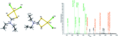
A series of six dialkyl/diaryldithiocarbamato (dtc) gold(III) complexes 1–6 of the composition [Au(Me2dtc)2]Cl (1), [Au(Me2dtc)Cl2] (2), [Au(Et2dtc)2]Cl (3), [Au(Et2dtc)Cl2] (4), [Au(Bn2dtc)2]Cl (5), [Au(Bn2dtc)Cl2] (6) (dtc = Dithiocarbamate, Me = Methyl, Et = Ethyl and Bn = Benzyl) was synthesised and characterized by elemental analysis, infrared and NMR spectroscopy, and single crystal X-ray analysis in the case of complexes 4 and 5. The anticancer potential of the complexes was evaluated against MCF7, A2780 and A2780R human cancer cell lines as well as against a healthy MRC5 cell line. The most promising data were obtained for complex 4. It showed similar cytotoxicity as cisplatin against A2780 cells (EC50 ≈ 9.5 μM vs. EC50 ≈ 10 μM), and significantly higher cytotoxicity as compared to cisplatin on A2780R and MCF7 cell lines, with EC50 ≈ 2.8 μM vs. EC50 ≈ 21 μM, and EC50 ≈ 2.2 μM vs. EC50 > 50 μM, respectively. The interactions of the representative complexes 3 and 4 with a mixture of physiological levels of L-cysteine (Cys) and reduced L-glutathione (GSH) were also studied.


1. Introduction

Platinum-based metallo-drugs have been used effectively and widely for chemotherapy of cancers patients for many decades.1–3 However, there are many concerns about the effectiveness of these drugs against different kinds of cancers.4 The use of platinum(II) anticancer drugs is limited due to severe side effects such as neurotoxicity, ototoxicity, anaemia, nausea and the acquired as well as intrinsic resistances exhibited by cancer cell lines.5,6 These undesirable and limiting factors of platinum(II) drugs are major impediments in their clinical use invariably against all kinds of cancers.7 These adverse effects and limits of platinum-based drugs have prompted scientists to search for new alternative anticancer agents connected with other metals such as gold-based complexes. To date, gold(III) complexes have been synthesized and evaluated for their potential anticancer activity with lower toxicity and fewer side effects against a wide panel of cancer cell lines.8–11

Gold(III) adopts a square-planar geometry and is expected to imitate the structural and electronic properties of platinum(II); These structural and electronic similarities of gold(III) complexes have attracted scientists and researchers attention for their outstanding cytotoxic activity.12–15 Recent studies have shown that several gold(III) complexes are highly cytotoxic against different tumor cells, including some cisplatin-resistant cancer cell lines.16–18

In this connection, a great interest in a new class of gold(III) complexes that contain dithiocarbamate ligands has been emerged as potential anticancer agents since 2000. Fregona and co-workers firstly prepared and characterized some novel gold(III) dithiocarbamate complexes containing N,N-dimethyldithiocarbamate and ethylsarcosinedithiocarbamate exhibiting the promising chemical and biological profile.19 Similarly, significant inhibitory effect of dibromo(N,N-dimethyldithiocarbamato)gold(III) in vivo was reported against growth of MDA-MB-231 breast tumor cells (BTC).20 Gold(I)-dithiocarbamato complexes, could inhibit the A549 (lung cancer), MCF7 (breast cancer), HeLa (cervical cancer) and chymotrypsin-like activity of purified 20S proteasome and 26S proteasome in human breast cancer MDA-MB-231 cells.21–23 Gold(I) complexes with sulfur ligands were employed clinically in the treatment of rheumatoid arthritis displayed some potency against various tumors but a greater potential is found in their analogues featuring a linear P–Au–S arrangement in which the thiolate ligand is derived from a biologically active thiol display high potency.24 F.K. Keter et al. synthesized new gold complexes that contain sulfur donor ligands (pyrazolyldithiocarbamate or 3,5-dimethylpyrazolyldithiocarbamate or indazolyldithiocarbamate) and found all complexes active against human cervical epithelial cancer cells (HeLa).25

This article follows our previous investigation of mixed ligand Au(I) complexes that contain dialkyl/diaryldithiocarbamate and phosphine ligands with excellent in vitro cytotoxicity against a panel of human cancer cells.22,26,27 Here, we have decided to use Au(III) acceptor and dialkyl/diaryldithiocarbamato and chlorido ligands. As we know that coordination and electronic features of the Au(I) center are different from Au(III) center. Herein, we report the synthesis, characterization, in vitro cytotoxicity of Au(III) complexes (Scheme 1) in normal human foetal lung cells (MRC5) and three different human cancer cell lines including breast (MCF7) and ovarian (A2780, A2780R), and cisplatin as control. The gold(III) complex 4 was also evaluated for its ability of interactions with L-cysteine (Cys) and reduced L-glutathione (GSH) by mass spectrometry.


image file: c5ra15123f-s1.tif
Scheme 1 Structural formulas of gold(III) complexes (1–6).

2. Experimental

2.1. Materials and methods

Sodium tetrachloroaurate(III) dihydrate, sodium dimethyldithiocarbamate monohydrate, sodium diethyldithiocarbamate trihydrate and sodium dibenzyldithiocarbamate hydrate were purchased from Sigma-Aldrich Co. St. Louis, Missouri, United States. All solvents including ethanol, and dichloromethane were purchased from Merck Darmstadt, Germany and used without further purification. All reactions were carried out under ambient conditions.

Elemental analyses of gold(III) complexes 1–6 were performed on Perkin-Elmer Series 11 (CHNS/O), Analyzer 2400. The solid state FTIR spectra of free ligands and their corresponding gold(I) complexes were recorded on a Perkin-Elmer FTIR 180 spectrophotometer or NICOLET 6700 FTIR using potassium bromide (KBr) pellets over the range 4000–400 cm−1. The electronic (UV/Vis) spectra were collected on a LAMBDA 40 spectrometer (Perkin-Elmer, USA).

1H and 13C NMR spectra were recorded on a LAMBDA 500 spectrophotometer operating at 500.01, 125.65, and 200.0 MHz, respectively; corresponding to a magnetic field of 11.74 T. Tetramethylsilane (TMS) was used as an internal standard for 1H and 13C. The 13C NMR spectra were obtained with 1H broadband decoupling, and the spectral conditions were: 32k data points, 0.967 s acquisition time, 1.00 s pulse delay and 45° pulse angle.

Electrospray-ionization (ESI) mass spectra of methanol solutions of the complexes were recorded on an LCQ Fleet Ion-Trap mass spectrometer using both the positive (ESI+) and negative (ESI−) mode of electrospray ionization (Thermo Scientific, USA).

2.2. Synthesis

2.2.1. Synthesis of [Au(Me2dtc)2]Cl (1). [Au(Me2dtc)2]Cl (1) was synthesized according to our previously reported procedure with minor modifications. NaAuCl4·2H2O, 0.5 mM (200 mg) and sodium dimethyldithiocarbamate dihydrate 1.0 mM (144 mg) in 20 mL of water and ethanol (1[thin space (1/6-em)]:[thin space (1/6-em)]1) were refluxed for 1 h. The complex appeared as bright yellow precipitate in the reaction medium. The precipitate was collected, washed with fresh distilled water (3 × 10 mL) and dried at room temperature for 24 h. The product appeared as bright yellow lumps. Yield: 93.09% (219.98 mg). FT-IR (KBr, νmax, cm−1): 2916 (w); 1576 (m), 1480 (s), 1243 (m), 1161 (w), 1093 (m), 961 (m), 925 (w), 555 (m), 473 (m). 1H NMR (500 MHz, DMSO-d6): δ = 3.37 (12H, s, 4 × CH3). 13C NMR (125.1 MHz, DMSO-d6): δ = 35.15 (CH3), 186.86 (NC[double bond, length as m-dash]S). Anal. calc. for C6H12ClN2S4Au (472.85): C, 15.24; H, 2.56; N, 5.92; S, 27.12%. Found: C, 15.31; H, 2.58; N, 5.95; S, 27.15%.
2.2.2. Synthesis of [Au(Me2dtc)Cl2] (2). [Au(Me2dtc)Cl2] (2) was synthesized according to the procedure as mentioned above for complex 1 with minor modifications.26–29 NaAuCl4·2H2O, 0.5 mM (200 mg) and sodium dimethyldithiocarbamate dihydrate 0.5 mM (72 mg) in 20 mL of water and ethanol (1[thin space (1/6-em)]:[thin space (1/6-em)]1) were refluxed for 1 h. The complex appeared as yellow precipitate in the reaction medium. The precipitate was collected, washed with fresh distilled water (3 × 10 mL) and dried at RT for 24 h. The product appeared as yellow crystalline powder. Yield: 86.87% (166.87 mg). FT-IR (KBr, νmax, cm−1): 2923 (w); 1571 (s), 1481 (m), 1241 (m), 1160 (w), 1095 (m), 966 (m), 920 (w), 550 (m), 472 (m). 1H NMR (500 MHz, DMSO-d6): δ = 3.35 (6H, s, 2 × CH3). 13C NMR (125.1 MHz, DMSO-d6): δ = 34.15 (CH3), 193.89 (NC[double bond, length as m-dash]S). Anal. calc. for C3H6Cl2NS2Au (388.10): C, 9.28; H, 1.56; N, 3.61; S, 16.52%. Found: C, 9.17; H, 1.58; N, 3.59; S, 16.55%.
2.2.3. Synthesis of [Au(Et2dtc)2]Cl (3). [Au(Et2dtc)2]Cl (3) was synthesized according to the procedure as mentioned above for complex 1 with minor modifications. NaAuCl4·2H2O, 0.5 mM (200 mg) and sodium diethyldithiocarbamate trihydrate 1.0 mM (226 mg) in 20 mL of water and ethanol (1[thin space (1/6-em)]:[thin space (1/6-em)]1) were refluxed for 1 h. The complex appeared as light brown lumps in the reaction medium. The solid product was collected, washed with fresh distilled water (3 × 10 mL) and dried at RT for 24 h. The product appeared as light brown lumps. Yield: 85.55% (226.13 mg). FT-IR (KBr, νmax, cm−1): 2977 (w), 2930 (w), 1550 (m), 1483 (s), 1285 (m), 1158 (w), 1089 (m), 998 (m), 907 (w), 550 (s), 480 (m). 1H NMR (500 MHz, DMSO-d6): δ = 1.29 (12H, d, 4 × CH3), 3.75 (8H, d, 4 × CH2). 13C NMR (125.1 MHz, DMSO-d6): δ = 12.15 (CH3), 46.60 (CH2), 193.87 (NC[double bond, length as m-dash]S). Anal. calc. for C10H20ClN2S4Au (528.96): C, 22.71; H, 3.81; N, 5.30; S, 24.25%. Found: C, 22.63; H, 3.85; N, 5.26; S, 24.33%.
2.2.4. Synthesis of [Au(Et2dtc)Cl2] (4). [Au(Et2dtc)Cl2] (4) was synthesized according to our previously reported procedure with minor modifications.26–29 NaAuCl4·2H2O, 0.5 mM (200 mg) and sodium diethyldithiocarbamate trihydrate 0.5 mM (113 mg) in 20 mL of water and ethanol (1[thin space (1/6-em)]:[thin space (1/6-em)]1) were refluxed for 1 h. The complex appeared as light yellow precipitate in the reaction medium. The precipitate was collected, washed with fresh distilled water (3 × 10 mL) and dried at RT for 24 h. The product appeared as light yellow semi crystalline cluster. Yield: 90.09% (187.27 mg). FT-IR (KBr, νmax, cm−1): 2970 (w), 2921 (w), 1556 (m), 1482 (s), 1280 (m), 1155 (w), 1078 (m), 991 (m), 909 (w), 550 (s), 481 (m). 1H NMR (500 MHz, DMSO-d6): δ = 1.28 (6H, d, 2 × CH3), 3.75 (4H, d, 2 × CH2). 13C NMR (125.1 MHz, DMSO-d6): δ = 12.13 (CH3), 46.62 (CH2), 193.88 (NC[double bond, length as m-dash]S). Anal. calc. for C5H10Cl2NS2Au (416.15): C, 14.43; H, 2.42; N, 3.37; S, 15.41%. Found: C, 14.33; H, 2.47; N, 3.31; S, 15.47%.
2.2.5. Synthesis of [Au(Bn2dtc)2]Cl (5). [Au(Bn2dtc)2]Cl (5) was synthesized according to the procedure as mentioned above for complex 3 with minor modifications. NaAuCl4·2H2O, 0.5 mM (200 mg) and sodium dibenzyldithiocarbamate hydrate 1.0 mM (296 mg) in 20 mL of methanol and water (9[thin space (1/6-em)]:[thin space (1/6-em)]1) were refluxed for 1 h. The complex appeared as dark brown precipitate in the reaction medium. The precipitate was collected, washed with fresh distilled water (3 × 10 mL) and dried at RT for 24 h. The product appeared as dark brown lumps. Yield: 88.51% (343.9 mg). FT-IR (KBr, νmax, cm−1): 3035 (w), 2993, 2916 (m), 1534 (s), 1491 (s), 1352 (m), 1227 (s), 1126 (m), 1071 (m), 977 (s), 800 (m), 533 (s), 468 (m). 1H NMR (500 MHz, DMSO-d6): δ = 5.03 (8H, s, 4 × CH2), 7.37 (20H, m, 4 × C6H5). 13C NMR (125.1 MHz, DMSO-d6): δ = 55.08 (CH2), 132.55 (C6H5), 199.18 (NC[double bond, length as m-dash]S). Anal. calc. for C30H28ClN2S4Au (777.23): C, 46.36; H, 3.63; N, 3.60; S, 16.50%. Found: C, 46.30; H, 3.64; N, 3.68; S, 16.59%.
2.2.6. Synthesis of [Au(Bn2dtc)Cl2] (6). [Au(Bn2dtc)Cl2] (6) was synthesized according to the procedure as mentioned above for complex 4 with minor modifications. NaAuCl4·2H2O, 0.5 mM (200 mg) and sodium dibenzyldithiocarbamate hydrate 0.5 mM (148 mg) in 20 mL of methanol and water (9[thin space (1/6-em)]:[thin space (1/6-em)]1) were refluxed for 1 h. The complex appeared as yellowish green precipitate in the reaction medium. The precipitate was collected, washed with fresh distilled water (3 × 10 mL) and dried at RT for 24 h. The product appeared as yellowish green powder. Yield: 95.01% (256.6 mg). FT-IR (KBr, νmax, cm−1): 3034 (w), 2990, 2923 (m), 1540 (s), 1495 (s), 1355 (m), 1229 (s), 1129 (m), 1079 (m), 979 (s), 808 (m), 537 (s), 458 (m). 1H NMR (500 MHz, DMSO-d6): δ = 5.01 (4H, s, 2 × CH2), 7.36 (10H, m, 2 × C6H5). 13C NMR (125.1 MHz, DMSO-d6): δ = 55.09 (CH2), 132.54 (C6H5), 199.16 (NC[double bond, length as m-dash]S). Anal. calc. for C15H14Cl2NS2Au (540.28): C, 33.35; H, 2.61; N, 2.59; S, 11.87%. Found: C, 33.43; H, 2.70; N, 2.53; S, 11.91%.

2.3. X-ray structure determination

Suitable crystals of complex 4 and 5 were obtained as orange rods and yellow plates by recrystallization of final products in mixture of chloroform and methanol (1[thin space (1/6-em)]:[thin space (1/6-em)]1) solution. The intensity data were collected at 173 K (−100 °C) on a Stoe Mark II-Image Plate Diffraction System30 equipped with a two-circle goniometer and using MoKα graphite monochromated radiation (λ = 0.71073 Å). The structure was solved by direct methods with SHELXS-2014.31 The refinement and all further calculations were carried out with SHELXL-2014.32 For complex 4, the C-bound H-atoms were included in calculated positions and treated as riding atoms: C–H = 0.99 and 0.98 Å for CH2, and CH3, respectively, with Uiso(H) = 1.5Ueq(C-methyl) and = 1.2Ueq(C) for other H-atoms. For complex 5, the C-bound H-atoms were included in calculated positions and treated as riding atoms: C–H = 0.95–0.99 Å with Uiso(H) = 1.2Ueq(C). The non-H atoms were refined anisotropically, using weighted full-matrix least-squares on F2 Table 1. A semi-empirical absorption correction was applied using the MULscanABS routine in PLATON.33 The summary of crystallographic data and structure refinement of complexes 4 and 5 is given in Table 1.

2.4. Cell cultures and in vitro cytotoxicity

Human breast adenocarcinoma cells (MCF7; ECACC no. 86012803), human ovarian carcinoma cells (A2780; ECACC no. 93112519), cisplatin-resistant human ovarian carcinoma cells (A2780R; ECACC no. 93112517) and human foetal lung cells (MRC5; ECACC no. 84101801) were purchased from European Collection of Cell Cultures (ECACC). The cells were cultured according to the ECACC instructions and they were maintained at 37 °C and 5% CO2 in a humidified incubator. The cells were treated with the tested complexes, and cisplatin, respectively, for 24 h, using multiwell culture plates of 96 wells. In parallel, the cells were treated with vehicle (DMF; 0.1%, v/v) and Triton X-100 (1%, v/v) to assess the minimal (i.e. the positive control), and maximal (i.e. the negative control) cell damage, respectively. The MTT assay was performed spectrophotometrically at 540 nm (TECAN, Schoeller Instruments LLC).

2.5. Mass spectrometry study of interactions of complexes 3 and 4 with L-cysteine and reduced glutathione

The interaction experiments between the selected representative complexes 3 and 4, and the mixture of physiological levels of L-cysteine (Cys) and reduced L-glutathione (GSH) were performed on a Thermo Scientific LTQ Fleet Ion-Trap mass spectrometer, using the positive ionization mode. The reaction system contained the physiological concentrations of L-cysteine (290 μM) and reduced glutathione (6 μM)54 and the tested complex (10 μM) in the methanol[thin space (1/6-em)]:[thin space (1/6-em)]water (1[thin space (1/6-em)]:[thin space (1/6-em)]1, v/v) mixture. The reference system was comprised of the solution of complex (10 μM) in the methanol[thin space (1/6-em)]:[thin space (1/6-em)]water (1[thin space (1/6-em)]:[thin space (1/6-em)]1, v/v) mixture. The flow injection analysis (FIA) method was used to introduce the reaction system (10 μL spikes) into the mass spectrometer, while the mixture of acetonitrile[thin space (1/6-em)]:[thin space (1/6-em)]water (9[thin space (1/6-em)]:[thin space (1/6-em)]1, v/v) was used as a mobile phase. The electro-spray ionization (ESI) source was set up as follows: source voltage was 4.9 kV, the vaporizer temperature was 160 °C, the capillary temperature was 275 °C, the sheath gas flow rate was 20 L min−1, and auxiliary gas flow rate was 5 L min−1. The system was calibrated according to the manufacturer specifications and no further tuning was needed.

3. Results and discussion

3.1. Characterization by FT-IR

Gold(III) dithiocarbamate complexes 1–6 were identified via the presence of certain absorbance peaks primarily ν(C–N) and ν(C–S). The infrared region 1480–1550 cm−1 is mostly related with the R2N–CSS ‘thioureide’ band in the IR spectra of dithiocarbamate complexes which defines the carbon–nitrogen bond order between a single bond at 1250–1350 cm−1 and a double bond at 1640–1690 cm−1.34

The thioureide band, ν(C–N) was detected at 1481, 1480, 1482, 1483, 1495 and 1491 cm−1 in complexes 1–6 respectively. Such frequency absorption bands lie in between those associated with single C–N and double C[double bond, length as m-dash]N bonds, hence the partial double bond character of ‘thioureide’ bond was confirmed for all gold(I) complexes.35 The presence of strong absorption band in the range of 1480–1550 in FTIR spectra clearly indicates the formation of dithiocarbamato gold(III) complexes.36,37 Similarly, C[double bond, length as m-dash]S stretching with medium intensity around 1070 and 970 cm−1 for complexes 1–6 is an additional evidence of the formation of target molecules. The common bands for sp3, sp2 and sp hybridized C–H stretches were observed within 2995–2915 cm−1 and above 3000 cm−1 respectively. These absorption bands are comparable to free sodium salt of diethyldithiocarbamate ligands.38

Table 1 Crystal data and structure refinement details for complexes 4 and 5
Complex 4 5
CCDC deposit no. 1403956 1403957
Chemical formula [C5H10NS2AuCl2] [(C15H14NS2)2Au]+·(AuCl2)
Molecular weight 416.13 1009.62
Crystal system, space group Monoclinic, P21/n Monoclinic, P21/c
Temperature (K) 173 173
a, b, c (Å) 17.2433 (8), 7.0535 (3), 17.2503 (10) 10.0213 (12), 18.6050 (17), 9.1040 (12)
β (°) 97.718 (4) 109.36 (1)
V3) 2079.07 (18) 1601.4 (3)
Z 8 2
Radiation type Mo Kα Mo Kα
μ (mm−1) 15.01 9.60
Crystal size (mm) 0.40 × 0.35 × 0.30 0.45 × 0.17 × 0.10
Tmin, Tmax 0.454, 1.000 0.401, 1.000
No. of measured, independent and observed [I > 2σ(I)] reflections 24[thin space (1/6-em)]350, 3929, 3519 10[thin space (1/6-em)]925, 3028, 1874
Rint 0.084 0.042
(sin[thin space (1/6-em)]θ/λ)max−1) 0.609 0.610
R[F2 > 2σ(F2)], wR(F2), S 0.022, 0.048, 0.92 0.019, 0.040, 0.77
No. of reflections 3929 3028
No. of parameters 205 185
Largest diff. peak and hole (e Å−3) Δρmax, Δρmin (e Å−3) 1.25, −1.94 1.03, −0.74


3.2. Characterization by NMR

The 1H NMR chemical shifts of all six complexes are given in the Section 2.2. Synthesis. Small downfield and upfield shifts for proton(s) of the coordinated dimethyl dithiocarbamate, diethyl dithiocarbamate and dibenzyldithiocarbamate have been seen in gold(III) complexes 1–6 in comparison to free dialkyl/diaryldithiocarbamate ligands.22,26,29,39–43

The 13C NMR spectra of complexes 1, 2 and 3–6 showed two and three resonances respectively (given in synthesis part of Experimental Section). There is an up-field chemical shift of NC[double bond, length as m-dash]S carbon of coordinated dialkyldithiocarbamate with respect to free dialkyl/diaryldithiocarbamate ligands for all gold(III) complexes. The 13C chemical shifts of NC[double bond, length as m-dash]S carbon of bonded dimethyl thiocarbamate, diethyl thiocarbamate and dibenzyl thiocarbamate are observed in the range 186–200 ppm in our synthesized complexes 1–6.22,26,29,39–43

3.3. X-ray crystallography

X-ray structure and crystal packing of complex 4 is shown in Fig. 1 and 2. The complex 5 crystallized with two independent molecules per asymmetric unit. The crystal structure contains two independent molecules in which Au(III) centre is tetracoordinated and surrounded by two chlorine atoms and both sulfur atoms of the diethyldithiocarbamate ligand in a cis position. The geometry around gold(III) ions is square planar (Cl–Au–Cl angle is 94.17(10) and 94.36(11)° for molecule 1 and molecule 2 respectively), although somehow distorted as a consequence of the restricting chelate angle of the dithiocarbamate ligand (S–Au–S 75.87(10) and 75.57(11)° for molecule 1, and molecule 2, respectively). This angle lies within the range found in a total of 76 entries in 41 crystal structures containing the fragment [Au(μ-S2CN)], which vary from 65.71(5) to 78.6(3)° (mean value of 75.87°).44,45
image file: c5ra15123f-f1.tif
Fig. 1 A view of the two crystallographically independent molecules of complex 4, together with the atom labelling scheme. The displacement ellipsoids are drawn at the 50% probability level.

image file: c5ra15123f-f2.tif
Fig. 2 The crystal packing of complex 4, as viewed along the a axis.

The Au1–S2 and Au2–S3 bond distances 2.294(2) and 2.293(2) Å are almost same in molecule 1 and molecule 2 respectively. Similarly Au1–S1 and Au2–S4 bond distances 2.310(3) and 2.312(3) Å are also very close in molecule 1, and molecule 2, respectively. The Au–Cl average bond lengths of 2.324(3) and 2.326(3) Å for molecule 1, and molecule 2, respectively, is slightly long for a gold(III) complex. This is due to the strong trans influence of the S donor ligands and is of the same order as the Au–Cl bond distances found in the related dithiocarbamate Au(III) complexes or in other complexes containing the fragment [AuCl2(μ-SCS)].46–48

The molecular structure of complex 5 is shown in Fig. 3 and 4. In ionic complex 5, one gold atom coordinates with two structurally equivalent dibenzyldithiocarmate ligand molecules in the S,S′-bidentate mode, thus forming the centrosymmetric [Au(Bn2dtc)2]+ cation. The second Au(I) atom in the centrosymmetric [AuCl2], is surrounded by two chloride anions. In one coordination sphere, gold(III) forms square-planar tetragonal chromophores: [AuS4] (low spin intra-orbital dsp2 hybrid state of the central gold(III) atom). Whereas, in second coordination sphere, gold(I) attains a linear geometry (Table 2).


image file: c5ra15123f-f3.tif
Fig. 3 A view of the molecular structure of complex 5, together with the atom labelling scheme. The displacement ellipsoids are drawn at the 50% probability level. The unlabelled atoms are generated by inversion symmetry [symmetry codes for Au1: −x, −y, −z, and for Au2: −x+1, −y, −z].

image file: c5ra15123f-f4.tif
Fig. 4 The crystal packing of complex 5, as viewed along the c axis.
Table 2 Selected bond lengths and bond angles of complexes 4 and 5
Bond lengths (Å) Bond angles (°)
Complex 4
Molecule 1
Au1–S2 2.294(2) S2–Au1–S1 75.87(10)
Au1–S1 2.310(3) S2–Au1–Cl1 172.47(11)
Au1–Cl1 2.323(3) S1–Au1–Cl1 96.71(10)
Au1–Cl2 2.325(3) S2–Au1–Cl2 93.25(11)
    S1–Au1–Cl2 169.11(12)
    Cl1–Au1–Cl2 94.17(10)
Molecule 2
Au2–S3 2.293(2) S3–Au2–S4 75.57(11)
Au2–S4 2.312(3) S3–Au2–Cl4 93.52(11)
Au2–Cl4 2.326(3) S4–Au2–Cl4 169.07(12)
Au2–Cl3 2.326(3) S3–Au2–Cl3 171.81(11)
    S4–Au2–Cl3 96.57(10)
    Cl4–Au2–Cl3 94.36(11)
[thin space (1/6-em)]
Complex 5
Au1–S2 2.3343(10) S2–Au1–S2i 180.0
Au1–S2i 2.3343(10) S2–Au1–S1i 104.56(4)
Au1–S1i 2.3396(10) S2i–Au1–S1i 75.44(4)
Au1–S1 2.3396(10) S2–Au1–S1 75.44(4)


The S1–Au1–S2, and S2–Au1–S1i bond angles in this centrosymmetric [Au(Bn2dtc)2]+ cation, are 75.44(4), and 104.56(4)° respectively. The Au1–S1 and Au1–S2 bond distances are 2.3396(10) and 2.3343(10) Å respectively. The Au–S bond length in 2 is nearly identical with the mean value of 2.3038 Å found in Au(III) complexes that contain dithiocarbamate ligands, in which the Au–S bond distances vary from 2.287(3) Å in [AuCl2{μ-S2CN(EtOH)2}] to 2.319(2) Å in [AuBr2{μ-S2CN(Me)(CH2CO2Et)}]. It also compares well with those observed in the [AuCl2(μ-SCS)] complexes.49–52

3.4. In vitro cytotoxicity

Normal human foetal lung cells (MRC5) and three different human cancer cell lines including breast (MCF7) and ovarian (A2780, A2780R) were used as experimental models for evaluation of in vitro cytotoxicity of the prepared complexes 1–6, see Table 3. The cells were incubated with the studied complexes for 24 h and the viability was assessed by a conventional MTT test. The cytotoxicity of complexes 1, 2, 5 and 6 was lower or comparable to that by cisplatin, regardless the particular cell line. Complex 3 displayed similar toxicity as cisplatin in both ovarian cancer cells A2780 and their cisplatin-resistant variant A2780R. On the other hand, complex 3 displayed in order of magnitude higher cytotoxicity against breast cancer cells MCF7 as compared to cisplatin (EC50 ≈ 3.9 μM vs. EC50 > 50 μM), but still keeping low toxicity against normal MRC5 cells (EC50 ≈ 19.7 μM). The most promising data were obtained for complex 4. While it showed similar cytotoxicity as cisplatin against A2780 cells (EC50 ≈ 9.5 μM vs. EC50 ≈ 10.0 μM), its cytotoxicity in cisplatin-resistant A2780R cells was drastically increased as compared to cisplatin (EC50 ≈ 2.8 μM vs. EC50 ≈ 21.0 μM). Moreover, complex 4 was much more toxic against breast cancer cells MCF7, as compared to cisplatin (EC50 ≈ 2.2 μM vs. EC50 > 50 μM), showing moderate toxicity against healthy MRC5 cells.
Table 3 In vitro cytotoxicity of the complexes 1–6 and cisplatin given as EC50 ± S.D. in μM
Complex A2780 A2780R MCF7 MRC5
1 16.8 ± 2,0 27.5 ± 2.3 19.4 ± 3.6 28.0 ± 1.4
2 7.3 ± 0.8 40.5 ± 4.2 21.5 ± 3.1 27.1 ± 2.9
3 6.3 ± 2.1 29.8 ± 5.8 3.9 ± 2.3 19.7 ± 6.4
4 9.5 ± 0.3 2.8 ± 0.1 2.2 ± 0.9 20.3 ± 0.8
5 27.1 ± 5.0 35.9 ± 2.3 >50 >50
6 25.1 ± 1.8 41.9 ± 1.2 >50 >50
Cisplatin 10.0 ± 1.6 21.0 ± 2.1 >50 >50


3.5. Interactions of complexes 3 and 4 with sulphur-containing biomolecules analysed by mass spectrometry

It is a known fact, that gold(III) complexes bind to sulfanyl-containing substances, such as amino acid L-cysteine (Cys) or small proteins, such as reduced L-glutathione (GSH), and high molecular weight proteins (e.g. albumin or globulins53), in the biologically relevant environments, such as blood or serum. The complexes formed by these interactions can be considered as transport intermediates and/or detoxication end-products in some cases. These processes are connected with changes in oxidation states and exchanges of ligands of the original complexes. In this work, we strived to uncover interaction potential of the selected complexes 3 and 4 (applied in the concentration of 10 μM, roughly corresponding to the determined EC50 values), in biologically relevant conditions using a mixture of Cys (at the 290 μM concentration) and GSH (at the 6 μM concentration).54 The complexes were chosen in connection with the fact that they contain the diethyldithiocarbamate moiety, however, on the other side each of them represents structurally different type of the complexes presented in this study, and moreover, they are also the most cytotoxic ones against the MCF7 cell line.

Based on the results of the mass spectrometry experiments, we may confirm that both the complexes 3 and 4 interact with the applied sulphur-containing substances in time-dependent manner by the ligand-exchange mechanism connected with the substitution of one diethyldithiocarbamato ligand for one cysteine and/or cystine (CysCys) molecule(s). (Note: CysCys represents an oxidation product of L-cysteine). This mechanism may be supported by the presence of the peaks at 465.03 m/z (for 4), 613.11 m/z (for 3 and 4), 704.90 m/z (for 4), 852.86 m/z (for 3 and 4), and 874.83 m/z (for 3 and 4), corresponding to the species of [Au(Et2dtc) + Cys]+, [Au(Et2dtc)2 + Cys]+, [Au(Et2dtc) + Cys + CysCys]+, [Au(Et2dtc)2 + Cys + CysCys]+, and [Au(Et2dtc)2 + Cys + CysCys + Na]+ species, respectively (see Fig. 5, and Fig. S1 in ESI).


image file: c5ra15123f-f5.tif
Fig. 5 The results of mass spectrometry study of interactions of complex 4 with physiological levels of L-cysteine (Cys) and reduced L-glutathione (GSH). Cystine (Cys–Cys) represents an oxidation product of cysteine. The peaks of Cys and GSH and their oxidized products are highlighted in green colour, while the peaks corresponding to the interaction adducts of complex 4 are given in orange colour.

Moreover, the performed experiments were also useful to reveal the solvolytic behaviour of the complexes, because the peaks at 116.10 m/z and 493.08 m/z revealed in the ESI + mass spectra of complexes 3 and 4, and they may be associated with the formation of the identical species of [Et2N–C[double bond, length as m-dash]S]+, and [Au(Et2dtc)2]+, respectively, see Fig. 6. In addition to that, the peak at 267.03 m/z was observed in the ESI-mass spectra of 3 and 4, and it may be related to the presence of the [AuCl2] species. These findings motivated us to use mass spectrometry to thorough study of all the complexes with the aim to find out the stability of them in methanol. Surprisingly, we have found that the dissolution of complexes 1, 3 and 5, having the general formula [Au(dtc)2]Cl, as well as complexes 2, 4 and 6, with the composition of [Au(dtc)Cl2], in methanol led to the formation of the [Au(dtc)2]+ and [AuCl2] species for 1, 3 and 5, and the [Au(dtc)2]+, [AuCl2] and [AuCl4] species for 2, 4 and 6, as demonstrated in Fig. 7 for the representative complexes 1 and 2. The reduction of Au(III) to Au(I), as seen from the [AuCl2] species depicted in Fig. 7, is compensated by oxidation of Me2dtc to [Me2N–CS–S–S–SC–NMe2].


image file: c5ra15123f-f6.tif
Fig. 6 The comparison of ESI + mass spectra of complexes 3 (left) and 4 (right) measured in methanol solutions, showing the presence of the peaks corresponding to the same cationic species [Et2N–C[double bond, length as m-dash]S]+ and [Au(Et2dtc)2]+.

image file: c5ra15123f-f7.tif
Fig. 7 The comparison of ESI-mass spectra of complexes 1 (left) and 2 (right) in methanol solutions, showing the presence of [AuCl2] and [AuCl4] species as well as species responding to oxidation of the dimethyldithiocarbamate moiety. The insets show the isotopic distributions for the corresponding species.

The above described findings may logically lead to the conclusion that mainly the [Au(dtc)2]+ species are responsible for interactions with sulphur-containing biomolecules (i.e. with Cys and GSH), and moreover, that these cationic species are also responsible for biological response, i.e. for the cytotoxicity of the complexes 1–6. The above said may be supported by cytotoxicity data given in Table 3, because the EC50 values between 1 and 2, between 3 and 4, and between 5 and 6, are close to each other, except for the complex 3 in the case of the A2780R cell line, however, this discrepancy may be explained by a possible biological impact of other species presented in the medium used for biological testing. Overall, it may be emphasized that the [Au(dtc)Cl2] complexes (2, 4 and 6) decompose most probably to two dominant species, i.e. to [Au(dtc)2]+ and [AuCl4]. This may be also supported by solution UV/Vis spectra of complexes 3 and 4 (measured in MeOH and DMF), (see Fig. S2 and S3 in ESI), from which can be seen that the positions of maxima of the d–d transitions for complex 3 and 4 are nearly identical both in the MeOH and DMF solution.

4. Conclusions

The synthesis and characterization of dialky/diaryldithiocarbamato gold(III) complexes 1–6 are reported. The in vitro cytotoxicity of the complexes has been evaluated against MCF7, A2780 and cisplatin-resistant A2780R human cancer cell lines as well as against healthy MRC5 cell line. The best cytotoxicity has been achieved for complex 4 against A2780R and MCF7, with the EC50 values of 2.8(1), and 2.2(9) μM, respectively, and with moderate toxicity on healthy MRC5 cell line (EC50 = 20.3(8) μM). Moreover, the results of interaction experiments of the representative complexes 3 and 4 with the mixture of physiological levels of L-cysteine (Cys) and reduced L-glutathione (GSH) revealed that the complexes interact with the applied sulfur-containing substances in time-dependent manner by the ligand-exchange mechanism connected with the substitution of one diethyldithiocarbamato ligand for one cysteine and/or cystine (CysCys) molecule(s). The results of mass spectrometry and UV/Vis spectroscopy experiments also revealed that the [Au(dtc)2]+ cations seem to be biologically relevant species influencing the resulting cytotoxicity of the complexes 1–6, owing to the findings that the complexes 2, 4 and 6, having the general composition [Au(dtc)Cl2], decompose dominantly to a mixture of [Au(dtc)2]+ and [AuCl4] species in MeOH and DMF solutions.

Acknowledgements

The project was funded by the National Plan for Science and Technology and Innovation (MAARIFAH)-King Abdul Aziz city for Science and Technology-through the Science and Technology unit at King Fahd University of Petroleum and Minerals (KFUPM)-the Kingdom of Saudi Arabia, Award No. 14-MED64-04. The financial support from the National Program of Sustainability I (LO1305) of the Ministry of Education, Youth and Sports of the Czech Republic is gratefully acknowledged.

References

  1. N. J. Wheate, S. Walker, G. E. Craig and R. Oun, Dalton Trans., 2010, 39, 8113 RSC .
  2. Y. Kidani, M. Noji and T. Tashiro, Gann, 1980, 71, 637 CAS .
  3. B. Rosenberg, L. van Camp and T. Krigas, Nature, 1965, 205, 698 CrossRef CAS PubMed .
  4. E. Wong and C. M. Giandomenico, Chem. Rev., 1999, 99, 2451 CrossRef CAS PubMed .
  5. V. G. Schweitzer, Otolaryngol. Clin., 1993, 26, 759 CAS .
  6. D. J. Stewart, Crit. Rev. Oncol. Hematol., 2007, 63, 12 CrossRef PubMed .
  7. S. Dhar and S. J. Lippard, Current Status and Mechanism of Action of Platinum-Based Anticancer Drugs, in Bioinorganic Medicinal Chemistry, ed. E. Alessio, Wiley-VCH Verlag GmbH & Co. KGaA, Weinheim, Germany, 2011, p. 79 Search PubMed .
  8. J. D. S. Chaves, J. L. Damasceno, M. C. F. Paula, P. F. de Oliveira, G. C. Azevedo, R. C. Matos, M. C. S. Lourenço, D. C. Tavares, H. Silva, A. P. S. Fontes, M. V. de Almeida, Biometals., 2015, 28, 845 Search PubMed.
  9. T. Gamberi, F. Magherini, T. Fiaschi, I. Landini, L. Massai, E. Valocchia, L. Bianchi, L. Bini, C. Gabbiani, S. Nobili, E. Mini, L. Messori and A. Modesti, Mol. BioSyst., 2015, 11, 1653 RSC .
  10. S. S. Al-Jaroudi, M. I. M. Wazeer, A. A. Isab and S. Altuwaijri, Polyhedron, 2013, 50, 434 CrossRef CAS PubMed .
  11. R. B. Bostancioglu, K. Isik, H. Genc, K. Benkli and A. T. Koparal, J. Med. Chem., 2012, 27, 458 CAS .
  12. L. Giovagnini, L. Ronconi, D. Aldinucci, D. Lorenzon, S. Sitran and D. Fregona, J. Med. Chem., 2005, 48, 1588 CrossRef CAS PubMed .
  13. A. Casini, M. A. Cinellu, G. Minghetti, C. Gabbiani, M. Coronnello, E. Mini and L. Messori, J. Med. Chem., 2006, 49, 5524 CrossRef CAS PubMed .
  14. D. Aldinucci, D. Lorenzon, L. Stefani, L. Giovagnini, A. Colombatti and D. Fregona, Anti-Cancer Drugs, 2007, 18, 323 CrossRef CAS PubMed .
  15. M. Coronnello, G. Marcon, S. Carotti, B. Caciagli, E. Mini, T. Mazzei, P. Orioli and L. Messori, Oncol. Res., 2001, 12, 361 Search PubMed .
  16. M. Monim-ul-Mehboob, M. Altaf, M. Fettouhi, A. A. Isab, M. I. M. Wazeer, M. N. Shaikh and S. Altuwaijri, Polyhedron, 2013, 61, 225 CrossRef CAS PubMed .
  17. A. Bindoli, M. P. Rigobello, G. Scutari, C. Gabbiani, A. Casini and L. Messori, Coord. Chem. Rev., 2009, 253, 1692 CrossRef CAS PubMed .
  18. L. Messori, F. Abbate, G. Marcon, P. Orioli, M. Fontani, E. Mini, T. Mazzei, S. Carotti, T. O'Connell and P. Zanello, Med. Chem., 2000, 43, 3541 CrossRef CAS PubMed .
  19. L. Ronconi, L. Giovagnini, C. Marzano, F. Bettio, R. Graziani, G. Pilloni and D. Fregona, Inorg. Chem., 2005, 44, 1867 CrossRef CAS PubMed .
  20. V. Milacic, D. Chen, L. Ronconi, K. R. Landis-Piwowar, D. Fregona and Q. P. Dou, Cancer Res., 2006, 66, 10478 CrossRef CAS PubMed .
  21. X. Zhang, M. Frezza, V. Milacic, L. Ronconi, Y. Fan, C. Bi, D. Fregona and Q. P. Dou, J. Cell. Biochem., 2010, 109, 162–172 CAS .
  22. M. Altaf, M. Monim-ul-Mehboob, A. A. Isab, V. Dhuna, G. Bhatia, K. Dhuna and S. Altuwaijri, New J. Chem., 2015, 39, 377 RSC .
  23. E. Barreiro, J. S. Casas, M. D. Couce, A. Sánchez, A. Sánchez-Gonzalez, J. Sordo and E. M. Vázquez-López, J. Inorg. Biochem., 2014, 89, 138 Search PubMed .
  24. E. R. Tiekink, Crit. Rev. Oncol. Hematol., 2002, 42, 225 CrossRef .
  25. F. K. Keter, I. A. Guzei, M. Nell, W. E. van Zyl and J. Darkwa, Inorg. Chem., 2014, 53, 2058 CrossRef CAS PubMed .
  26. M. Altaf, M. Monim-ul-Mehboob, A. A. A. Seliman, A. A. Isab, V. Dhuna, G. Bhatia and K. Dhuna, J. Organomet. Chem., 2014, 765, 68 CrossRef CAS PubMed .
  27. M. Altaf, M. Monim-ul-Mehboob, A. A. A. Seliman, M. Sohail, M. I. M. Wazeer, A. A. Isab, L. Li, V. Dhuna, G. Bhatia and K. Dhuna, Eur. J. Med. Chem., 2015, 95, 464 CrossRef CAS PubMed .
  28. M. Altaf, M. Monim-ul-Mehboob, A. A. Isab, V. Dhuna, G. Bhatia, K. Dhuna and S. Altuwaijri, New J. Chem., 2015, 39, 377 RSC .
  29. S. S. Al-Jaroudi, M. Monim-ul-Mehboob, M. Altaf, M. Fettouhi, M. I. M. Wazeer, S. Altuwaijri and A. A. Isab, New J. Chem., 2014, 38, 3199 RSC .
  30. Stoe & Cie, X-Area & X-RED32, GmbH, Darmstadt, Germany, 2009 Search PubMed .
  31. G. M. Sheldrick, Acta Crystallogr., 2008, A64, 112 CrossRef PubMed .
  32. A. L. Spek, Acta Crystallogr., 2009, D65, 148 CrossRef PubMed .
  33. C. F. Macrae, I. J. Bruno, J. A. Chisholm, P. R. Edgington, P. McCabe, E. Pidcock, L. Rodriguez-Monge, R. Taylor, J. van de Streek and P. A. Wood, J. Appl. Crystallogr., 2008, 41, 466 CrossRef CAS .
  34. A. J. Odola and J. A. O. Woods, J. Chem. Pharm. Res., 2011, 3, 865 CAS .
  35. F. Jian, Z. Wang, Z. Bai, X. You, H. Fun, K. Chinnakali and L. A. Razak, Polyhedron, 1999, 18, 3401 CrossRef CAS .
  36. A. Jayaraju, M. M. Ahamad, R. M. Rao and J. Sreeramulu, Pharma Chem., 2012, 4, 1191 CAS .
  37. J. Chatt, L. A. Duncanson and L. M. Venanzi, Nature, 1956, 177, 1042 CrossRef CAS PubMed .
  38. C. J. Pouchert, Aldrich Library of FT-IR Spectra, Aldrich Chemical Company, Milwaukee, 2nd edn, 1997, vol. 1 Search PubMed .
  39. C.-M. Che and R. W.-Y. Sun, Chem. Commun., 2011, 47, 9554 RSC .
  40. P. Calami, A. Carotti, T. Guerri, L. Messori, E. Mini, P. Orioli, G. P. Speroni and J. Inorg, Biochem., 1997, 66, 103 Search PubMed .
  41. R. K. Johnson, C. K. Mirabelli, L. F. Faucette, F. L. McCabe, B. M. Sutton, D. L. Bryan, G. R. Girard and D. T. Hill, Proc. Am. Assoc. Cancer Res., 1985, 26, 254 Search PubMed .
  42. C. K. Mirabelli, L. F. Faucette, F. L. McCabe, B. M. Sutton, D. L. Bryan, G. R. Girard, D. T. Hill, J. O. Bartus, S. T. Crooke and R. K. Johnson, J. Med. Chem., 1987, 30, 2181 CrossRef CAS .
  43. L. Ronconi, L. Giovagnini, C. Marzano, F. Bettio, R. Graziani, G. Pilloni and D. Fregona, Inorg. Chem., 2005, 44, 1867 CrossRef CAS PubMed .
  44. F. Jian, L. Lu, X. Wang, S. S. Raj, I. A. Razak and H.-K. Fun, Acta Crystallogr., Sect. C: Cryst. Struct. Commun., 2000, 56, 939 Search PubMed .
  45. R. V. Parish, B. P. Howe, J. P. Wright, J. Mack, R. G. Pritchard, R. G. Buckley, A. M. Elsome and S. P. Fricker, Inorg. Chem., 1996, 35, 1659 CrossRef CAS PubMed .
  46. C. J. L. Lock, M. A. Turner, R. V. Parish and G. Potter, Acta Crystallogr., Sect. C: Cryst. Struct. Commun., 1988, 44, 2082 CrossRef .
  47. G. Mazzone, N. Russo and E. Sicilia, Organometallics, 2012, 31, 3074 CrossRef CAS .
  48. T. F. Carlson, W. H. Watson, Private communication to the Cambridge Structural Database, deposition number CCDC 160743..
  49. J. Vicente, M.-T. Chicote, P. Gonzalez-Herrero and P. G. Jones, Inorg. Chem., 1997, 36, 5735 CrossRef CAS PubMed .
  50. D. Jentsch, P. G. Jones, C. Thone and E. J. Schwarzmann, J. Chem. Soc., Chem. Commun., 1989, 1495 RSC .
  51. J. Vicente, M. T. Chicote, P. Gonzalez-Herrero, P. G. Jones, M. G. Humphrey, M. P. Cifuentes, M. Samoc and B. Luther-Davies, Inorg. Chem., 1999, 38, 5018 CrossRef CAS PubMed .
  52. D. Saggioro, M. P. Rigobello, L. Paloschi, A. Folda, S. A. Moggach, S. Parsons, L. Ronconi, D. Fregona and A. Bindoli, Chem. Biol., 2007, 14, 1128 CrossRef CAS PubMed .
  53. C. F. Shaw, M. T. Coffer, J. Klingbeil and C. K. Mirabelli, J. Am. Chem. Soc., 1988, 110, 729 CrossRef CAS .
  54. G. Salemi, M. C. Gueli, M. D'Amelio, V. Saia, P. Mangiapane, P. Aridon, P. Ragonese and I. Lupo, J. Neurol. Sci., 2009, 30, 361 CrossRef PubMed .

Footnote

Electronic supplementary information (ESI) available. CCDC 1403956 and 1403957. For ESI and crystallographic data in CIF or other electronic format see DOI: 10.1039/c5ra15123f

This journal is © The Royal Society of Chemistry 2015
Click here to see how this site uses Cookies. View our privacy policy here.