Plasmon induced Au particle and surface oxidation co-decorated BiOIO3 heteronanostructures with highly promoted photocatalysis and photoelectrochemical properties

Hongwei Huang*, Ke Xiao, Na Tian, Yuxi Guo and Yihe Zhang*
Beijing Key Laboratory of Materials Utilization of Nonmetallic Minerals and Solid Wastes, National Laboratory of Mineral Materials, School of Materials Science and Technology, China University of Geosciences, Beijing 100083, China. E-mail: hhw@cugb.edu.cn; zyh@cugb.edu.cn; Tel: +86-10-82332247

Received 21st July 2015 , Accepted 4th September 2015

First published on 8th September 2015


Abstract

Plasmon induced Au nanoparticle and surface oxidation induced co-decorated BiOIO3 heterostructured nanocomposites have been developed via a facile in situ photosynthesis route. The structural and optical properties of the as-prepared photocatalysts were systematically characterized by XRD, XPS, TEM, SEM, UV-Vis DRS and PL. Fascinatingly, the introduction of Au nanoparticles induced not only an enhanced photoabsorption in the visible region, but also the microstructural variation of BiOIO3. The oxidative effect of HAuCl4 resulted in the formation of Bi4+/Bi5+, which led to the increased specific surface area of the products. The photocatalysis and photoelectrochemical properties of the samples were investigated by monitoring the photodecomposition of Rhodamine B (RhB) and photocurrent generation under UV-visible light illumination. The results revealed that Au@BiOIO3 presents drastically enhanced photoreactivity compared with the pristine BiOIO3. The highly improved photochemical properties are ascribed to the synergic contribution of the highly promoted generation and separation of charge carriers induced by the surface plasmon resonance (SPR) effect of Au particles, surface chemical state change, as well as the significantly high surface area that provides more reactive sites. These results are corroborated by the electrochemical impedance spectra (EIS), bode-phase spectra, PL spectra, active trapping and DMPO-assisted ESR measurements. This study not only provides evidence for the feasibility of metallic Au as a SPR co-catalyst of bismuth-based materials, but also furnishes new insights into the multiple effects for enhancing the photochemical properties.
1. Introduction

Increasing wastewater emissions from industry, in particular organic pollutants, into the river are broadly recognized to be one of the primary causes of environmental deterioration. The development of semiconductor photocatalytic materials as a green purification technique appears to be a potential way to solve this crisis,1–3 and thus various types of photocatalysts, such as TiO2, ZnO, CdS, and g-C3N4, have been developed.4–9 In spite of the advantages of the photocatalysis process including sustainability by harnessing solar energy, it suffers from poor efficiency, which originates from the high recombination rate of electrons and holes in the photocatalysts. This is a major barrier that needs to be overcome in the development of high-performance photocatalysts.

BiOIO3 has recently been reported to be a new non-centrosymmetrical bismuth-based material that can be used for photocatalysis.10–12 The unique crystal configuration of BiOIO3, which is composed of (Bi2O2)2+ layers and polar (IO3) groups, is very beneficial for charge transfer in its three-dimensional (3D) structure. Nevertheless, BiOIO3 cannot effectively absorb visible light due to its relatively short absorption edge around 400 nm, which greatly confines the practical application from the point of view of reclaiming solar energy. On the other hand, BiOIO3 has been shown to be a good cooperator for enhancing the photocatalytic activity of other semiconductor materials.13–17 Noble metal nanoparticles (NPs), such as Pt, Ag, and Au have recently attracted considerable attention because they exhibit a strong surface plasmon resonance (SPR) effect, originating from the collective coherent oscillation of superficial electrons.18–20 The SPR effect provides the NPs with the advantages of intensive visible-light absorption, electron injection, charge separation promotion, and resonant energy transfer.21–24 Therefore, they provide a potential way to overcome the limited efficiency of semiconductors, especially wide-band-gap (WBG) semiconductors. For instance, it has been demonstrated that the decoration of Au NPs on WBG TiO2 (ref. 25 and 26) and ZnO27,28 endowed them with visible-light photocatalytic activity by effectively extending their photoabsorption to longer wavelengths. Besides, local generation of hole–electron pairs in these semiconductors is induced by the resonant energy delivery of localized plasmonic oscillations in metallic Au NPs, reinforcing the photochemical properties. Given the advantages of Au nanoparticles, the solar-energy conversion efficiency of BiOIO3 might be elevated by coupling Au NPs, and thus enhanced photochemical properties may be realized.

In this study, we report the successful fabrication of Au-decorated BiOIO3 (Au@BiOIO3) heteronanostructures via a facile in situ photosynthesis route. Introduction of Au NPs not only results in significantly enhanced visible-light absorption, but also induces the microstructural alteration of BiOIO3 along with the increase in specific surface area. Both factors benefit the photochemical reaction. The photochemical properties of Au@BiOIO3 are investigated in terms of Rhodamine B (RhB) decomposition and photocurrent generation under simulated solar light illumination. In comparison with pristine BiOIO3, Au@BiOIO3 displays a significant enhancement in photoreactivity, which is attributed to the cooperative effect of the boosted generation of charge carriers, efficient separation of electron–hole pairs, and increased surface area. To confirm the abovementioned results, the behavior of charge carriers is surveyed in detail and the underlying photocatalysis mechanism is also proposed.

2. Experimental section

2.1 Synthesis

All the reagents were of AR grade and were used without further purification. The Au@BiOIO3 composites were prepared by the following processes: first, BiOIO3 precursors were prepared by a hydrothermal method according to a previous study.14 Subsequently, BiOIO3 was ultrasonically dispersed in HAuCl4 solution and then exposed to UV light for 30 min to obtain Au@BiOIO3. The composites with HAuCl4 molar proportions of 1%, 2% and 5% are denoted as 1%, 2% and 5% Au@BiOIO3, respectively. The schematic illustration of the formation of the Au@BiOIO3 composite is shown in Scheme 1.
image file: c5ra14405a-s1.tif
Scheme 1 Schematic illustration of the formation of the Au@BiOIO3 composite.

2.2 Characterization

The as-prepared samples were characterized by X-ray diffraction (XRD) with monochromatized Cu Kα (λ = 1.5406 nm). The X-ray photoelectron spectroscopy (XPS) operating at 150 W irradiation of Al Kα X-ray was employed to investigate the surface chemical properties of the samples. The scanning electron microscopy (SEM) was conducted on an S-4800 scanning electron microscope (Hitachi) to examine the general morphology of the photocatalysts. Transmission electron microscopy (TEM and HRTEM; H-800 Hitachi) was used to specify the microstructure of the samples. Specific surface area was measured by the BET nitrogen adsorption method with a 3020 Micromeritics instrument. UV-Vis diffuse reflectance spectra (DRS) were recorded on a Cary 5000 (America Varian) spectrophotometer. The photoluminescence (PL) emission spectra were obtained using a Hitachi F-4600 fluorescence spectrophotometer. Paramagnetic species were spin trapped by electron spin resonance (ESR) on a Bruker A300E spectrometer with 5,5-dimethyl-1-pyrroline N-oxide (DMPO) as trapping agent, and a quanta-Ray Nd:YAG laser system was used as the light source (λ = 420 nm).

2.3 Photodegradation test

The photocatalytic properties were studied by the degradation of Rhodamine B (RhB) in an aqueous solution (50 mL, 1 × 10⁻⁵ mol L−1) under UV-visible light irradiation using a 500 W Xe lamp. To exclude the adsorption effect, an adsorption–desorption equilibrium between the photocatalyst (50 mg) and RhB was allowed to be achieved in darkness by stirring the abovementioned suspensions for 30 min. After the photoreaction, about 3 mL supernatants were sampled at 20 min interval and analyzed by recording the variation of the absorption spectra on a Cary 5000 UV-Vis spectrophotometer.

2.4 Active species trapping experiment

As scavengers of the hole (h+), hydroxyl radical (˙OH) and superoxide radical (˙O2−), respectively,29,30 disodium ethylenediaminetetraacetate (EDTA), isopropyl alcohol (IPA) and 1,4-benzoquinone (BQ) were added to the RhB solution to study the function of the active species. Generally, 50 mg photocatalyst with different scavengers (1 mmol) were dispersed in the RhB aqueous solution, and the following processes are similar to those in the abovementioned RhB photodegradation experiment.

2.5 Photoelectrochemical experiments

Photoelectrochemical measurements were performed on an electrochemical analyzer (CHI 660E, Shanghai) in a standard three-electrode system with a Pt wire as the counter electrode and a Hg/Hg2Cl2 (in saturated KCl solution) electrode as the reference electrode. The photocatalysts coated onto the ITO glass served as the working electrode, and an aqueous solution of Na2SO4 (0.1 M) was used as the electrolyte. Irradiation was conducted with a Xe arc lamp. The working electrodes were prepared via a dip-coating method: briefly, 15 mg of photocatalyst were suspended in 1 mL ethanol to obtain a slurry. Subsequently, the mixture was dip-coated on a 20 mm × 40 mm indium-tin oxide (ITO) glass. Then, the electrodes were exposed to air for 1 day to remove ethanol, and subsequently dried at 80 °C for 1 day.

3. Results and discussion

3.1 Phase structure

The XRD patterns of the pure BiOIO3, 1% Au@BiOIO3, 2% Au@BiOIO3 and 5% Au@BiOIO3 samples are shown in Fig. 1. All the samples show similar diffraction peaks that correspond to the orthorhombic BiOIO3 (ICSD # 262019), and the strongest peak was assigned to the (121) plane of BiOIO3.12,13 There were no obvious peaks of Au that can be indexed, which may be due to its low content. The XRD results demonstrate that the introduction of Au did not significantly change the crystalline phase and growth orientation of BiOIO3.
image file: c5ra14405a-f1.tif
Fig. 1 XRD patterns of BiOIO3 and Au@BiOIO3 composites.

3.2 XPS analysis

XPS was conducted to examine the surface chemical composition and oxidation state of pristine BiOIO3 and 2% Au@BiOIO3. Both survey spectra contain the peaks of I 3p, I 3d, O 1s, Bi 4p, Bi 4d, Bi 4f and C 1s. Besides, an obvious Au 4f peak was observed in the spectra of Au@BiOIO3 (Fig. 2a). Fig. 2b shows the high-resolution XPS of I 3d. A shift in the binding energy of I 3d occurred in Au@BiOIO3, which indicates that the coordination environment of I5+ in Au@BiOIO3 may have changed. The two samples show almost the same O 1s spectra (Fig. 2c), which are composed of the peaks of lattice O at 530.34 eV and hydroxyl groups (–OH) adsorbed on the surface (531.21 eV).31 The Bi 4f high-resolution XPS is displayed in Fig. 2d. The pristine BiOIO3 shows the characteristic peaks of Bi3+4f5/2 at 164.01 eV and Bi3+4f7/2 at 158.70 eV. In contrast to pristine BiOIO3, two sets of new peaks appeared at 164.84/159.58 eV and 165.61/160.27 eV, which correspond to the characteristic states of Bi4+ and Bi5+.32,33 This implies that some Bi3+ ions were oxidized to Bi4+ and Bi5+ due to the strong oxidation ability of HAuCl4, consistent with the change in binding energy of I 3d. Fig. 2e shows the high-resolution Au XPS with binding energies of 87.49 and 83.83 eV, which can be assigned to the spin–orbit splitting of Au 4f and Au 0 states,34 respectively, confirming the existence of Au particles in the Au@BiOIO3 composite.
image file: c5ra14405a-f2.tif
Fig. 2 XPS of BiOIO3 and Au@BiOIO3: (a) typical survey spectra; high-resolution XPS spectra of (b) I 3d, (c) O 1s, (d) Bi 4f, and (e) Au 4f.

3.3 Morphological investigation

The morphological properties of the samples were investigated by SEM. The pure BiOIO3 consists of slabs or particles with sizes ranging from 500 nm to 2 μm, and their surfaces were very smooth (Fig. 3a and b). With the introduction of Au, it is obvious that Au nanoparticles were deposited on the surface of BiOIO3, and the amount of Au particles assembled on BiOIO3 gradually increased from 1% to 5% Au@BiOIO3 (Fig. 3c–f). Interestingly, the surfaces of the BiOIO3 crystals became rough after the deposition of metallic Au, which indicates that the surface structures of BiOIO3 were destroyed because of the oxidative effect of HAuCl4. This is in accordance with the presence of Bi4+/Bi5+ observed from the XPS results. Specific surface area measurements confirmed this result. The surface areas of BiOIO3, 1% Au@BiOIO3, 2% Au@BiOIO3 and 5% Au@BiOIO3 were measured to be 2.6, 2.9 and 3.4 and 3.6 m2 g−1, respectively, indicating that the surface area of BiOIO3 increased after the oxidation reaction. This enhancement is considered to be favorable for photocatalytic process.
image file: c5ra14405a-f3.tif
Fig. 3 SEM images of (a and b) BiOIO3; (c and d) 2% Au@BiOIO3; (e) 1% Au@BiOIO3; and (f) 5% Au@BiOIO3.

The TEM images of the pure BiOIO3, 1%, 2% and 5% Au@BiOIO3 samples are shown in Fig. 4. It can be seen from Fig. 4a that the pristine BiOIO3 samples are composed of nano-crystals of good quality with neat edges. Fig. 4b–f show the morphologies of the 1%, 2% and 5% Au@BiOIO3. After Au deposition, uniform black dots with an average size of 10 nm were homogeneously dispersed on the surface of BiOIO3. With increasing HAuCl4 concentration, more and more Au particles were assembled on BiOIO3, indicating the successful synthesis of Au@BiOIO3 samples. Moreover, a change in morphology of BiOIO3 was also observed, confirming that BiOIO3 was oxidized by HAuCl4. These results correspond well to those of XPS and SEM.


image file: c5ra14405a-f4.tif
Fig. 4 TEM images of (a) BiOIO3; (b) 1% Au@BiOIO3; (c and d) 2% Au@BiOIO3; and (e and f) 5% Au@BiOIO3.

3.4 Optical properties

Fig. 5 displays the DRS spectra of the pure BiOIO3 and Au@BiOIO3 samples. The bare BiOIO3 crystals exhibit an absorption edge around 400 nm. For the as-obtained Au@BiOIO3 composites, their photoabsorption in the visible region is greatly enhanced with the increased amounts of Au. Moreover, all the Au@BiOIO3 samples show SPR peaks at approximately 556 nm as a result of the Au plasmonic oscillation. The band gap energies of BiOIO3 were obtained by the following equation:35,36
 
αhv = A(hvEg)n (1)
where Eg, α, h and v indicate band gap energy, absorbance, Planck's constant and photon frequency, respectively. Because BiOIO3 is reported to be an indirect-allowed semiconductor,12,14 the optical transitions for the BiOIO3 are directly allowed, and its n value is 2. The band gap of BiOIO3 was determined to be 3.05 eV (Fig. 5b).

image file: c5ra14405a-f5.tif
Fig. 5 (a) UV-Vis diffuse reflectance spectra of the pure BiOIO3 and Au@BiOIO3 and (b) band gap of BiOIO3.

3.5 Photocatalytic and photoelectrochemical properties

Fig. 6a displays the photocatalytic activities of the pure BiOIO3 and Au@BiOIO3 samples in the degradation of RhB on illumination with UV-visible light. Pristine BiOIO3 presents relatively poor photocatalytic activity, and about 60% of RhB was degraded within 60 min. All the Au@BiOIO3 composites exhibit largely enhanced photocatalytic activities compared to the pure BiOIO3, revealing the positive effect of Au nanoparticles in promoting photocatalytic activity. Although UV-visible light is the light source used in this study, the degradation efficiency of Au@BiOIO3 is considerably higher than the literature data of similar photocatalysts.14,15 With an increase in the Au content from 1% to 5%, the photocatalytic activity of Au@BiOIO3 first increases, reaching a maximum at 2% Au content, and then decreases with further increase in the Au content. This may be explained as follows: owing to the SPR effect of Au particles and the significantly high surface area providing more reactive sites, the photocatalytic activity of Au@BiOIO3 for degrading RhB first increases with increasing Au content. However, excessive Au loading amounts may favor the formation of recombination centers of electrons and holes, reducing the photoreactivity. As depicted in Fig. 6a and c, almost 100% of RhB can be decomposed by 2% Au@BiOIO3, with the absorption spectra vanishing after 60 min of illumination. Fig. 6b shows that the apparent rate constant k for 2% Au@BiOIO3 is as high as 4.3 times that of pristine BiOIO3 (0.109 min−1).
image file: c5ra14405a-f6.tif
Fig. 6 (a) Photocatalytic degradation curves of RhB. (b) Apparent rate constants for the RhB degradation over pure BiOIO3 and Au@BiOIO3 under UV-Vis light irradiation. (c) Temporal absorption spectral patterns of RhB during the photodegradation process. (d) Comparison of transient photocurrent responses of pure BiOIO3 and 2% Au@BiOIO3 under UV-Vis light irradiation ([Na2SO4] = 0.1 M).

The photoelectrochemical properties of BiOIO3 and Au@BiOIO3 electrodes were disclosed by monitoring the transient photocurrent response.37 As shown in Fig. 6d, both BiOIO3 and Au@BiOIO3 present obvious current switches with good reproducibility via on–off cycles and immediate response once the light is on, demonstrating that the photoresponse is reversible and the electrodes are stable. In contrast to pristine BiOIO3 with a photocurrent density of 2.03 μA, Au@BiOIO3 presents an improved photoelectrochemical property, where the current response (4.66 μA) is about 2.3 times higher than that of BiOIO3. The highly strengthened photocurrent intensity also bodes efficient separation of photo-induced electron–hole pairs.

3.6 Photocatalytic mechanism

Electrochemical impedance spectroscopy (EIS) was utilized to indicate the interfacial charge transfer efficiency.38,39 The EIS Nyquist plots of the pure BiOIO3 and 2% Au@BiOIO3 samples are shown in Fig. 7a. The overall semicircle radius of the 2% Au@BiOIO3 appears to be smaller compared with that of pure BiOIO3, revealing that a lower interfacial resistance occurred in the charge transfer process of Au@BiOIO3.28 This could be attributed to the SPR effect of Au, which greatly facilitates the charge transfer process and contributes to the enhanced photochemical properties. Moreover, compared with the pristine BiOIO3, the shift of the characteristic frequency peak to a lower frequency was observed for the 2% Au@BiOIO3 electrode, as shown in Fig. 7b. The peak shift from high frequency to low frequency indicates a more swift electron transport process, because the frequency (f) is closely related to the lifetime (τ) of the injected electrons according to the equation: τ ≈ 1/(2πf).40,41 The minimum inverse frequencies of Au@BiOIO3 and BiOIO3 were determined to be 1546 and 3016 Hz, respectively. The electron lifetime of the 2% Au@BiOIO3 (103 μs) is estimated to be about 2 times higher than that of the pure BiOIO3 (52.8 μs). The rapid charge transfer could effectively suppress the charge recombination and enhance the photoconversion efficiency.
image file: c5ra14405a-f7.tif
Fig. 7 (a) EIS Nyquist plots (inset indicates the simulated circuit) and (b) Bode-phase spectra of pure BiOIO3 and 2% Au@BiOIO3 with light on–off cycles under the irradiation of UV-Vis light ([Na2SO4] = 0.1 M).

To further understand the fate of photoexcited electrons and holes, PL emission spectra were used to investigate the separation efficiency of charge carriers.42,43 Fig. 8 shows the PL spectra of the as-prepared BiOIO3 and Au@BiOIO3 samples. They all present emission peaks between 400 and 500 nm, corresponding to the characteristic peaks of bismuth compounds. Compared with pure BiOIO3, Au deposition results in obviously decreased emission intensities, suggesting that the SPR effect of Au plays an important role in hindering the charge recombination. This is consistent with the results of RhB degradation and photoelectrochemical measurements.


image file: c5ra14405a-f8.tif
Fig. 8 PL spectra of BiOIO3 and Au@BiOIO3.

Because the photocatalytic mechanism is mainly determined by the types of reactive species, we used radical trapping experiments and ESR to detect the main active species produced in the photocatalytic process. As illustrated in Fig. 9a, the degradation efficiency of RhB is significantly affected by adding benzoquinone (BQ, 1 mM) as a scavenger of ˙O2, demonstrating that ˙O2 is the most crucial reactive species for degrading RhB.29,30 It appears that h+ and ˙OH does not play any part in RhB degradation, because the degradation efficiencies were not reduced by the addition of isopropyl alcohol (IPA, a scavenger of ˙OH) and ethylenediaminetetraacetate (EDTA, a scavenger of h+). With the addition of EDTA to the degradation system, holes can be trapped; at the same time, it also leads to an efficient separation of electrons and holes. Because ˙O2 radicals are the most crucial active species, the photocatalytic activity is supposed to be drastically promoted by the generation of ˙O2 radicals. Nonetheless, as revealed by the results of the active species trapping experiments (Fig. 9a), the degradation rate of RhB is only slightly improved by adding EDTA, and the difference can be neglected. This observation may suggest that holes also participate in the degradation reaction, but the influence on the degradation rate is far smaller than that of ˙O2. The DMPO assisted ESR spin-trapping method was also utilized to capture the reactive species over BiOIO3 and Au@BiOIO3 under illumination of visible light,44,45 as shown in Fig. 9b. For BiOIO3, no obvious signals were generated under visible light. However, on exposure to irradiation, Au@BiOIO3 exhibited obvious signal peaks, which were the characteristic peaks of the DMPO-˙O2 adducts.46 These results demonstrate that much more ˙O2 as active species were generated in Au@BiOIO3, contributing to its enhanced photocatalytic properties.


image file: c5ra14405a-f9.tif
Fig. 9 (a) Photocatalytic degradation curve of RhB over Au@BiOIO3 photocatalysts alone, and with the addition of EDTA, IPA, or BQ. (b) DMPO spin-trapping ESR spectra of BiOIO3 and Au@BiOIO3 under visible light.

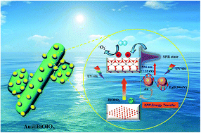
On the basis of the abovementioned experimental results, the tentative reaction mechanism of the highly enhanced photocatalysis and photoelectrochemical properties of Au@BiOIO3 was postulated as follows (Scheme 2): our previous study demonstrated that the bottom of the CB of BiOIO3 consists of O 2p, Bi 6p and I 5p orbitals, and the top of the VB is mainly composed of O 2p orbitals.12 On excitation with UV-Vis light, only a few e–h+ pairs were generated over the pure BiOIO3, and thereby its as-exhibited photocatalytic activity was moderate. After the introduction of Au nanoparticles (NPs), the photoactivity of BiOIO3 was greatly improved. First, the photogenerated electrons of Au metal NPs can be excited from the Fermi level (0.94 eV) to about −1.29 eV by its characteristic absorption at 556 nm (∼2.23 eV),47 and then transferred to the CB of BiOIO3 through Schottky energy potential. This process induces more electrons to produce abundant superoxide radicals (˙O2), which are beneficial for the enhanced photocatalysis activity. Second, the near-field resonant energy transfer, resulting from the SPR effect of Au NPs, can greatly boost the steady-state population of energetic charge carriers in adjacent BiOIO3, and simultaneously strengthen the electric field in the BiOIO3 crystal, making charge separation more efficient.23,34 Furthermore, the Au deposition process enables a largely increased specific surface area on BiOIO3, and alters the surface chemical state by modifying the microstructure, which favors the formation of more reactive sites; this further propels the enhancement of photocatalysis and photoelectrochemical properties of BiOIO3.


image file: c5ra14405a-s2.tif
Scheme 2 Schematic diagrams of the charge transfer for (a) BiOIO3 and (b) Au@BiOIO3 under UV-Vis light irradiation.

4. Conclusion

In summary, we have deposited metallic Au particles on BiOIO3 to fabricate heterostructured Au@BiOIO3 hybrid photocatalysts by a simple in situ UV-light reduction method. It is interesting to find that HAuCl4 as a Au source not only provides the SPR of Au to enhance the photoabsorption in the visible region, but also leads to the enlarged specific surface area of the products by modifying the microstructure of BiOIO3 due to the strong oxidative ability of HAuCl4, which was confirmed by the appearance of Bi4+/Bi5+. In contrast to the pristine BiOIO3, Au@BiOIO3 exhibits significantly boosted photoreactivity pertaining to RhB degradation and photocurrent response. The results from the measurements of charge separation and transfer behavior revealed that the synergic contributions from the SPR effect induced efficient separation of electron–hole pairs, boosted the generation of charge carriers and increased surface area, and thus are responsible for highly promoted photocatalysis and photoelectrochemical properties. This study has furnished new perspectives on the in situ construction of Au@semiconductor hybrid materials with multiple contributions to enhancing the photochemical properties.

Acknowledgements

This study was supported by the National Natural Science Foundations of China (Grant No. 51302251), the Fundamental Research Funds for the Central Universities (No. 2652013052 and No. 2652015296).

Notes and references

  1. A. Kubacka, M. Fernández-García and G. Colón, Chem. Rev., 2012, 112, 1555 CrossRef CAS PubMed.
  2. H. L. Wang, L. S. Zhang, Z. G. Chen, J. Q. Hu, S. J. Li, Z. H. Wang, J. S. Liu and X. C. Wang, Chem. Soc. Rev., 2014, 43, 5234 RSC.
  3. L. Shang, T. Bian, B. Zhang, D. H. Zhang, L. Z. Wu, C. H. Tung, Y. D. Yin and T. R. Zhang, Angew. Chem., Int. Ed., 2014, 53, 250 CrossRef CAS PubMed.
  4. J. G. Yu, J. X. Low, W. Xiao, P. Zhou and M. Jaroniec, J. Am. Chem. Soc., 2014, 136, 8839 CrossRef CAS PubMed.
  5. C. T. Yip, H. T. Huang, L. M. Zhou, K. Y. Xie, Y. Wang, T. H. Feng, J. S. Li and W. Y. Tam, Adv. Mater., 2011, 23, 5624 CrossRef CAS PubMed.
  6. A. B. Djurisic, X. Chen, Y. H. Leung and A. Man Ching Ng, J. Mater. Chem., 2012, 22, 6526 RSC.
  7. Q. Li, B. D. Guo, J. G. Yu, J. R. Ran, B. H. Zhang, H. J. Yan and J. R. Gong, J. Am. Chem. Soc., 2011, 133, 10878 CrossRef CAS PubMed.
  8. X. C. Wang, X. F. Chen, A. Thomas, X. Z. Fu and M. Antonietti, Adv. Mater., 2009, 21, 1609–1612 CrossRef CAS PubMed.
  9. F. Dong, Z. Y. Wang, Y. H. Li, W. K. Ho and S. C. Lee, Environ. Sci. Technol., 2014, 48, 10345 CrossRef CAS PubMed.
  10. S. D. Nguyen, J. Yeon, S.-H. Kim and P. S. Halasyamani, J. Am. Chem. Soc., 2011, 133, 12422 CrossRef CAS PubMed.
  11. W. Wang, B. Huang, X. Ma, Z. Wang, X. Qin, X. Zhang, Y. Dai and M.-H. Whangbo, Chem.–Eur. J., 2013, 19, 14777 CrossRef CAS PubMed.
  12. H. W. Huang, Y. He, R. He, X. X. Jiang, Z. S. Lin, Y. H. Zhang and S. C. Wang, Inorg. Chem. Commun., 2014, 40, 215 CrossRef CAS PubMed.
  13. F. Dong, T. Xiong, Y. J. Sun, Y. X. Zhang and Y. Zhou, Chem. Commun., 2015, 51, 8249 RSC.
  14. H. W. Huang, S. B. Wang, N. Tian and Y. H. Zhang, RSC Adv., 2014, 4, 5561 RSC.
  15. H. W. Huang, L. Y. Liu, Y. H. Zhang and N. Tian, RSC Adv., 2015, 5, 1161 RSC.
  16. N. Tian, H. W. Huang, Y. H. Zhang, Y. He and Y. X. Guo, RSC Adv., 2014, 4, 42716 RSC.
  17. H. W. Huang, L. Y. Liu, Y. H. Zhang and N. Tian, J. Alloys Compd., 2015, 619, 807 CrossRef CAS PubMed.
  18. E. M. Larsson, C. Langhammer, I. Zorić and B. Kasemo, Science., 2009, 326, 1091 CrossRef CAS PubMed.
  19. S. Linic, P. Christopher and D. B. Ingram, Nat. Mater., 2011, 10, 911 CrossRef CAS PubMed.
  20. C. Burda, X. Chen, R. Narayanan and M. A. El-Sayed, Chem. Rev., 2005, 105, 1025 CrossRef CAS PubMed.
  21. R. Costi, G. Cohen, A. Salant, E. Rabani and U. Banin, Nano Lett., 2009, 9, 2031 CrossRef CAS PubMed.
  22. Z. Liu, W. Hou, P. Pavaskar, M. Aykol and S. B. Cronin, Nano Lett., 2011, 11, 1111 CrossRef CAS PubMed.
  23. D. Jiang, W. Z. Wang, S. M. Sun, L. Zhang and Y. L. Zheng, ACS Catal., 2015, 5, 613 CrossRef CAS.
  24. S. J. Guo and S. H. Sun, J. Am. Chem. Soc., 2012, 134, 2492 CrossRef CAS PubMed.
  25. M. Murdoch, G. I. N. Waterhouse, M. A. Nadeem, J. B. Metson, M. A. Keane, R. F. Howe, J. Llorca and H. Idriss, Nat. Chem., 2011, 3, 489 CAS.
  26. V. Subramanian, E. E. Wolf and P. V. Kamat, J. Am. Chem. Soc., 2004, 126, 4943 CrossRef CAS PubMed.
  27. M. N. Tahir, F. Natalio, M. A. Cambaz, M. Panthofer, R. Branscheid, U. Kolb and W. Tremel, Nanoscale., 2013, 5, 9944 RSC.
  28. Y. Chen, D. Zeng, K. Zhang, A. Lu, L. Wang and D.-L. Peng, Nanoscale., 2014, 6, 874 RSC.
  29. L. Q. Ye, J. Y. Liu, Z. Jiang, T. Y. Peng and L. Zan, Appl. Catal., B, 2013, 142, 1 Search PubMed.
  30. H. W. Huang, K. Liu, K. Chen, Y. L. Zhang, Y. H. Zhang and S. C. Wang, J. Phys. Chem. C, 2014, 118, 14379 CAS.
  31. N. Tian, Y. H. Zhang, H. W. Huang, Y. He and Y. X. Guo, J. Phys. Chem. C, 2014, 118, 15640 CAS.
  32. S. Sajjad, S. A. K. Leghari, F. Chen and J. L. Zhang, Chem.–Eur. J., 2010, 16, 13795 CrossRef CAS PubMed.
  33. X. Ding, K. Zhao and L. Z. Zhang, Environ. Sci. Technol., 2014, 48, 5823 CrossRef CAS PubMed.
  34. H. H. Yin, K. Yu, C. Q. Song, R. Huang and Z. Q. Zhu, ACS Appl. Mater. Interfaces, 2014, 6, 14851 CAS.
  35. Y. Ohko, K. Hashimoto and A. Fujishima, J. Phys. Chem. A, 1997, 101, 8057 CrossRef CAS.
  36. C. S. Pan and Y. F. Zhu, Environ. Sci. Technol., 2010, 44, 5570 CrossRef CAS PubMed.
  37. H. W. Huang, Y. He, Z. S. Lin, L. Kang and Y. H. Zhang, J. Phys. Chem. C, 2013, 117, 22986–22994 CAS.
  38. L. M. Sun, L. Xiang, X. Zhao, C. J. Jia, J. Yang, Z. Jin, X. F. Cheng and W. L. Fan, ACS Catal., 2015, 5, 3540 CrossRef CAS.
  39. H. W. Huang, J. J. Wang, F. Dong, Y. X. Guo, N. Tian, Y. H. Zhang and T. R. Zhang, Cryst. Growth Des., 2015, 15, 534 CAS.
  40. R. Kern, R. Sastrawan, J. Ferber and S. R. Luther, Electrochim. Acta, 2002, 47, 4213 CrossRef CAS.
  41. S. M. Sun, W. Z. Wang and L. Zhang, J. Phys. Chem. C, 2013, 117, 9113 CAS.
  42. S. F. Chen, Y. F. Hu, S. G. Meng and X. L. Fu, Appl. Catal., B, 2014, 150, 564 CrossRef PubMed.
  43. H. W. Huang, X. Han, X. W. Li, S. C. Wang, P. K. Chu and Y. H. Zhang, ACS Appl. Mater. Interfaces, 2015, 7, 482 CAS.
  44. F. Dong, Q. Y. Li, Y. J. Sun and W. K. Ho, ACS Catal., 2014, 4, 4341 CrossRef CAS.
  45. H. W. Huang, X. W. Li, J. J. Wang, F. Dong, P. K. Chu, T. R. Zhang and Y. H. Zhang, ACS Catal., 2015, 5, 4094–4103 CrossRef CAS.
  46. J. L. Wang, Y. Yu and L. Z. Zhang, Appl. Catal., B, 2013, 136, 112 CrossRef PubMed.
  47. J. Y. Zhang, Y. H. Wang, J. Zhang, Z. Lin, F. Huang and J. G. Yu, ACS Appl. Mater. Interfaces, 2013, 5, 1031 CAS.

This journal is © The Royal Society of Chemistry 2015
Click here to see how this site uses Cookies. View our privacy policy here.