Flexible periodical micro- and nano-structuring of a stainless steel surface using dual-wavelength double-pulse picosecond laser irradiation

Mindaugas Gedvilas*, Justinas Mikšys and Gediminas Račiukaitis
Center for Physical Science and Technology, Savanoriu Ave. 231, LT-02300, Vilnius, Lithuania. E-mail: mgedvilas@ftmc.lt

Received 18th July 2015 , Accepted 21st August 2015

First published on 21st August 2015


Abstract

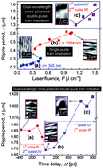
The picosecond laser-induced ripple formation on the stainless steel surface upon irradiation with linearly-polarized single-pulse and dual-wavelength cross-polarized double-pulse trains in air was studied experimentally. The characteristic switching of the ripple period and orientation were observed depending on the inter-pulse delay in the dual-wavelength cross-polarized double-pulse train irradiation experiments.


Birnbaum was the first to discover laser-induced periodic surface structures (LIPSS), also known as ripples, on the surface of various semiconductors, using a focused ruby laser beam in 1965.1 Since that time, the ripples have been investigated in numerous scientific works experimentally and theoretically.2–5 The period of the periodical or quasi-periodical structures is considered one of the most important ripple defining characteristics.5 There are two main types of ripples grown after an ultra-short laser irradiation. The coarse or low spatial frequency ripples (LSFRs) have a period close to the wavelength of the laser irradiation and direction, perpendicular to the polarization of the laser irradiation. The fine or high spatial frequency ripples (HSFRs), with a period much smaller than the irradiation wavelength, have a direction parallel to the polarization vector.5 The most commonly used theoretical model of ripple formation is the interference model,6 which states that ripples are being formed via the interference between the incident and the surface scattered light.

These laser-induced periodic micro- and nano-ripple structures have found many applications during the last decade. These include the control of surface wetting,7–9 light extraction and harvesting,10,11 chemical and mechanical alteration of materials,12 colouring surfaces,13–16 nano-textured surfaces for bio-sensors,17–19 micro-optical elements20,21 and light absorption enhancement.22–25

Irradiation with sequences of a high number of ultra-short laser pulses is required for the formation of fine and coarse ripples on a stainless steel surface.26 Usually, the metal surface is irradiated with single-pulse trains.27 However, in some studies, double-pulse trains with the same wavelength have been used for ripple formation28,29 and also for laser micro-fabrication.30–33 In the more complicated approaches, dual-wavelength double-pulse trains with a time delay in between pulses have been used for various laser processing experiments.34–38 In the last half-decade, much research on the ripple formation upon the double-pulse femtosecond laser irradiation of silicon and fused silica has been done using parallel-39–43 and cross-polarizations.44–46 There are only a few papers dedicated to laser-induced ripple formation using dual-wavelength double-pulse femtosecond laser irradiation37,47 and only two with cross-polarizations.45,48 However, the time delay between the IR and UV pulses was varied only in the range of a few picoseconds.45,47 No similar research has been performed on metallic surfaces.

In this work, the experimental results of the picosecond laser-induced ripple formation on a stainless steel surface after irradiation with the linearly-polarized single-pulse trains and dual-wavelength cross-polarized double-pulse trains are presented. The morphology of the periodical micro- and nano-ripples was investigated using scanning electron microscopy (SEM). The periodicity and orientation of the structures were analysed using two-dimensional (2D) Fast Fourier Transformation (FFT). The characteristic coarse or LSFR ripple structures with wavelength-related periods and polarization-perpendicular orientations were observed after the single-pulse train irradiation at IR (λ = 1064 nm) and UV (λ = 355 nm) wavelengths. The ripple period dependence on the laser fluence was found for both single- and double-pulse irradiation regimes. Three distinguishable ranges of time delays between the IR and UV laser pulses with the characteristic switching of the ripple period and their orientation were found in the dual-wavelength cross-polarized double-pulse train irradiation experiments. The possible formation mechanisms are discussed.

Stainless steel – grade 304 plates with thicknesses of 400 μm were chosen as the sample material for the laser-induced ripple formation.

The principal scheme of the experimental setup for the dual-wavelength cross-polarized double-pulse train irradiation is presented in Fig. 1. An industrial-grade diode-pumped picosecond laser (Atlantic, Ekspla) with a pulse duration of τ = 10 ps, repetition rate of frep = 100 kHz and wavelength of IR irradiation λ = 1064 nm was used in the experiments. The incident laser light was separated into two beams using a beam splitter cube (BSC). The time delay, Δt, between the pulses of the fundamental and third harmonics was varied by changing the optical path length of two delay lines DL1 and DL2 with retro-reflector prisms RP1 and RP2, respectively:

 
Δt = 2Δx/c (1)
where Δx = x2x1 is the position difference of the retro-reflector prisms, x1 and x2 are the absolute positions of the retro-reflector prisms RP1 and RP2, and c = 3.0 × 10⁸ m s−1 is the speed of light in air.


image file: c5ra14210e-f1.tif
Fig. 1 The experimental setup for the dual-wavelength (355 nm and 1064 nm) cross-polarized double-pulse laser irradiation. LASER is a picosecond laser source; BSC is the beam splitter cube; DL1 and DL2 are the delay lines with movable retro-reflector prisms RP1 and RP2; M1, M2, M3, and M4 are the high reflective mirrors; SHC is the second harmonic crystal; THC is the third harmonic crystal; M5 is the harmonic beam splitter mirror; FL is the focusing lens, and S is the sample.

The third harmonic was generated on the left side of the scheme using the second and third harmonic crystals. Both beams were brought together using the harmonic beam splitter mirror, M5, and were focused on the surface of the sample using the focusing lens, FL.

Two regimes of irradiation were used in the experiments: the single-pulse train irradiation and dual-wavelength double-pulse train irradiation (Fig. 2). The single-pulse train irradiation was used with two laser wavelengths: either λ = 1064 nm (Fig. 2a) or λ = 355 nm (Fig. 2b). In the dual-wavelength double-pulse train irradiation scheme, the dual-wavelength (λ = 1064 nm and λ = 355 nm) pulse pair with the time delay, Δt, between them were used for the irradiation of the samples. The negative time delay of Δt < 0 represents the situation when the first pulse was at the IR wavelength in the double-pulse pair and the UV one was the second (Fig. 2c). The positive time delay Δt > 0 represents the opposite situation when the UV pulse is the first in the double-pulse pair and the IR pulse was the second one (Fig. 2d). The temporal distance between the repetitive laser pulses or double-pulse pairs was 1/frep = 10 μs. The number of pulses N = 1, 10, 100 and 1000 were used in the irradiation pulse trains.


image file: c5ra14210e-f2.tif
Fig. 2 Principal scheme of the irradiation regimes in the intensity versus time representation: (a) and (b) – single-pulse train irradiation; (c) and (d) – dual-wavelength double-pulse train irradiation.

A laser beam with the Gaussian transverse spatial intensity distribution was used in the experiments:

 
F(r) = F0[thin space (1/6-em)]exp(−2r2/w02) (2)
where F0 is the peak laser fluence in the central part of the Gaussian beam, r is the radial distance from the center axis of the beam, and w0 is the spot radius on the sample at the 1/e2 level. The peak laser fluence in the center of the beam was evaluated using F0 = 2Ep/(πw02), where Ep is the laser pulse energy. The peak laser fluence for the IR irradiation was F0 IR = 1.1 J cm−2 and for the UV one was F0 UV = 0.45 J cm−2. The ripple period was measured across the laser irradiated spot. The radial distance, r, from the center of the Gaussian beam was converted to the local laser fluence, F(r), using eqn (2).

Ripple formation is highly dependent on the number of laser pulses applied to the surface of stainless steel.26,49 Usually, a high number of pulses is required for a regular ripple formation using ultra-short laser pulses.26,27,50 In our work, the first experiments were conducted to find the optimal number of laser pulses for ripple formation in our experimental conditions. The number of laser pulses was changed, keeping the laser fluence fixed at the single-pulse and dual-wavelength cross-polarized double-pulse train irradiations. The SEM micrographs of the ripples formed using different pulse counts in the irradiation trains and the 2D-FFT of the images are given in Fig. 3. The ripple formation starts after the first 1–10 laser pulses (Fig. 3a, b, e and f). However, this is only the initial stage of the ripple formation, and the higher contrast image is taken from the experiment exposed to N = 100 laser pulses (Fig. 3c and g). When the exposure time was further increased to the 1000 pulse regime, unwanted bubbles started to form in the laser processed area for both wavelengths of irradiation (Fig. 3d and h).


image file: c5ra14210e-f3.tif
Fig. 3 The SEM micrographs of the ripples formed on the stainless steel surface by laser irradiation: (a)–(h) single-pulse trains; (i)–(l) dual-wavelength cross-polarized double-pulse trains. The processing parameters: (a)–(d) wavelength of irradiation λ = 355 nm, laser fluence in the center of the beam F0 UV = 0.45 J cm−2; (e)–(h) λ = 1064 nm, F0 IR = 1.1 J cm−2; (i)–(l) time delay Δt = 600 ps, 1st pulse UV, 2nd pulse IR, laser fluences are the same as in (a)–(h). The numbers N = 1, 10, 100, 1000 on the top right corners of the images represent the number of laser pulses used in the irradiation trains. The vertical and horizontal arrows in (a), (e) and (i) indicate the orientation of the polarization of the IR and UV laser beams for each row of images. Inserts on the bottom left corners of the images are four times enlarged micrographs of the center of the pictures. Inserts in the middle right of the micrographs are the 2D-FFTs of the images. The scale bars, given in (d), (h) and (l), are the same for all images.

The main criteria for choosing the optimal processing regime were the SEM images with the highest possible contrast, the characteristic ripple period and their orientation over the main part of the laser spot and the distinguishable and sharp maximum peaks in the 2D-FFT of the SEM images. The processing with irradiation exposure using 100 laser pulses was chosen as an optimal number of laser pulses for further investigations.

The ripple period dependence on the laser fluence was investigated for both single- and double-pulse irradiation experiments. The ripple period was evaluated from the 2D-FFT of the SEM micrographs. The free and open source software Gwyddion was used for image analysis and evaluation of the ripple period. 2D-FFT was performed to the SEM micrographs and the ripple period was measured from the distance between the peaks in the spatial frequency domain. The dependence of the ripple period on the laser fluence for the single-pulse and dual-wavelength double-pulse train irradiation is given in Fig. 4. The ripple period grows linearly from 215 nm to 310 nm with the increasing laser fluence from 0.05 J cm−2 to 0.45 J cm−2 applied for the 355 nm wavelength irradiation (Fig. 4a). The ripple period linearly grows from 420 nm to 620 nm with the increasing laser fluence from 0.25 J cm−2 to 1.05 J cm−2 applied for the 1064 nm wavelength irradiation (Fig. 4b). The ripple period also grows linearly from 1.3 μm to 1.6 μm with the increasing laser fluence from 1.1 J cm−2 to 1.5 J cm−2 applied for the dual-wavelength (UV and IR) cross-polarized double-pulse train irradiation with the inter-pulse time delay of Δt = 600 ps (Fig. 4c).


image file: c5ra14210e-f4.tif
Fig. 4 The ripple period dependence on the laser fluence. The bottom part of the graph represents the single-pulse train irradiation with (a) UV (λ = 355 nm), and (b) IR (λ = 1064 nm) wavelengths. The top part of the graph (c) represents the dual-wavelength cross-polarized double-pulse train irradiation with the time delay of Δt = 600 ps between the UV and IR pulses. The number of laser pulses in the trains was N = 100. The inserts are the SEM micrographs of the laser-induced ripples.

The mechanism for the self-formation of periodic grating structures on a metal surface using ultra-short laser pulses has been proposed by S. Sakabe et al.51–53 A parametric process involving the interaction of laser light and surface plasma waves as well as the excitation of the surface solid-state plasma, suggesting the increase of the ripple period, Λ, with the laser fluence within the range between 0.5λ and 0.85λ, was demonstrated theoretically and experimentally. In our case the ripple periods, ΛUV and ΛIR, increased with the laser fluence within the range from 0.62λUV to 0.87λUV and from 0.39λIR to 0.58λIR for the 355 nm and 1064 nm wavelengths of irradiation, respectively. Our experimental results are consistent with this model.

The SEM images of the laser-irradiated areas with the dual-wavelength cross-polarized double-pulse trains are presented in Fig. 5. The regular ripples were formed on the steel surface that was firstly irradiated with the IR pulse and after 360 ps with the UV pulse (negative time delay Δt = −360 ps) (Fig. 5a). The ripple period was Λ = 850 nm. Fig. 5b shows the sample surface that firstly interacted with the UV pulse, and after the positive time delay of Δt = 30 ps, with the IR pulse. The period of ripples, in this case, was Λ = 700 nm. The anti-clockwise inclination of the ripple orientation by an angle of ∼14° after the switching from negative to positive time delays was observed. The similar rotation of the grating direction in the dual-wavelength cross-polarization irradiation experiments on the semiconductor surface depending on the pulse energy ratio between the IR and UV pulses has been observed by T. Q. Jia et al.37 The rotation of the ripple orientation with the increase of the UV pulse energy was observed at zero delay time between pulses. The rotation in our experiment might be related to the interaction of the surface plasma waves induced by the electrical field of the IR and UV laser beams. Fig. 5c shows the quasi-periodical ripple formation with the period of Λ = 1.4 μm when the IR beam reached the sample the second after a time delay of Δt = 600 ps. The dependence of the ripple period on the time delay between the third and fundamental harmonics is given in Fig. 6.


image file: c5ra14210e-f5.tif
Fig. 5 The SEM micrographs of the stainless steel surface irradiated with the dual-wavelength (1064 nm and 355 nm) cross-polarized double-pulse train at different time delays between the laser pulses: (a) 1st pulse IR (1064 nm), 2nd pulse UV (355 nm), time delay Δt = −360 ps, ripple period Λ ≈ 850 nm; (b) 1st pulse UV, 2nd pulse IR, time delay Δt = 30 ps, ripple period Λ ≈ 700 nm; (c) 1st pulse UV, 2nd pulse IR, time delay Δt = 600 ps, quasi-periodical ripple formation with period Λ = 1.4 μm. The number of pulses N = 100 was used in the irradiation trains and the laser fluence in the central area of the micrographs: F0 UV = 0.45 J cm−2 and F0 IR = 1.1 J cm−2. Inserts on the left of the images are micrographs enlarged four times from the center of the picture. Inserts on the middle right of the micrographs are the 2D-FFTs of the images. The scale bars on (c) are the same for all images.

image file: c5ra14210e-f6.tif
Fig. 6 The ripple period dependence on the time delay between the IR and UV laser pulses. (a) The experimental points on the left side of the graph are for the case when the IR laser pulse reaches the sample surface first. (b) The points on the central part of the graph represent the case when the UV laser pulse reaches the sample surface first. (c) The points on the right side of the graph show the quasi-periodical ripple formation when Δt > 600 ps. The inserts are the SEM micrographs of the laser-induced ripples.

The characteristic coarse ripple formation with the wavelength-related periods and the polarization-perpendicular orientations was observed when single-pulse trains were applied to a stainless steel surface at the 1064 nm and 355 nm wavelengths of irradiation. The ripple period linearly increased with the applied laser fluence for both wavelengths in the single-pulse train irradiation regimes and also in the double-pulse irradiation experiments.

In conclusion, three distinguishable regimes of the periodical and quasi-periodical ripple formation were observed in the dual-wavelength cross-polarized double-pulse train irradiation experiments with different time delays between the UV and IR laser pulses. Characteristic change of the ripple period and their orientation was observed depending on the pulse order in the train. The decrease in the ripple period and the anti-clockwise rotation of their orientation was observed when the time delay switched from negative to positive. Switching from the highly-periodical to the quasi-periodical ripples was observed with time delays higher than 600 ps.

Notes and references

  1. M. Birnbaum, J. Appl. Phys., 1965, 36, 3688–3689 CrossRef CAS PubMed.
  2. J. Bonse, J. Krüger, S. Höhm and A. Rosenfeld, J. Laser Appl., 2012, 24, 042006 CrossRef.
  3. M. Huang, Y. Cheng, F. Zhao and Z. Xu, Ann. Phys., 2013, 525, 74–86 CrossRef CAS PubMed.
  4. A. Y. Vorobyev and C. Guo, Laser Photonics Rev., 2013, 7, 385–407 CrossRef CAS PubMed.
  5. R. Buividas, M. Mikutis and S. Juodkazis, Prog. Quantum Electron., 2014, 38, 119–156 CrossRef PubMed.
  6. J. E. Sipe, J. F. Young, J. S. Preston and H. M. van Driel, Phys. Rev. B: Condens. Matter Mater. Phys., 1983, 27, 1141–1154 CrossRef CAS.
  7. M. Silvennoinen, J. Kaakkunen, K. Paivasaari, P. Vahimaa and T. Jaaskelainen, J. Laser Micro/Nanoeng., 2010, 5, 97–98 CrossRef CAS.
  8. L. Zhang, X. W. Cao, R. Y. Xiang, S. G. Li, L. Wang and H. C. Sun, Adv. Mater. Res., 2014, 1004–1005, 1311–1315 CrossRef CAS.
  9. V. Zorba, L. Persano, D. Pisignano, A. Athanassiou, E. Stratakis, R. Cingolani, P. Tzanetakis and C. Fotakis, Nanotechnology, 2006, 17, 3234–3238 CrossRef CAS.
  10. A. Y. Vorobyev, V. S. Makin and C. Guo, Phys. Rev. Lett., 2009, 102, 234301 CrossRef CAS.
  11. V. Zorba, P. Tzanetakis, C. Fotakis, E. Spanakis, E. Stratakis, D. G. Papazoglou and I. Zergioti, Appl. Phys. Lett., 2006, 88, 081103 CrossRef PubMed.
  12. X. Yu, Y. Liao, F. He, B. Zeng, Y. Cheng, Z. Xu, K. Sugioka and K. Midorikawa, J. Appl. Phys., 2011, 109, 053114 CrossRef PubMed.
  13. B. Dusser, Z. Sagan, H. Soder, N. Faure, J. P. Colombier, M. Jourlin and E. Audouard, Opt. Express, 2010, 18, 2913–2924 CrossRef CAS PubMed.
  14. J. Yao, C. Zhang, H. Liu, Q. Dai, L. Wu, S. Lan, A. V. Gopal, V. A. Trofimov and T. M. Lysak, Appl. Surf. Sci., 2012, 258, 7625–7632 CrossRef CAS PubMed.
  15. A. Y. Vorobyev and C. Guo, Appl. Phys. Lett., 2008, 92, 041914 CrossRef PubMed.
  16. J. Long, P. Fan, M. Zhong, H. Zhang, Y. Xie and C. Lin, Appl. Surf. Sci., 2014, 311, 461–467 CrossRef CAS PubMed.
  17. R. Buividas, P. R. Stoddart and S. Juodkazis, Ann. Phys., 2012, 524, L5–L10 CrossRef CAS PubMed.
  18. C.-H. Lin, L. Jiang, Y.-H. Chai, H. Xiao, S.-J. Chen and H.-L. Tsai, Opt. Express, 2009, 17, 21581–21589 CrossRef CAS PubMed.
  19. E. D. Diebold, N. H. Mack, S. K. Doorn and E. Mazur, Langmuir, 2009, 25, 1790–1794 CrossRef CAS PubMed.
  20. M. Beresna, M. Gecevičius, P. G. Kazansky and T. Gertus, Appl. Phys. Lett., 2011, 98, 201101 CrossRef PubMed.
  21. E. Brasselet, G. Gervinskas, G. Seniutinas and S. Juodkazis, Phys. Rev. Lett., 2013, 111, 193901 CrossRef.
  22. P. Gečys, A. Vinčiūnas, M. Gedvilas, A. Kasparaitis, R. Lazdinas and G. Račiukaitis, J. Laser Micro/Nanoeng., 2015, 10, 129–133 CrossRef.
  23. A. Žukauskas, M. Malinauskas, A. Kadys, G. Gervinskas, G. Seniutinas, S. Kandasamy and S. Juodkazis, Opt. Express, 2013, 21, 6901–6909 CrossRef PubMed.
  24. A. Y. Vorobyev, A. N. Topkov, O. V. Gurin, V. A. Svich and C. Guo, Appl. Phys. Lett., 2009, 95, 121106 CrossRef PubMed.
  25. T. Y. Hwang, A. Y. Vorobyev and C. Guo, Opt. Express, 2011, 19, A824–A829 CrossRef CAS PubMed.
  26. B. Liu, W. Wang, G. Jiang, X. Mei, K. Wang, J. Wang and Z. Wang, J. Laser Appl., 2014, 26, 012001 CrossRef.
  27. X. Jun, W. Feng, J. Lan, Z. Liangliang and L. Yongfeng, Laser Phys., 2015, 25, 056103 CrossRef.
  28. V. Hommes, M. Miclea and R. Hergenröder, Appl. Surf. Sci., 2006, 252, 7449–7460 CrossRef CAS PubMed.
  29. J. Kim, S. Na, S. Cho, W. Chang and K. Whang, Opt. Lasers Eng., 2008, 46, 306–310 CrossRef PubMed.
  30. A. C. Forsman, P. S. Banks, M. D. Perry, E. M. Campbell, A. L. Dodell and M. S. Armas, J. Appl. Phys., 2005, 98, 033302 CrossRef PubMed.
  31. K. Sugioka, M. Iida, H. Takai and K. Micorikawa, Opt. Lett., 2011, 36, 2734–2736 CrossRef PubMed.
  32. S. Wu, D. Wu, J. Xu, Y. Hanada, R. Suganuma, H. Wang, T. Makimura, K. Sugioka and K. Midorikawa, Opt. Express, 2012, 20, 28893–28905 CrossRef PubMed.
  33. S. Wu, D. Wu, J. Xu, H. Wang, T. Makimura, K. Sugioka and K. Midorikawa, Opt. Express, 2013, 21, 24049–24059 CrossRef PubMed.
  34. W. Zhao, W. Wang, X. Mei, G. Jiang and B. Liu, Opt. Laser Technol., 2014, 58, 94–99 CrossRef CAS PubMed.
  35. X. Yu, Q. Bian, B. Zhao, Z. Chang, P. B. Corkum and S. Lei, Appl. Phys. Lett., 2013, 102, 101111 CrossRef PubMed.
  36. B. Tan, K. Venkatkrishnan, N. R. Sivakumar and G. K. Gan, Opt. Laser Technol., 2003, 35, 199–202 CrossRef.
  37. T. Q. Jia, H. X. Chen, M. Huang, F. L. Zhao, J. R. Qiu, R. X. Li, Z. Z. Xu, X. K. He, J. Zhang and H. Kuroda, Phys. Rev. B: Condens. Matter Mater. Phys., 2005, 72, 125429 CrossRef.
  38. M. Sakamoto, T. Tachikawa, M. Fujitsuka and T. Majima, Chem. Phys. Lett., 2006, 420, 90–94 CrossRef CAS PubMed.
  39. A. Rosenfeld, M. Rohloff, S. Höhm, J. Krüger and J. Bonse, Appl. Surf. Sci., 2012, 258, 9233–9236 CrossRef CAS PubMed.
  40. T. Y. Derrien, J. Krüger, T. Itina, S. Höhm, A. Rosenfeld and J. Bonse, Appl. Phys. A: Mater. Sci. Process., 2014, 117, 77–81 CrossRef.
  41. S. Höhm, A. Rosenfeld, J. Krüger and J. Bonse, Appl. Surf. Sci., 2013, 278, 7–12 CrossRef PubMed.
  42. S. Höhm, A. Rosenfeld, J. Krüger and J. Bonse, J. Appl. Phys., 2012, 112, 014901 CrossRef PubMed.
  43. M. Barberoglou, D. Gray, E. Magoulakis, C. Fotakis, P. A. Loukakos and E. Stratakis, Opt. Express, 2013, 21, 18501–18508 CrossRef CAS PubMed.
  44. M. Rohloff, S. K. Das, S. Höhm, R. Grunwald, A. Rosenfeld, J. Krüger and J. Bonse, J. Appl. Phys., 2011, 110, 014910 CrossRef PubMed.
  45. S. Höhm, M. Herzlieb, A. Rosenfeld, J. Krüger and J. Bonse, Opt. Express, 2015, 23, 61–71 CrossRef PubMed.
  46. S. Höhm, M. Rohloff, A. Rosenfeld, J. Krüger and J. Bonse, Appl. Phys. A: Mater. Sci. Process., 2013, 110, 553–557 CrossRef.
  47. S. Höhm, M. Herzlieb, A. Rosenfeld, J. Krüger and J. Bonse, Appl. Phys. Lett., 2013, 103, 254101 CrossRef PubMed.
  48. S. Höhm, M. Herzlieb, A. Rosenfeld, J. Krüger and J. Bonse, Appl. Surf. Sci., 2015, 336, 39–42 CrossRef PubMed.
  49. J.-W. Yao, C.-Y. Zhang, H.-Y. Liu, Q.-F. Dai, L.-J. Wu, S. Lan, A. V. Gopal, V. A. Trofimov and T. M. Lysak, Opt. Express, 2012, 20, 905–911 CrossRef PubMed.
  50. L. Qi, K. Nishii and Y. Namba, Opt. Lett., 2009, 34, 1846–1848 CrossRef.
  51. S. Sakabe, M. Hashida, S. Tokita, S. Namba and K. Okamuro, Phys. Rev. B: Condens. Matter Mater. Phys., 2009, 79, 033409 CrossRef.
  52. S. Sakabe, M. Hashida, S. Tokita, S. Namba and K. Okamuro, Phys. Rev. B: Condens. Matter Mater. Phys., 2015, 91, 159902 CrossRef.
  53. M. Hashida, Y. Ikuta, Y. Miyasaka, S. Tokita and S. Sakabe, Appl. Phys. Lett., 2013, 102, 174106 CrossRef PubMed.

This journal is © The Royal Society of Chemistry 2015
Click here to see how this site uses Cookies. View our privacy policy here.