
Multifunctional core–shell Co–SiO2 nanowires via electrodeposition and sol–gel techniques
Laura Martín-Garcíaa,
Sandra Ruiz-Gómezb,
Manuel Abuínbc,
Yaiza Montañab,
Noemi Carmonab and
Lucas Pérez*bde
aInstituto de Química Física Rocasolano (IQFR), CSIC, C/Serrano 119, 28006 Madrid, Spain
bDept. Física de Materiales, Universidad Complutense de Madrid, 28040 Madrid, Spain. E-mail: lucas.perez@fis.ucm.es
cCEI Campus Moncloa, UCM-UPM, 28040 Madrid, Spain
dInstituto de Sistemas Optoelectrónicos y Microtecnología, Universidad Politécnica de Madrid, 28040 Madrid, Spain
eUnidad Asociada IQFR (CSIC)-UCM, Madrid, 28040, Spain
First published on 30th October 2015
Abstract
In this work, we propose a new strategy for the synthesis of multifunctional nanowires using a combination of sol–gel and electrodeposition techniques, based on a two-step procedure. First of all, nanotubes of SiO2 are synthesized via a sol–gel technique using polycarbonate membranes as templates. Homogenous nanotubes are obtained after centrifugation and thermal annealing. Afterwards, a ferromagnetic cobalt core is grown using potentiostatic electrodeposition. Finally, the core–shell Co–SiO2 nanowires are released by dissolving the template using wet-etching. These nanodevices can be used for many detection and sensing purposes. As a proof of concept, we have developed a pH nanosensor by including a pH-sensitive organic dye in the SiO2 shell. The sensing principle is based on the optical response of the organic dye towards pH when added to a solution. The magnetic core allows the recovery of the nanosensors after use. These nanowires can therefore be used as recoverable pH nanosensors. By changing the dye molecule to another molecule or receptor, the procedure described in the paper can be used to synthesize nanodevices for many different applications.
1 Introduction
One-dimensional nanostructures have been a focus in nanotechnology in recent years. Their geometry and the possibility of controlling their composition, together with their large surface area, are the main reasons for the potential applications of nanowires in many different areas such as gas1–3 and chemical sensing4,5 or biomedical applications,6–9 among others. The possibility of making core–shell structures adds new functionalities to these one-dimensional structures. In particular, the shell can be functionalized with different molecules to interact with the media in which the nanostructure is immersed while the core can be used for detection purposes, interacting with the nanostructure by changing the electrical properties10 or using magnetic fields.8Porous anodic aluminium oxide (AAO)11 or polycarbonate (PC)12 membranes have been widely used to grow nanowires with a controlled size and morphology, usually using electrochemical deposition in the case of metallic nanowires. After the synthesis, the nanoporous membrane used as the template can be dissolved and the nanowires are dispersed in different media to have individual access to the nanowires and measure the properties of a single nanowire.13 Different studies have shown that a similar approach can be used to obtain arrays of core–shell ferromagnetic/oxide nanowires using AAO membranes as templates.14–16 In this case, a combination of sol–gel and electrodeposition techniques is used to synthesize the nanostructures. This combination appears to be a good approach to make nanodevices due to the possibilities that sol–gel technology offers to make functionalized-sensing coatings17–19 and the possible use of the core to manipulate or detect the nanosensors. However, there are no reports up to now on the use of these structures as individual nanodevices. This is probably due to wet-etching, needed to release the devices from the alumina template, and normally based on highly basic solutions, which affects the molecules added to the shell and degrades the functionality of the shell. A different approach is therefore necessary to use these core–shell one-dimensional structures as nanosensors. Recently, nanodevices based on arrays of core–shell nanostructures made using PC templates have also been reported20 but, to our best knowledge, there is not a report on the use of released core–shell electrodeposited nanowires as individual nanosensors.
In this work, we describe a route for the synthesis of core–shell one-dimensional structures that can be released from the templates without degrading the shell and, therefore, that can be used individually as nanosensors. As we will show, this approach is possible using PC instead of AAO templates in the synthesis of the nanodevices. As a proof of concept, we report the preparation of a recoverable, environmentally friendly pH nanosensor composed of a functionalised SiO2 shell and a ferromagnetic Co core.
2 Materials and methods
2.1 Synthesis of the nanosensors
We have used a template method for the synthesis of the nanodevices in a two-step process: the sol–gel synthesis of a functionalized SiO2 shell followed by the electrodeposition of a ferromagnetic Co core. We used polycarbonate templates (PC), from GE Water & Process Technologies, with a 6 μm pore length and a diameter of 100 nm, dimensions that define the geometry of the nanosensors. However, both parameters depend on the choice of the template and can be varied just by choosing a different one. In order to compare our nanosensors with previously reported results, we have followed a similar fabrication procedure using anodic aluminum oxide templates (AAO), purchased from Whatman.The sol–gel was prepared from TEOS (tetraethyl orthosilicate, Si(OC2H5)4), ethanol absolute (CH3CH2OH), distilled water and hydrochloric acid (HCl). Chlorophenol red (C19H12Cl2O5S) and thymol blue (C27H30O5S) were used to functionalize the SiO2 shell to use the device as a pH sensor. To prepare the sol, TEOS (1 M), ethanol (64 M), water (4 M), HCl (0.01 M) and chlorophenol red or thymol blue (0.005 M) were mixed at room temperature. The solution was stirred during the preparation and kept under stirring for 40 min after preparation. To synthesize the SiO2 nanotubes inside the pores, the templates were immersed in the sol under sonication for 60 s and centrifuged at 7000 rpm for 60 s. This resulted in the elimination of the air bubbles trapped into the pores and the excess sol. Then, the sol was densified by heating at 60 °C for 72 h in a conventional oven.
The Co core was grown inside the SiO2 nanotubes via electrochemical deposition, from a sulphate-based electrolyte—0.1 M cobalt(II) sulphate (CoSO4·7H2O)—with 0.1 M boric acid (H3BO3) as the supporting electrolyte. Prior to electrodeposition, a Au layer was thermally evaporated on one side of the templates as the working electrode. Electrodeposition was carried out in a Teflon cell, using a Pt mesh (99.9%) as the anode and a BaSi Microanalytics Ag/AgCl electrode as the reference. Cobalt was grown under a potential of −1.1 V vs. Ag/AgCl.
After the growth, the templates should be removed to study and use the nanowires individually. The Au layer was removed via wet etching using a mixture of 0.1 M I2 and 0.6 M KI in water. Afterwards, we used dichloromethane (CH2Cl2) to dissolve the polycarbonate template. Finally, the nanosensors were washed in acetone and ethanol, and transferred into deionized water.
Fig. 1 summarizes the procedure followed for the synthesis of the individual nanodevices. The starting point is a nanoporous template (panel a). SiO2 tubes are formed inside the nanopores of the template via a sol–gel process (panel b), tubes that include the dye molecules. Afterwards, one side of the structure is metallized with a Au layer (panel c). Using this layer as the working electrode, the tubes are filled with Co using electrodeposition (panel d). Finally, the Au layer is chemically removed (panel e) and the template is dissolved to release the nanowires (panel f). Following this procedure, a 1D core–shell nanostructure with a functionalized oxide shell and a metallic core is obtained. In this work, we have made pH-sensitive nanosensors by incorporating chlorophenol red and thymol blue in the SiO2 shell but the functionality of the nanosensors can be simply varied by choosing the appropriate molecule for each particular application.
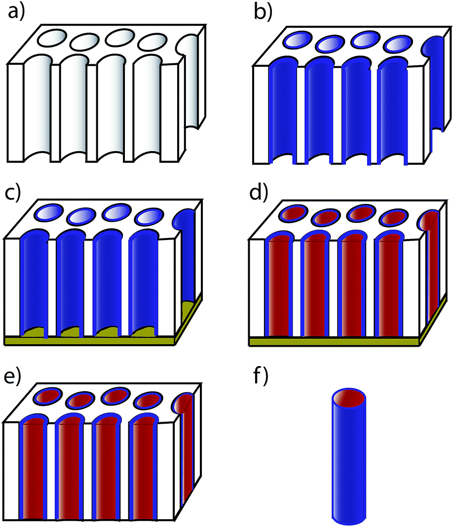 | ||
| Fig. 1 Schematics of the procedure for the synthesis of the core–shell nanodevices. A PC template (a) is used to make functionalized oxide nanotubes using a sol–gel process (b). One side of the structure is metallized (c) and the tubes are filled with a metal via electrodeposition (d). Finally, the metallized layer is removed (e) and the nanowires are released from the template (f). | ||
2.2 Characterization
The morphology of the nanowires was studied with a JEOL Scanning Electron Microscope (SEM) JEM 6335 F. The crystalline structure was studied using Grazing Incidence X-Ray Diffraction (GIXRD) with a PANalytical X-ray diffractometer with Cu Kα radiation. A Lake Shore Vibrating Sample Magnetometer (VSM) was used for the magnetic characterization of the ferromagnetic core. Optical absorption spectra were measured over the wavelength range 350–700 nm with a Shimadzu UV spectrophotometer.3 Results and discussion
3.1 Characterization of the nanosensors
One of the main problems faced during the synthesis of the nanodevices is the preparation of the nanotubes: the sol should fully penetrate the nanopores, creating a thin layer that covers the wall of each nanopore forming a nanotube, but leaving an empty space to allow the electrodeposition of the metal inside the nanotube to form the core. In this sense, the centrifugation process after immersing the template in the sol is extremely important to ensure that the pores are open. Otherwise, the pores are completely filled with the sol, producing a compact layer that impedes the electrodeposition of the ferromagnetic core. Only when the pores are fully open can the electrolyte fill the pores, the electrodeposition process take place and the metal core be electrodeposited inside the SiO2 nanotubes. In this sense, the occurrence of a cathodic current during the electrodeposition process is evidence that the pores are open and, therefore, that the dilution of the sol is adequate.In fact, the evolution of the electrodeposition current with time gives very useful information about the homogeneity of the pores. Fig. 2a shows a typical current vs. time curve measured during the electrodeposition process of the Co core of the nanosensors; three different zones can be observed. In the first few seconds (zone I) there is a sudden drop of the current, corresponding to the charging of the Helmholtz double layer. Afterwards (zone II), the nucleation of the Co NWs starts on the Au electrode. Finally, the value of the current is stabilized in zone III, which corresponds to the growth of the NWs. In an electrodeposition process, when the steady state is reached, the current density is constant. Therefore, if the current is also constant, as shown in the figure, the growth area should be constant along the wire, meaning that the diameter of the grown nanowires is the same for the whole growth period. This is clear evidence that the shell is homogeneous. In fact, the thickness of the SiO2 shell, measured using SEM, is 20 ± 2 nm along the nanowire.
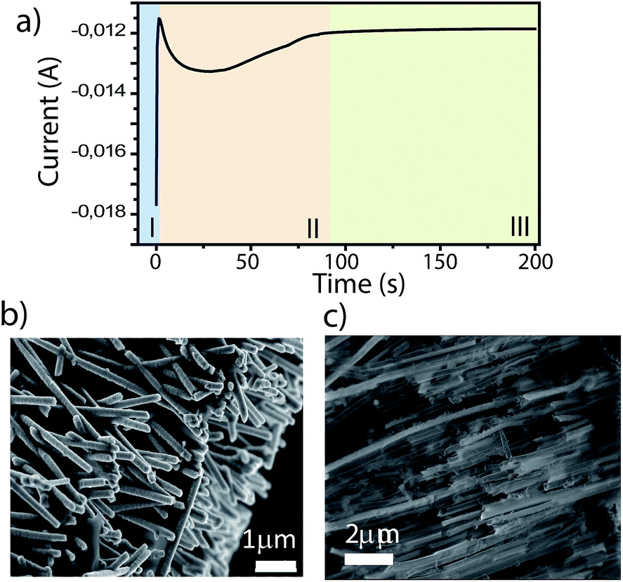 | ||
| Fig. 2 (a) Evolution of the cathodic current during the electrodeposition process. Three zones can be distinguished: charging of the double layer (I), nucleation (II) and growth (III). SEM images of the nanostructure after removing the PC (b) and the AAO (c) templates. | ||
After the growth, we need to release the nanowires from the template to use them as sensors using CH2Cl2 to dissolve the polycarbonate. Fig. 2b shows a SEM image of the nanowires after removing the template. It can be seen that, after being released from the template, their surface is smooth, without being etched by the solvent. For comparison, we have repeated the same procedure using an AAO template. A SEM image of the nanostructures after partially dissolving the AAO template is shown in Fig. 2c. The nanowires are rough because the NaOH solution needed to remove the template not only dissolved the template but also partially etched the SiO2 shell, changing its morphology. In particular, the shell loses its functionality because the pH-sensitive molecules inside the SiO2 are also dissolved and the released nanowires cannot act as sensors anymore. Therefore, although according to previous work, the combination of sol–gel and electrodeposition techniques seems to be a good approach for synthesizing core–shell nanowire arrays using AAO templates, a different approach should be used for individual core–shell functionalized nanostructures. We have shown that the use of PC templates preserves the shell, and is a better approach for the fabrication of these sensors.
X-ray diffraction was performed after removing the template to check that both SiO2 and Co are present in the sample. The diffractogram shown in Fig. 3a reveals reflections that can be indexed as either Co or SiO2. The crystal structure of Co nanowires is hexagonal-close-packed (hcp). The space-group is P63/mmc. This stable structure appears in other Co nanowires grown using different techniques.21
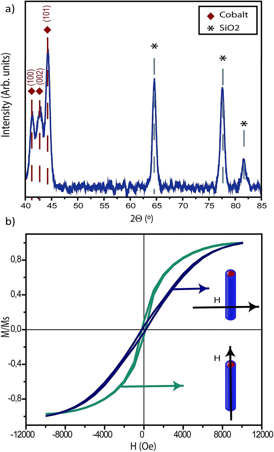 | ||
| Fig. 3 (a) XRD pattern of a collection of the nanowires in which the peaks of both SiO2 and Co can be identified. (b) Hysteresis loops measured on an array of nanosensors before removing the template. The hysteresis loops were measured with the applied field along the axis of the nanowires and in a perpendicular direction. | ||
Hysteresis loops have been measured at room temperature to test the magnetic characteristics of the nanosensors. In this case, the measurements were done before removing the template, to have an array of oriented sensors. The hysteresis loops, measured with the applied field in two perpendicular directions, along the axis of the nanosensors and perpendicular to it, show the characteristic behavior of an elongated magnetic system (see Fig. 3b), with an easy axis along the direction of the length of the nanosensors. When the magnetic field is applied in a direction perpendicular to the axis, the magnetization clearly rotates towards the direction of the applied field, as it corresponds to a hard magnetization axis.22
3.2 Proof of concept: pH nanosensors
We have synthesized the pH nanosensors using thymol blue and chlorophenol red as the pH sensing molecules. Thymol blue (thymolsulphonephthalein) is a crystalline powder that present transitions from red to yellow at pH 1.2–2.8 and from yellow to blue at pH 8.0–9.6. Chlorophenol red is an indicator dye that changes color from yellow to violet in the pH range from 4.8 to 6.7. After the preparation procedure summarized in Fig. 1, these molecules are embedded in the SiO2 shell, changing color and, therefore, changing the color of the nanosensor, when they are placed in a water-based solution with different pH values. In order to check it, the nanosensors were resuspended in deionized water, and in acidic (diluted H2SO4) and basic (diluted KOH) solutions. Fig. 4 shows the change in color of the nanosensors, which can be detected by the naked eye. The nanosensors made using thymol blue show a clear transition from red in acidic solution (panel a) to yellow in water (panel b) and to blue in basic solution (panel c), as expected. Those made with chlorophenol red show a transition from yellow in acidic solution (panel f) to violet in basic solution (panel h). It is also shown in the figure that the transition starts at a pH close to pH = 6 (panel g).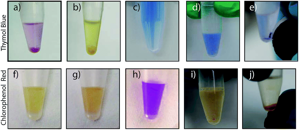 | ||
| Fig. 4 The change in color produced by the nanosensors can be detected by the naked eye in nanosensors with thymol blue at pH = 2 (a), pH = 7 (b) and pH = 12 (c) and in nanosensors made with chlorophenol red at pH = 2 (f), pH = 6 (g) and pH = 12 (h). (d), (e), (i) and (j) show the possibility of collecting the nanowires using a magnet, leaving a clean solution and making the nanosensors recoverable. | ||
The nanowires can be collected and recovered using a magnet, taking advantage of their Co core. Fig. 4d shows a homogeneous solution of nanosensors with thymol blue in basic solution. When a magnet is placed close to the solution, all nanosensors are collected (see Fig. 4b), which is reflected as a blue small spot in the solution, close to the magnet, leaving a clean transparent solution. The nanowires can be dispersed again in the solution via sonication, recovering a homogeneously colored solution again. This procedure was repeated five times to prove its reproducibility. Fig. 4i and j show the same procedure carried out using nanosensors made with chlorophenol red, in this case, in acidic solution.
The change in the optical response of the nanosensors as a function of pH was also measured using spectrophotometry. For that, the nanosensors were dispersed in five solutions going from pH 2 to pH = 12. Fig. 5 shows the response of the sensor for these pH values. All spectra clearly show that the nanosensors respond to pH as expected. This optical response to pH, combined with the possibility of recovering the nanosensors, shows the potential use of these nanowires as recoverable pH nanosensors.
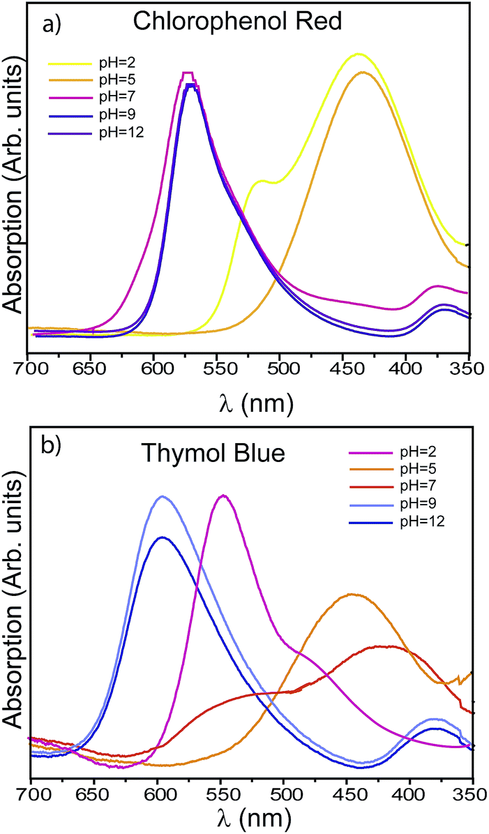 | ||
| Fig. 5 Optical response of a water-based diluted solution at different pHs containing nanosensors with chlorophenol red (a) and thymol blue (b). The changes in the wavelength of the absorption peaks at the different pH values with both molecules are clearly observed. | ||
Once the performance of the nanodevice has been demonstrated, it should be noted that by changing the dye molecule for another molecule or receptor or the metal forming the core, this procedure can be used to synthesize nanosensors for many different applications. For example, Ag or Au can be grown as the core to enhance the fluorescence of a molecule acting as a pH sensor, improving the sensitivity, as already reported in nanoparticles.23 Multifunctional devices could be also produced by including different molecules in the shell, getting recoverable and guidable nanodevices with potential uses in biomedical technology.24 Finally, the silica tube can be only partially filled, allowing the use of these nanodevices as scaffolds for on-command drug delivery, or as nanodevices that can be guided towards a target using the magnetic field and/or the functionalization of the shell.25
4 Conclusions
To sum up, we have shown that a combination of sol–gel and electrodeposition techniques can be used to make core–shell nanodevices provided that polycarbonate templates are used as templates for the synthesis. The use of polycarbonate templates for the synthesis of the nanodevices allows them to be released from the template and used as individual devices. We have checked the procedure by fabricating a pH nanosensor with a magnetic core. The nanosensor is sensitive to pH, like its bulk counter-part, and it can be recovered from dissolution by using a magnet, which allows the nanodevice to be reused.Acknowledgements
We thank the Spanish National Center of Electron Microscopy for SEM measurements. This work was funded by the Spanish Ministerio de Economía y Competitividad (MINECO) under grants MAT2011-28751-C02 and MAT2012-38045-C04-03. Manuel Abuín acknowledges the UCM Campus de Excelencia Internacional (PICATA Program) and Laura Martín-García acknowledges the MINECO (FPI program) for their pre-doctoral fellowships.References
- X. Chen, C. K. Y. Wong, C. A. Yuan and G. Zhang, Sens. Actuators, B, 2013, 177, 178–195 CrossRef CAS.
- P. Offermans, M. Crego-Calama and S. H. Brongersma, Nano Lett., 2010, 10, 2412–2415 CrossRef CAS PubMed.
- O. Lupan, V. V. Ursaki, G. Chai, L. Chow, G. A. Emelchenko, I. Tiginyanu, A. N. Gruzintsev and A. N. Redkin, Sens. Actuators, B, 2010, 144, 56–66 CrossRef CAS.
- G. Ghini, C. Trono, A. Giannetti, G. L. Puleo, L. Luconi, J. Amadou, G. Giambastiani and F. Baldini, Sens. Actuators, B, 2013, 179, 163–169 CrossRef CAS.
- A. Lapresta-Fernandez, T. Doussineau, A. J. Moro, S. Dutz, F. Stiniger and G. J. Mohr, Anal. Chim. Acta, 2011, 707, 164–170 CrossRef CAS.
- W. U. Wang, C. Chen, K. Hui Lin, Y. Fang and C. M. Lieber, Proc. Natl. Acad. Sci. U. S. A., 2005, 102, 3208–3212 CrossRef CAS PubMed.
- J. in Hahm and C. M. Lieber, Nano Lett., 2004, 4, 51–54 CrossRef.
- M.-M. Song, H. Bi and Y. Zhang, J. Appl. Phys., 2012, 111, 07B302 Search PubMed.
- D. T. Mitchell, S. B. Lee, L. Trofin, N. Li, T. K. Nevanen, H. Soderlund and C. R. Martin, J. Am. Chem. Soc., 2002, 124, 11864–11865 CrossRef CAS PubMed.
- J. H. He, Y. Y. Zhang, J. Liu, D. Moore, G. Bao and Z. L. Wang, J. Phys. Chem. C, 2007, 111, 12152–12156 CAS.
- C. T. Sousa, D. C. Leitao, M. P. Proenca, J. Ventura, A. M. Pereira and J. P. Araujo, Appl. Phys. Rev., 2014, 1, 031102 Search PubMed.
- M. E. Toimil-Molares, Beilstein J. Nanotechnol., 2012, 3, 2860 Search PubMed.
- N. Marcano, S. Sangiao, M. Plaza, L. Perez, A. Fernandez-Pacheco, R. Córdoba, M. C. Sánchez, L. Morellón, M. R. Ibarra and J. M. de Teresa, Appl. Phys. Lett., 2010, 96, 082110 CrossRef.
- Z. Ye, H. Liu, W. Isabel Schultz, D. G. Naugle and I. Lyuksyutov, Nanotechnology, 2008, 19, 325303 CrossRef PubMed.
- Q. Lin, L. Yao, G. Jiang, C. Jin, W. Liu and W. Cai, J. Mater. Sci. Technol., 2004, 20, 684–686 CAS.
- T. Dastagir and H. Yu, J. Appl. Phys., 2014, 115, 17B526 CrossRef.
- N. Carmona, E. Herrero, J. Llopis and M. A. Villegas, Sens. Actuators, B, 2007, 126, 455–460 CrossRef CAS.
- C. T. Sousa, C. Nunes, M. P. Proenca, D. C. Leitao, J. L. F. C. Lima, S. Reis, J. P. Araujo and M. Lucio, Colloids Surf., B, 2012, 94, 288–295 CrossRef CAS PubMed.
- P.-D. Nguyen, D. T. X. Nguyen, S. J. Son and J. Min, J. Nanosci. Nanotechnol., 2014, 14, 8719–8723 CrossRef CAS PubMed.
- B. L. Fiser, A. R. Shields, M. R. Falvo and R. Superfine, J. Micromech. Microeng., 2015, 25, 025004 CrossRef CAS PubMed.
- K. Gandha, K. Elkins, N. Poudyal, X. Liu and J. P. Liu, Sci. Rep., 2014, 4, 5345 CAS.
- L. Sun, Y. Hao, C. L. Chien and P. C. Searson, IBM J. Res. Dev., 2005, 49, 79–102 CrossRef CAS.
- Z. Bai, R. Chen, P. Si, Y. Huang, H. Sun and D.-H. Kim, ACS Appl. Mater. Interfaces, 2013, 5, 5856–5860 CAS.
- Y.-S. Li and C. L. Haynes, Chem. Mater., 2009, 21, 3979–3986 CrossRef.
- N. Mas, D. Arcos, L. Polo, E. Aznar, S. Sanchez-Salcedo, F. Sancenon, A. Garcia, M. D. Marcos, A. Baeza, M. Vallet-Regi and R. Martinez-Mañe, Small, 2014, 10, 4859–8464 CrossRef CAS PubMed.
| This journal is © The Royal Society of Chemistry 2015 |