Metal oxide nanostructures incorporated/immobilized paper matrices and their applications: a review

Indu Chauhan , Sudiksha Aggrawal , Chandravati and Paritosh Mohanty *
Department of Applied Science and Engineering, IIT Roorkee, Saharanpur Campus, Saharanpur-247001, U.P., India. E-mail: paritosh75@gmail.com; Fax: +91-132-271-4311; Tel: +91-132-271-4338

Received 11th July 2015 , Accepted 14th September 2015

First published on 14th September 2015


Abstract

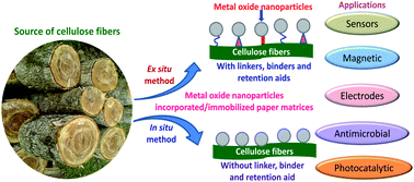
Metal oxide nanostructures of TiO2, ZnO, Fe2O3/Fe3O4, Bi2O3, CeO2, ITO, SiO2, MoO2, and WO3 have shown great potential for applications in various fields such as piezoelectric, magnetic, gas sensors, and dye sensitized solar cells due to their unique optical, electronic, conductivity, catalytic and antimicrobial properties. However, recovery and reuse of these nanostructures pose big threats from cost and environmental perspectives. Thus, various substrates have been employed for incorporating or immobilizing them but finding suitable substrate is still a big challenge. Paper, being a natural biopolymer, has been used recently to incorporate/immobilize various metal oxide nanostructures. The metal oxide nanostructures are normally adhered to the cellulose matrices through weak interactions such as van der Waals force and hence, usually have retention related issues. This was circumvented to a great extent by using suitable linkers, binders, or retention aids for the incorporation/immobilization of the nanostructures in paper matrices. Although these reagents improve retention, as well as some of the properties, they ultimately add cost to the final product. Additionally, these retention aids and linkers hinder accessibility of active surface sites of metal oxide nanostructures for their various applications. Very recently our group developed an in situ single step hydrothermal method to immobilize metal oxide nanostructures such as TiO2, ZnO, and Bi2O3 without using any binder, linker or retention aid. In this review a comprehensive account of the development of methodology for incorporation/immobilization of metal oxide nanostructures is discussed. Furthermore, how the immobilization of nanostructures evolved without using any binder, linker or retention aid is thoroughly discussed based on the chemistry of the cellulose and metal oxides. Applications of nanostructure immobilized paper matrices are highlighted and critical challenges are discussed along with directions for future research.


image file: c5ra13601f-p1.tif

Indu Chauhan

Indu Chauhan has recently received her Ph.D. (June 2015) from Department of Applied Science and Engineering, IIT Roorkee, India. She obtained her M.Tech. (2012) from IIT Roorkee and B.Tech. (2010) from Deenbandhu Chhotu Ram University of Science and Technology, India. Her research area includes synthesis of metal oxide nanostructures, nanostructures immobilized paper matrices for high-end applications with recent focus on colorimetric detection and deactivation of microbes by functionalized paper matrices.

image file: c5ra13601f-p2.tif

Sudiksha Aggrawal

Sudiksha Aggrawal is a Ph.D. student in Department of Applied Science and Engineering, IIT Roorkee, India. She obtained her M.Tech. (2015) from IIT Roorkee and B.Tech. (2013) from Deenbandhu Chhotu Ram University of Science and Technology, India. Her area of interest is incorporation of metal oxides in paper matrices for antibacterial applications and fabrication of paper matrices with various organic, inorganic and biomolecules for the detection and deactivation of microbes.

image file: c5ra13601f-p3.tif

Chandravati

Chandravati received her M.Tech. from Department of Paper Technology, IIT Roorkee, India. She did B.Tech. (2012) from Gautam Buddh Technical University, India. Her area of interest is nanotechnology in paper products and bio-nanocomposites.

image file: c5ra13601f-p4.tif

Paritosh Mohanty

Dr Paritosh Mohanty is an Assistant Professor in the Department of Applied Science and Engineering at IIT Roorkee, India. He received his B.Sc. (1995) and M.Sc. (1997) in Chemistry from the Utkal University, India and Ph.D. (2004) in Materials Science from IIT Kharagpur, India. After completing postdoctoral research at KAIST, South Korea and Lehigh University, USA, he joined as a faculty at IIT Roorkee in 2011. His research program is centered on creating advanced functional materials for protecting the environments and generating renewable energy.


1. Introduction

From ancient times until our modern age, metal oxides have played important roles in developing technologies that have helped society progress.1–5 Based on needs, metal oxides were used for making ceramic potteries in ancient times and now find uses with microelectronic circuits, sensors, piezoelectric devices, fuel cells, and catalysts. With the development of nanotechnology, applications of metal oxides have increased few fold because of enhancements from many of their physical and chemical properties as well as observations of new phenomena.6–10 Recently, considerable research has been focused on development of methodology to control structure, microstructure, and crystallographic phases of metal oxide nanostructures.11–20

Rapid increases in pollutant levels and recurrence of infectious diseases caused by various microbes in the environment pose a serious threat to human health.21–26 These were primarily caused by increases in industrialization because of high demands for various products. Thus, there was a need for development of low cost and efficient systems for purification of water and air through degradation of pollutants.21 Metal oxides such as TiO2, ZnO, Fe2O3/Fe3O4, Bi2O3, and CuO have attracted great attention as photocatalysts that use solar light to degrade organic compounds as well as for their antimicrobial activities.26–33 Most of these metal oxides have high oxidizing power when illuminated with light. They also can generate hydroxyl radicals and superoxide ions which can oxidize organic compounds and kill microbes (Scheme 1).26–30 In addition, most of them are cost effective, chemically, and thermally stable. Photocatalysis, being a surface and interface phenomenon, increases upon increasing the possible surface to volume ratio of material by synthesizing low dimensional materials.34–37 However, one of the major challenges of using these nanostructures is to recover them from a treated solution by centrifugation, filtration, or sedimentation.38


image file: c5ra13601f-s1.tif
Scheme 1 Schematic diagram representing photoinduced photocatalytic and antibacterial actions of metal oxides.32

This problem was circumvented by immobilizing or incorporating the nanostructures on various substrates such as ceramics, glass, polymers, cellulose fibers, and paper.39–48 However, each substrate has its own benefits and disadvantages.49–53 For example, ceramics are harder, bio-inert, and chemically and thermally stable.54,55 But, they also are difficult to process, weak in tension strength, and fragile.56 Glass as a substrate also has similar advantages and disadvantages.57 Polymers, such as polypropylene (PP) and polymethyl methacrylate (PMMA), can also advantageously be used as a substrate. These substrates can be used at high temperatures, adapt to different kinds of climates, are non-corrosive and have excellent moisture barrier properties.58 However, they are very fragile and easily dissolved in organic solvents. In addition, other substrates have finite resources, and are expensive and non-biodegradable. Moreover, in some cases improper disposal of substrates has led to secondary environmental pollution.59–61

Paper could be the best alternative to ceramics, glass and polymer substrates because of its biodegradability, cost effectiveness and great abundance.62–68 Using paper as a substrate is not new62–73 but with the development of nanotechnology, using paper matrices for incorporation and/or immobilization of nanostructures is progressing at a rapid pace. Nanostructure modified paper matrices have been used for the high-end applications.27,62–65,74–77 However, paper has also some disadvantages such as low mechanical strength and mechanical properties deteriorate under wet conditions.

In this review, various methodologies adopted to incorporate/immobilize metal oxide nanostructures in paper matrices are discussed along with the chemistry of the immobilization processes. Development of methodology for incorporation/immobilization with time is discussed by choosing the best available examples. Nanostructures were incorporated/immobilized in paper matrices by broadly two ways: the traditional way of incorporation/immobilization using binders, linkers, or retentions aids and more advanced ways of immobilization of nanostructures in the absence of any of these retention aids. A discussion of incorporation/immobilization of individual metal oxide nanostructures, along with investigated applications, is summarized along with their advantages and disadvantages. At the end, both methods are compared along with the challenges and future prospects.

2. Incorporation/immobilization of metal oxide nanostructures in paper matrices

Traditionally, commercial powders of sizes in the range of 0.2–20 μm were used as fillers in paper industries to improve smoothness, thermal resistance, gloss, optical, and other properties of paper matrices.78–80 Usually, these fillers adhered to the cellulose surface by weak interaction such as hydrogen bonding and van der Waals forces. Hence, binders, linkers and retention aids were added to improve retention in the final products.81–83 With the advancement of nanoscience and nanotechnology, applications in research of paper and paper products have not been ignored. Researchers working in areas of paper and paper products have also explored various possibilities. Several metal oxide nanostructures have been incorporated into paper products and the effect of these nanostructures on overall properties and applications have been investigated.63–65,67,81,83 In most of these reports, various additives have been used to improve the adherence of the nanostructures in paper matrices. In this section, detailed methodology used to incorporate/immobilize individual metal oxide nanostructures in paper matrices along with investigated properties and applications are discussed.

2.1 TiO2 nanostructures incorporated/immobilized paper matrices

The seminal work of Matsubara et al. in 1995 marks the real beginning of the research area of incorporation of nanostructures in paper matrices.64 Commercial TiO2 powder (2.0 to 10.0 wt% with respect to 100 g m−2 weight of paper matrices) with particle sizes of around 7 nm were used to incorporate polyacrylamide and polyamine as binders in paper matrices with the help of Al(OH)3.65,81,83 The standard handsheet-making method was used to prepare the paper matrices.65,81,83 Also, paper matrices modified with TiO2 nanoparticles have been explored for photocatalytic applications. Previously, in 1972, Fujishima and Honda demonstrated the potential of TiO2 as a photocatalyst to split water and several other research groups have successfully utilized this concept to degrade many other organic compounds.30,84–92 Moreover, traditionally, TiO2 has been used as one of the most common fillers in the paper industry due to its novel optical properties. Because of its photostability, abundance, and affordable price, TiO2 is a popular choice in photocatalytic reactions.93,94

Although Matsubara et al. incorporated smaller nanoparticles, agglomerated TiO2 nanostructures of size ca. 100 nm with a random distribution were observed (Fig. 1A) in the final paper matrices.64 Agglomeration was mainly due to using flocculants and retention aids that were intended to improve percent of retention. Importantly, the paper matrices degraded gaseous acetaldehyde (CH3CHO) with an initial conc. of 10 ppm in the presence of UV-light (Fig. 1B). As expected, the decomposition rate of CH3CHO increased with the increase in TiO2 content. The quantum yield of paper matrices modified with TiO2 nanoparticles was compared with one of the most photoactive commercial TiO2 powders, Degussa P-25. A higher quantum yield of 90% was reported using the paper matrix with 10 wt% TiO2 as compared with 43% in the Degussa P-25 powder.64


image file: c5ra13601f-f1.tif
Fig. 1 (A) SEM image of paper matrix incorporated with 5 wt% of TiO2 powder. (B) Decomposition of gaseous acetaldehyde by: (a) TiO2-free paper, paper matrices incorporated with: (b) 2 wt%, (c) 5 wt% and (d) 10 wt% of TiO2 powder.64

No further developments were made in this research area until 2003 when Tanaka's group reported several articles on incorporation of TiO2 nanoparticles in paper matrices and studied their photocatalytic activities.95–99 They employed various retention aids, flocculants, and binders such as polyacrylamide, ceramic fibers, zeolites-Y, and polydiallyldimethylammonium chloride (PDADMAC) along with TiO2 powder to understand effects on overall performance with paper matrices. In all of these reports, the paper matrices were also prepared by the standard handsheet-making procedure (Fig. 2).81,100 In all these reports a substantial improvement on the retention percentage was recorded from using binders and flocculants. However, the nanoparticles became agglomerated in each of the cases.81 Moreover, when ceramic fibers were used, the nanostructures normally adhered to the ceramic fibers surface rather than the cellulose fibers surface as shown in Fig. 3. Using ceramic fibers in addition to TiO2 nanoparticles improved the durability of the paper matrices as compared to the TiO2 incorporated paper matrices without ceramics fibers (Fig. 4a).81 However, using ceramic fiber took a toll on the applications. The paper matrix without incorporated TiO2 ceramic fibers decomposed gaseous CH3CHO (250 ppm) completely in 60 min as compared to 80 min taken by the paper matrices prepared by incorporating the TiO2-flocculants and TiO2-ceramic fibers (Fig. 4b). However, the authors concluded that paper matrices incorporated with both TiO2 and ceramic fibers were promising materials when both photocatalytic ability and durability are considered.81


image file: c5ra13601f-f2.tif
Fig. 2 Schematic diagram of procedure followed to prepare paper matrices incorporated with TiO2 powder.81

image file: c5ra13601f-f3.tif
Fig. 3 SEM images of the paper matrix prepared with: (a) flocculants only, (b) TiO2 powder only, (c) TiO2 and flocculants, (d) TiO2 and ceramic fibers, (e) mixture of TiO2, ceramic fibers, and flocculants. Figure d′ is the corresponding EDX-mapping image of image d.81

image file: c5ra13601f-f4.tif
Fig. 4 (a) Changes in relative tensile strength under UV irradiation: pulp matrix with flocculants (×), TiO2 matrix without flocculants (●), TiO2 matrix with flocculants (▲), and TiO2 matrix with ceramic fibers (■). (b) Photocatalytic decomposition of acetaldehyde by paper matrices incorporated with: (×) flocculants only, (●) TiO2 without flocculants, (▲) TiO2 with flocculants, and (■) TiO2 with ceramic fibers under UV light irradiation.81

Additionally, Kitaoka's group demonstrated a synergistic effect in photocatalytic activity of TiO2-incorporated paper matrices by using different types of zeolites such as A-, F-, or Y-types.83,101 The TiO2-zeolite-incorporated paper matrices were prepared following the handsheet-making procedure. Bisphenol-A (BPA) (100 μM, 50 mL) was chosen as a model compound for studying photocatalytic activity under UV light irradiation. The key feature of this work was the effect of zeolite types on overall degradation of BPA. The Y-type zeolite matrix reportedly has shown the best adsorption of BPA from solution.101 This was mainly due to the larger pore size of Y-type zeolites (ca. 0.7 nm) compared to A- and F-type zeolites.101 Synergistic photocatalytic activity of TiO2-Y-type zeolite paper matrices was attributed to higher adsorption by Y-type zeolites which facilitates degradation by TiO2 because the TiO2 nanoparticles mostly adhered to zeolite rather than cellulose matrices.101 The same group further documented that a higher porosity in resulting paper matrices have a major role in photocatalytic activity by reducing irradiation time to around 40%.101

Raillard et al. studied the effect of water vapour on kinetics of the photo-oxidation of ketones (acetone and 2-butanone) by TiO2 nanostructures supported on a nonwoven paper matrix in the presence of UV light.102 SiO2 and the organic mixture were used as binders and retention aid, respectively. Photocatalytic activity of acetone was inhibited in the presence of water vapor whereas it improved slightly for 2-butanone with a concentration higher than 2 g m−3. This was attributed to the competitive adsorption of the ketones and water vapor on the catalyst surface.

The role of pH on photocatalytic activity of TiO2-coated paper was reported by Aguedach et al.103 A size press machine was used to coat TiO2 nanoparticles (anatase, surface area >320 m2 g−1, crystallites size 5–10 nm) on non-woven paper matrices in presence of colloidal SiO2 as a binder. Two dyes, RY145 and RB5 of conc. 40 ppm, were degraded under irradiation of a 25 W mercury fluorescent lamp.

At pH 3, both RY145 and RB5 dyes were degraded substantially; however, at pH > 4.5, the rate of degradation reduced (Fig. 5). The pH dependency was attributed to a lower isoelectric point at lower pH that leads to formation of a TiOH2+ group on the surface of a TiO2/SiO2 photocatalyst-coated paper. Moreover, both the RY145 and RB5 dyes are weakly acidic and hence, would be in the neutral electrical form of ϕ-SO3H. An attractive electrostatic force between TiOH2+ and ϕ-SO3H was responsible for the higher degradation rate. At pH > 4.5, repulsive electrostatic forces existing between TiO and ϕ-SO3 prevented the adsorption of dyes to be degraded on the coated paper surface. In order to further support their concept, various salts such as NaCl, KCl, CaCl2, LiCl, and Ca(NO3)2 were added to enhance ionic strength of the solution. This in turn improved the degradation rate in the order of Ca2+ > K+ > Na+ > Li+, irrespective of the anions used.103 Similar dye degradation by the TiO2-coated non-woven fibers was performed by Barka et al. by varying pH and temperature. As expected a higher degradation was documented at lower pH (<3) and higher temperature (>30 °C).104,105


image file: c5ra13601f-f5.tif
Fig. 5 Effect of pH on the decomposition of aqueous solutions of: (a) RB5 and (b) RY145.103

The role of zeolites on efficiency of photocatalysts was further documented by Fatemi and co-workers very recently.107 TiO2 nanostructures co-doped with N and Fe (N–Fe–TiO2) were supported on a 13X zeolite using a sol–gel method.107 These supported nanostructures were incorporated in the paper matrices by the standard handsheet-making procedure using CPAM as a retention aid. The photocatalytic efficiency of the supported catalyst shows a 2.1 fold enhancement for the degradation of CH3CHO in visible light compared to unsupported TiO2 catalyst incorporated in paper matrices.107

Coating TiO2 nanostructures on filter paper surfaces was reported recently by Ko et al. and El-Sherbiny et al.67,108 In both cases binders and retention aids were used in addition to TiO2 powder. The paper matrices showed improvement in optical and photocatalytic activities. Moreover, Ko et al. investigated the effect of the crystallographic phase of TiO2 on photocatalytic properties. A paper matrix coated with an optimum ratio of anatase to rutile (0.29[thin space (1/6-em)]:[thin space (1/6-em)]0.71) showed the best optical properties. In contrast, best photocatalytic activity was shown by a paper matrix coated with a 0.7[thin space (1/6-em)]:[thin space (1/6-em)]0.3 ratio of anatase and rutile against toluene degradation.108 This was attributed to low conduction band energy in a rutile phase with lower photocatalytic oxidation capability than anatase.

Recently, Wang et al. used an interesting approach to improve photocatalytic and antibacterial activities of TiO2-incorporated paper matrices.106 TiO2 nanobelts synthesized by a hydrothermal method were further acid-corroded by treating the nanobelts with 0.02 M H2SO4 (AC TiO2). Further, these TiO2 and AC TiO2 nanobelts were decorated with Ag nanostructures by treating with an aqueous solution of AgNO3 in the presence of UV light. Paper matrices were made by using a vacuum filtration method.106 Both the photocatalytic and antibacterial activities of Ag nanostructures decorated with AC TiO2 nanobelts that incorporated paper matrices showed better results compared to all other paper matrices. This was attributed to heterostructures formed between Ag nanostructures and TiO2 nanobelts. Photocatalytic activity was tested against MO and MB, and the antibacterial activity was tested against Escherichia coli (E. coli) using the zone of inhibition method as shown in Fig. 6.106


image file: c5ra13601f-f6.tif
Fig. 6 Antibacterial activity by the zone of inhibition method shown by the paper matrices incorporated with: (a) TiO2 nanobelts, (b) AC TiO2 nanobelts, (c) Ag/TiO2 nanobelts, and (d) Ag/AC TiO2 nanobelts, respectively.106

Very recently our research group also reported the incorporation of TiO2 nanowires in paper matrices by the handsheet-making procedure and studied its optical properties. TiO2 nanowires were hydrothermally synthesized using a commercial TiO2 powder as a precursor at 200 °C for 24 h and then calcined at 400 °C for 3 h.109 The agglomerated and highly dense bundles of TiO2 nanowires, of length 1 to 7 μm, were dispersed in paper matrices (Fig. 7b). To circumvent this problem, a triblock copolymer, P123 (Pluronic 123), was used whose hydrophilic end interacted with the TiO2 nanowires forming a layer over the surface of TiO2 nanowires, thus preventing its agglomeration. These dispersed TiO2 nanowires of diameter ca. 20 to 40 nm were then incorporated into the paper matrices (Fig. 7c). However, neither of the paper matrices showed appreciable photocatalytic or antibacterial activities. This may be due to hindrance of accessible reactive sites of TiO2 nanowires.109


image file: c5ra13601f-f7.tif
Fig. 7 FESEM images of paper matrices incorporated: (a) without TiO2 NWs, (b) with TiO2 NWs, and (c) with TiO2 NWs incorporated along with surfactant P123.109 Inset of (a) shows a digital camera picture of the blank paper.

As discussed earlier, in almost all cases TiO2 was incorporated in the paper matrices in addition to flocculants, retention aids, ceramic fibers, zeolites, and silica and that hinders nanostructure agglomeration, interaction with cellulose fibers, and binds TiO2 nanostructures to paper matrices. Although the additives help TiO2 nanostructures to be retained in paper matrices, it simultaneously hinders access of total activated TiO2 surface. In order to overcome this, very recently, Ye et al. reported the use of bio-molecules to conjugate TiO2 nanostructures with cellulose fibers of paper matrices.110 This process involves biotinylation of the surface of TiO2 nanostructures followed by bioconjugation with streptavidin that has been functionalized on the cellulose surface as shown in Fig. 8.


image file: c5ra13601f-f8.tif
Fig. 8 Immobilization of TiO2 nanostructures onto paper by bioconjugation of fiber–CBM2a-strep-tag II–streptavidin with biotinylated TiO2 nanostructures.110

Photocatalytic activity of biotinylated TiO2 immobilized paper matrices was investigated against reactive black dye 5 (RB5, 23 ppm) in the absence, as well as in the presence, of UV light (23 W m−2) and these results were compared with three control specimens (Table 1).110 No regular trend in degradation of the RB5 dye was reported from any of the paper matrices in the absence of UV light (Fig. 9a). However, in the presence of UV light, around 10% degradation of RB5 was reported by control-3 paper matrix after 12 h of UV light irradiation (Fig. 9b). On the other hand, a complete degradation of RB5 dye was reported for the biotinylated TiO2 nanostructures immobilized paper matrix with 12 h of UV light exposure (Fig. 9b). These observations indicate that photocatalytic efficiency of TiO2-containing paper matrices can be improved by immobilizing individual TiO2 nanostructures on the paper surface using a linker such as streptavidin (which should not restrict the accessibility of the TiO2 surface).110

Table 1 Composition of different filter paper matrices prepared to study their photocatalytic activity110
Experiment Paper CBM2a-strep-tag-II Streptavidin TiO2
Immobilization experiment Whatman no. 1 Applied Applied Biotinylated TiO2
Control-1 Whatman no. 1 Not applied Applied Biotinylated TiO2
Control-2 Whatman no. 1 Applied Applied Untreated TiO2
Control-3 Whatman no. 1 Applied Applied APTS-modified TiO2


image file: c5ra13601f-f9.tif
Fig. 9 Photocatalytic activity of prepared paper matrices in the: (a) absence and (b) presence of UV light. [Reprinted with permission from ref. 110 © 2009 RSC Publisher.]

The incorporation/immobilization of TiO2 not only improves photocatalytic, optical, and antibacterial applications but also has a large role in improving the application as an electrode in lithium ion batteries.111 This was first reported recently by Zhao et al.111 Hierarchical TiO2 nanostructure layers were coated on filter paper using a dip coating method using a solution of tert-butyl titanate (C16H36O4Ti), ethanol (EtOH) and acetic acid at room temperature (RT). The coated papers were dried at 60 °C overnight and calcined at 450 °C for 2 h by keeping the paper between the two plates of glass (designated as TiO2-0). For comparison, hierarchical TiO2 thin film also was prepared with an additional hydrolysis treatment at 95–100 °C for 3 h in the presence of deionized water after impregnation for 2 h (designated as TiO2-h) as shown schematically in Fig. 10. The anatase TiO2 with a trace amount of brookite TiO2 was formed in both samples (TiO2-0 and TiO2-h) with an average particles size of 8–20 nm. Electrochemical performance of obtained TiO2-coated paper electrodes (TiO2-0 and TiO2-h) as lithium intercalation electrodes was examined using a galvanostatic charge and discharge half cell configuration. In the TiO2-h, discharge capacity reached 239 mA h g−1 compared to 71 mA h g−1 for the TiO2-0 electrode (Fig. 11). The hydrolysis step increases surface area of TiO2 by generating mesopores which results in improving kinetics of electron transport.111


image file: c5ra13601f-f10.tif
Fig. 10 Schematic diagram representing the fabrication of TiO2-0 and TiO2-h thin films.111

image file: c5ra13601f-f11.tif
Fig. 11 The first charge/discharge curves of cells with TiO2-0 (black) and TiO2-h (red) film electrodes at 0.5 °C.111

Recently, our research group developed an in situ hydrothermal method to immobilize metal oxide nanostructures on cellulose fibers of paper matrices without using any retention aids, binders, or linkers.19 The TiO2 nanoparticles of diameter ca. 40 to 250 nm were immobilized over the surface of cellulose fibers by hydrothermally treating commercial TiO2 powder in the presence of cellulose fibers at 150 °C for 20 h. With this method around 40% of the TiO2 nanoparticles were retained in the paper matrices. However, in the absence of cellulose fibers, TiO2 nanowires of diameter ca. 89–90 nm were obtained. This shows that cellulose plays an important role in manipulating the microstructure of nanoparticles.19

In order to understand the incorporation/immobilization of nanostructures in paper matrices, it is very important to understand the chemistry of cellulose fibers in paper matrices. As a biopolymer, interaction between cellulose fibrils depends on the number of molecular contacts and number of molecular binding sites.112 Several bonding mechanisms could be envisioned in cellulose fibers due to the possibility of various types of bonds such as, inter- and intra-molecular H-bonding, van der Waals interaction and entanglement of fibers.113–126 In cellulosic material, H-bonds usually form when a hydroxyl (OH) group interacts with an electronegative atom such as oxygen. In paper, the OH groups are engaged mostly in molecular bonding and some are associated with the adsorbed water molecule.115,116 However, the formation of fibers from fibrils is primarily due to both intermolecular and intramolecular H-bonding between cellulose chains. The various bonds possible between cellulose chains are shown schematically (Fig. 12). The H-bonds between fibrils gives structural rigidity to the fibers, whereas, bonds between the fibers holds together the fiber network of paper.115,116 More precisely, each glucose unit in a cellulose molecule can form three hydrogen bonds: two bonds (O6–H⋯O3 and O3⋯H–O6) form between chains (cellulose molecules) and the third bond forms within the chain (O2–H⋯O6) (Fig. 12).113–126


image file: c5ra13601f-f12.tif
Fig. 12 Schematic representation showing various bonds formed in a cellulose chain. The inter- and intramolecular H-bonding formations between two chains of cellulose molecules are denoted by thick and thin dotted lines, respectively. Arrows show the donor–acceptor–donor directions.116

In a wet state, cellulose fibrils are more flexible and hence, more intermolecular H-bonding can be formed.115,116 Certain chemical groups, such as carboxylic groups, dissociate in water promoting fiber swelling and contribute to improved flexibility and conformability to the fiber wall.126–129 High content of carboxylic acid groups on fiber surfaces may also increase inter-fiber bond strength.128,130 However, the fibrils put constraints on formation of H-bonds in the dry state. Besides H-bonds, van der Waals forces are important in inter-fiber bonding. The cohesion of a wet paper web comes from van der Waals forces. Covalent and ionic bonds may also form between fibers when additives are added.127,131 Water does not influence covalent bonds much as compared to H-bondings.127 Because of this, in many earlier reports retention aids, binders, and linkers were used to incorporate/immobilize metal oxide nanostructures in paper matrices.42–45 These additives in turn improve connectivity between metal oxides and cellulosic materials. However, the two major drawbacks of using additives are the additional cost and hindrance of the active surface for applications.21,31–45 However, in the developed in situ hydrothermal method, the formation of a covalent bond between the cellulose fibers and metal oxide nanostructures was achieved; this helps in retaining the high percent of nanoparticles in paper matrices. Another advantage of the in situ method is accessibility of active sites for the applications. These active sites in the nanostructures could easily be hindered by addition of retention aids.

A possible nucleation, growth, and immobilization mechanism of TiO2 nanoparticles on the cellulose fibers surface was also proposed. As discussed, hydroxyl groups present both on the surface of cellulose fibers and the surface of TiO2 nanoparticles interact with each other through H-bonding (Fig. 13).19 Dehydration leads to the formation of permanent covalent bonds between TiO2 and cellulose fibers as shown in Fig. 13. This helps to generate TiO2 nuclei which grow on the cellulose fibers surface. The mechanism of covalent bond formation was confirmed from spectroscopic investigations such as XPS and FTIR.19


image file: c5ra13601f-f13.tif
Fig. 13 Mechanism for the nucleation, growth, and immobilization of TiO2 nanospheres on the surface of cellulose fibers of paper matrices.19

image file: c5ra13601f-f14.tif
Fig. 14 Photocatalytic degradation of: (a) methyl orange and (b) formaldehyde by paper matrices immobilized with 2.5, 9.0, 13.0 and 21.0 wt% TiO2 nanospheres.20

These TiO2-immobilized paper matrices also have interesting antibacterial and photocatalytic activities.19 The photocatalytic activity was studied by degrading a synthetic organic dye, methyl orange, (0.03 mM) in the presence of both UV-light and sunlight. Around 95% of methyl orange was degraded in 24 and 3 h on exposure to UV light and sunlight, respectively. Almost a complete growth reduction of E. coli bacteria by these paper matrices was achieved within 9 h of visible light exposure (Table 2).

Table 2 Antibacterial activity of TiO2-decorated paper matrices against E. coli19a
Sample ID Bacterial count (CFU) on illumination with fluorescent light (0.276 J cm−2) for different exposure times
3 h 6 h 9 h
a Viable bacteria were monitored by the plate count method when counting the colony forming units (CFUs).
0.0 wt% TiO2 56 × 105 54 × 105 62 × 105
1.0 wt% TiO2 52 × 105 25 × 105 10 × 105
3.5 wt% TiO2 49 × 105 23 × 105 10 × 105
6.0 wt% TiO2 25 × 105 20 × 105 3 × 10⁵
10.0 wt% TiO2 2 × 10⁵ 2 × 10⁵ No growth


The covalent bonding concept was further proved by using an organo-metallic precursor of TiO2 i.e., titanium isopropoxide, Ti{OCH(CH3)2}4. Similar hydrothermal treatment at a lower temperature of 80 °C for 14 h resulted in the immobilization of TiO2 nanoparticles of even smaller dimension of 10 to 20 nm on the cellulose surface. The organometallic precursor has a profound role in the immobilization process.20

The retention of as high as 90% was achieved when the organometallic precursor was used compared to only 40% when TiO2 powder was used as a precursor. Moreover, the immobilized TiO2 nanoparticles are of much smaller dimensions. The only drawback of using the organometallic precursor is its cost which is higher than commercial TiO2 powder. The photodegradation of methyl orange and formaldehyde were 100 and 82%, respectively, using these paper matrices within just 3 h of sunlight exposure (Fig. 14).20

2.2. ZnO nanostructures incorporated/immobilized paper matrices

ZnO is considered to be a very important industrial material because of its application in various fields including catalysis, electronics, and biotechnology.132–134 It is a wide band gap semiconductor with band-gap energy of 3.15–3.35 eV. Depending upon the crystal structure and microstructure, the band-gap energy could also be tuned.135–137 Major applications of ZnO nanostructures are in the fields of nanolasers, gas sensors, piezoelectric nanogenerators, and dye-sensitized solar cells.138–141 Recently, a number of research reviews have been published on the synthesis, fabrication and application of ZnO nanostructures.138–141 Wang and co-workers did substantial work on various ZnO nanostructures and demonstrated piezoelectric behavior in nanobelts and nanowires.142,143 Thus, incorporated/immobilized ZnO nanostructures on paper matrices have drawn immense interest in several research groups working in these areas.27,62,63,66,68,144–148

Ghule et al. in 2006 for the first time coated ZnO nanoparticles of diameters ca. 20 nm on paper matrices using an ultrasound wave assisted method as shown schematically in Fig. 15.66 The ZnO loading in the paper matrices depends on the sonication time which was varied from 5 to 30 min. However, at the beginning of the sonication (within 5 min), maximum loading (14.3 wt%) could be achieved and there was only marginal increase in the loading (17.7 wt%) after 30 min of sonication. Thus, the authors suggested a sonication time of 10–20 min was enough to have substantial coating of ZnO nanoparticles in paper matrices.66 The antibacterial activity of a ZnO nanostructures-coated paper matrix was investigated against E. coli in the presence of UV light. Around 99.99% reduction in E. coli growth in 24 h by the ZnO nanostructures coated paper matrices was reported.66,67


image file: c5ra13601f-f15.tif
Fig. 15 Schematic diagram representing the coating of ZnO nanostructures on the surface of a paper matrix by the sonication method.66

Growth of ZnO nanorods (80 nm width and 600 nm length) on a paper surface was reported by Dutta and co-workers by a two-step seed and growth method using hydrothermal synthesis.62,68 The first step involved the formation of seed particles using Zn(CH3COO)2·2H2O and NaOH at 70 °C and the second step was the growth of nanorods hydrothermally at 90 °C for 10 h using Zn(NO3)2·6H2O as the ZnO source and hexamethylenetetramine (CH2)6N4 as a structure directing agent on the paper surface (Fig. 16).68


image file: c5ra13601f-f16.tif
Fig. 16 Scanning electron micrographs showing (a) porous structure of the paper, (b) ZnO nanorods growing in the paper pores, (c) ZnO nanorods on the top surface of the paper, and (d) close up view of the ZnO nanorods.68

These nanorod-incorporated paper matrices have shown improved photocatalytic and antibacterial activities. Photocatalytic activity of prepared paper sheets was tested by degrading methyl blue and methyl orange dyes (initial conc. 10 μM) in presence of visible light (963 W m−2).

Around 93% and 30% of methyl blue and methyl orange was degraded by ZnO nanorods-coated paper matrices in 120 min, respectively.68 Furthermore, the ZnO nanorods coated paper matrices were used to study antibacterial activity against E. coli by the zone of inhibition method. The paper matrices showed an enhanced antibacterial activity in the presence of visible light (11 W m−2) compared to the dark. The maximum inhibition zone of 3.2 cm2 was observed in the dark under illumination for 72 h; however, the inhibition zone area increased to 4.4 and 4.9 cm2 under illumination of visible light for 24 and 48 h, respectively (Fig. 17).68


image file: c5ra13601f-f17.tif
Fig. 17 Antibacterial activity of ZnO paper sheets in light and dark conditions.

The same group further extended the antimicrobial study of ZnO nanorods incorporated in paper matrices using the zone of inhibition method.62 Both gram positive (S. aureus) and gram negative (E. coli) bacteria as well as a common airborne fungus (A. niger) were deactivated using these paper matrices. The zone of inhibition very much depends on the light source. When halogen and fluorescent lamps were used, the zone of inhibition was 239% and 163% for E. coli, 102% and 70% for S. aureus, and 224% and 183% for A. niger, respectively, in an area that was the area of the paper sample. A representative zone of inhibition was shown in Fig. 18 for the A. niger.62


image file: c5ra13601f-f18.tif
Fig. 18 Growth of A. niger in the presence of (a) untreated paper and (b) paper with ZnO nanorods, after 72 h of growth.62

Recently, Khatri et al. reported the incorporation of saccharide capped-ZnO nanoparticles in paper matrices and studied the antimicrobial activities as well as antibody immobilization.63 The saccharide capped-ZnO nanoparticles were synthesized by a microwave assisted method using carbohydrates (glucose, G; sucrose, S; starch, St and alginic acid, AA), Zn(CH3COO)2·2H2O and NaOH solution at 800 W for 30 sec.63 The average particle sizes of 30.9 nm, 28.3 nm, 23.6, and 19.0 nm were reported for G-ZnO, S-ZnO, St-ZnO, and AA-ZnO, respectively. These nanoparticles were incorporated in paper matrices during paper making by the handsheet making procedure. Among all the paper matrices, the one incorporated with AA-ZnO nanoparticles showed the best performance by reducing around 86.06 and 81.71% of E. coli and S. aureus bacterial growth, respectively. Furthermore, the AA-ZnO paper matrix showed antifungal activity by preventing degradation of cellulose fibers by G. trabeum fungus (Fig. 19a and b). Additionally, an RBC agglutination test reported 96.0% retention of antibodies immobilized on AA-ZnO paper matrix (Fig. 19c). This best performance was attributed to the smaller size of the nanoparticles compared to the other saccharide capped ZnO nanoparticles.


image file: c5ra13601f-f19.tif
Fig. 19 (a) Fungal degradation of paper matrices, (b) resistance to fungal attack by AA-ZnO NPs paper matrix, and (c) RBC agglutination test done on AA-ZnO NPs paper matrix.63

Martins et al. demonstrated the coating of ZnO nanoparticles and a nanofibrillated cellulose (NFC) composite on paper matrices and studied the antibacterial activity.27 The surface modified NFC by poly(diallyldimethylammonium chloride) and poly(sodium 4-styrenesulfonate) was mixed with ZnO nanoparticles of size ∼40.7 nm to form the nanocomposites (NFC/ZnO). These NFC/ZnO nanocomposites were then coated on the paper surface using a size press machine. Antibacterial activity of the prepared paper matrices was studied against S. aureus, B. cereus, and K. pneumonia in the dark as well as under irradiation of solar light (8000 Lux). In the dark, the NFC/ZnO paper matrices showed some antibacterial activity, however, it was enhanced greatly under irradiation with solar light (Fig. 20).27


image file: c5ra13601f-f20.tif
Fig. 20 Antibacterial activity of coated papers against S. aureus, B. aureus, and K. pneumoniae in the presence and in the absence of light.27

Miura et al. prepared catalytic cellulosic papers for conversion of 4-nitophenol to 4-aminophenol. The paper matrices were prepared by synthesizing gold nanoparticles preloaded into commercial ZnO whiskers and these were then incorporated into paper matrices.144


image file: c5ra13601f-f21.tif
Fig. 21 Time dependent absorption spectra (a) 4-nitophenol profile, (b) for the catalytic reduction of 4-nitophenol over paper matrix incorporated with gold nanoparticles preloaded into the commercial ZnO whiskers.144

The reduction of 4-nitophenol to 4-aminophenol was monitored using a UV-visible spectrophotometer as shown in Fig. 21.144 In these spectra, the reduction in absorbance of the characteristic peak at 400 nm of 4-nitrophenol was seen with the emergence of new peak 293 nm, which was the characteristic absorption of 4-aminophenol. Further, the reaction followed pseudo-first-order kinetics (Fig. 21). The reusability of the paper strips was also tested and it was observed that, after five cycles, the paper catalyst was stable in aqueous media.144 Ajayan and co-workers prepared a flexible piezoelectric strain sensor composed of ZnO-paper nanocomposite by a solvothermal method.145 Gimenez et al. further prepared a photoconductive UV sensor with ZnO coated paper.146 Optical characteristics and conductivity of paper products with ZnO nanocrystals were studied. Graphite layers were formed by a pencil line over the surface to make it conducting. The crystals were uniformly distributed over the surface with a size of around a few hundred nanometres. Paper shows more linear photoconductivity as compared to glass.146 Paper only with graphite electrodes measured current values on the order of 10−9 in dark and when they were exposed to UV, there was a slight increase in the current. When paper with ZnO was tested the same way, the current measured was on the order of 10−8 in dark (Fig. 22).146


image file: c5ra13601f-f22.tif
Fig. 22 Current versus voltage measurements of paper matrices with and without ZnO. Behaviour of paper matrix with ZnO (solid lines) is shown under dark conditions and under UV at 5 and 10 cm from the sample. The effect of UV light on a paper matrix without ZnO (circles) is too small to be appreciated in the plot.146

Similarly, Manekkathodi et al. prepared ZnO nanowires (dia. 60–70 nm and length of 60–70 nm) (Fig. 23) with paper for cheap and flexible electronics.147 Photoemission at 379 nm and a broad emission from 475–620 nm were observed in cathode luminescence (CL).


image file: c5ra13601f-f23.tif
Fig. 23 (a) Representative FESEM image of aligned ZnO nanorods. (b) TEM image of a single nanorod. (c) HRTEM image taken from the edge of the ZnO nanorod. Inset: Corresponding SAED pattern.147

There was an increase in current with an increase in bias voltage both in the dark and under UV illumination. The increase was in the order of 80–85 times (IUV/Idark) which is due to electron hole generation by photoexcitaion.147 The photocurrent can be switched from the ON to OFF state with a sensitivity of 60 (ImaxImin)/Imin, at a low bias voltage of 5 V. Conductivity in the dark was lower than that with illumination by photons, due to generation of electron holes. The electron trapping effects are comparatively slower than relaxation, which is due to fast discharge and recapture of chemisorbed oxygen molecules.147

Following the similar in situ hydrothermal procedure as discussed above for the immobilization of TiO2 nanoparticles, ZnO nanowires of diameter ca. 130 to 500 nm were immobilized on the surface of cellulose fibers at 140 °C for 14 h by a hydrothermal method using ZnCl2 as the ZnO precursor (Fig. 24).148 Different ZnO contents were used to check the efficacy in the antibacterial application. The retention percent of the ZnO nanowires could be as high as 70% of the initial ZnO concentration used. The mechanism of the immobilization of the ZnO nanowires was very similar to the above discussed mechanism for immobilization of TiO2 nanoparticles. However, it was interesting to see that, unlike in most of the reports on nanowires immobilization in paper matrices, the ZnO nanowires in this case were aligned along the cellulose fiber axis. This further supports the mechanism as the covalent bond was formed between cellulose and ZnO throughout the nanowires length and not only the base of the nanowires (Fig. 25).


image file: c5ra13601f-f24.tif
Fig. 24 FE-SEM images of paper matrices (a) 0.0Z-P, (b) 2.0Z-P, (c) 7.0Z-P, (d) 10.0Z-P, and (e) 18.0Z-P. (f) FESEM image of the ZnO nanowires synthesized in absence of cellulose fibers. Insets show the high magnification FESEM images of the respective paper.148

image file: c5ra13601f-f25.tif
Fig. 25 Mechanism for the nucleation and growth of ZnO NWs on a cellulose fiber surface: (a) cellulose surface with hydroxyl groups, (b) interaction of Zn2+ with hydroxyl groups on a cellulose surface, (c) nucleation of ZnO, and (d) growth of the ZnO nanowires.148

Further, the antibacterial activity was studied by inhibiting growth of E. coli in the presence of visible light. Around 99.99% reduction in E. coli growth was observed with the paper matrices after just 9 h of visible light irradiation (Fig. 26).145


image file: c5ra13601f-f26.tif
Fig. 26 Images of antibacterial activity of ZnO-immobilized paper matrices under visible light illumination of: (a) 3, (b) 6, and (c) 9 h.148 (d) Digital camera picture of ZnO-immobilized paper matrix used to study the antibacterial activity.148

2.3. Paper matrices incorporated/immobilized with iron oxide nanostructures

Carrazana-Garcia et al. reported the incorporation of magnetic nanostructures in cellulose fibers using a lumen loading method.149 Synthesis of the nanostructures in a pulp suspension with vigorous stirring forced the nanostructures to go into the lumen of the cellulose fibers. Ferrite or cobalt ferrite nanostructures of diameter 20–90 nm were synthesized by treating a M2+ chlorides (M = Fe or Fe + Co) solution with NaOH in the pulp suspension. The modified cellulose fibers were used to prepare the paper matrices with the help of a Buchner funnel. At a low Co conc., the major phase was FeOOH; however, at an increased Co conc., glassy grains of a spinel phase with different degrees of Fe substitution was formed. As usual, the coercivity (Hc) of the magnetic paper was proportional to Co content (Fig. 27).149
image file: c5ra13601f-f27.tif
Fig. 27 Magnetization vs. temperature graph for three ferrite samples with different Co/Fe molar contents: (I) 33.3%, (II) 20%, and (III) 0%.149

image file: c5ra13601f-f28.tif
Fig. 28 (a) Increase in magnetic properties of paper matrices with an increase in the degree of loading and (b) hysteresis loops of paper containing magnetic nanostructures (MP6) and cobalt ferrite nanostructures (MP7).150

Preparation of magnetic paper using a chemical co-precipitation method was reported by Chai et al.150 A mixture of FeCl2·4H2O and FeCl3 was added in a cellulose fiber suspension in an inert atmosphere under continuous stirring. Different paper matrices were made by varying the stirring speed from 200 to 2000 rpm for 1 h and also by varying temperature. Furthermore, FeCl2·4H2O was replaced by CoCl2·6H2O to make CoFe2O4. Depending upon the synthesis conditions, nanostructures of sizes 33 nm to 100 nm were incorporated into the lumen of the cellulose fibers. The magnetic properties of these paper matrices showed a linear relationship with the degree of loading (Fig. 28a) and the maximum Hc of 29.6 Oe was observed with a loading of 37.3% (Fig. 28b). Interestingly, the Hc could further be increased up to 219.4 Oe in the CoFe2O4 incorporated paper matrices (Fig. 28b).150

In an attempt to make use of the paper matrices for body warmers, Kumamoto et al. investigated the possibilities of preparing steam generating paper matrices by incorporating commercial iron powder, activated carbon, vermiculite, and sodium polyacrylate into paper matrices.74 A polyaminopolyamide-epichlorohydrin (PAE) resin and carboxymethylated cellulose (CMC) were added as retention aids and the paper matrices were prepared by the handsheet making procedure. The steam generating behaviour of the prepared paper matrices were studied by treating with NaCl (Fig. 29a).74 It was reported that the an optimal weight ratio of 86[thin space (1/6-em)]:[thin space (1/6-em)]6:8 for the iron (Fig. 29b) cellulose fibers and activated carbon showed the best steam generating behaviour of the most stable temperature profile and longest duration time (>10 h) at 40–41 °C, making it suitable for body warmers (Fig. 29b).


image file: c5ra13601f-f29.tif
Fig. 29 (a) Thermographic image of a steam-generating sheet and (b) temperature profile of the paper sheet prepared with different component ratios.74

2.4. Other metal oxide nanostructures immobilized/incorporated in paper matrices

Ornatska et al. prepared ceria nanostructures-based paper matrices for colorimetric detection of glucose (Fig. 30).151 Glucose oxidase and ceria nanostructures were co-immobilized on filter paper using a salinization procedure for the construction of a glucose sensor.151 Initially, Whatman no. 1 filter paper was soaked in a colloidal solution of ceria for 10 min, and then dried at 70 °C. Then the ceria paper was dipped in 5% APTS (aminopropyltrimethoxysilane) solution in ethanol and then dried. The silane treatment was used for stabilization of ceria nanostructures on the filter paper because without using silica, the binding was very weak and there was a problem of leaching. APTS helps form siloxane bridges through hydrogen bonding between the hydroxyl rich surface of cellulose and hydroxylated ceria. Amino functionalities were also provided by APTS for the grafting of glucose oxidase.151 For co-immobilization of glucose oxidase on a ceria paper strip, freshly prepared ceria papers were soaked in 1% chitosan solution (in 0.5% succinic acid), followed by treatment with glutaraldehyde. Then, glucose oxidase (10 μL of 9 mg mL−1) was applied on the paper strips. Phosphate buffer at pH 7.4 was used to rinse the paper strips and then they were air dried.151
image file: c5ra13601f-f30.tif
Fig. 30 Colorimetric bioassay for detection of glucose based on ceria paper.151

Ceria based paper matrices can detect H2O2 in a concentration range of 2.5 to 100 mM. The color change was observed from white-yellowish to dark orange (hydroxylated Ce4+). When ceria came in contact with H2O2 there was a change in oxidation state of ceria from Ce3+ → Ce4+ as H2O2 acts as both oxidising and reducing agent.151 H2O2 is released when glucose oxidase comes in contact with glucose, hence ceria based paper was indirectly sensing the concentration of glucose.151 The H2O2 sensing activity of the ceria based paper was not pH dependent as the group has also tested the activity of paper at different ranges of pH ranging from 2 to 10.5 and the colorimetric response was found to be the same. The minimum detection limit of glucose was found to be 0.5 mM with an assay time of 10 min. Ceria paper had shown excellent storage capacity even at RT after storing it for 79 days. The paper had also shown reusability; when it was used 10 times there was no significant loss in the activity.151

Conducting paper using indium tin oxide (ITO) was developed by Peng et al.152 Initially, polyethyleneimine (PEI) was coated on wood fibers and then poly(sodium 4-styrenesulfonate) was deposited using a layer-by-layer assembly method. Further, several bilayers of ITO were coated followed by preparation of a paper sheet by traditional paper making methods.152 Zeta potential of both the wood fibers and ITO nanostructures was measured to evaluate surface charge intensity. Aggregation of ITO nanoparticles led to the formation of interconnected conductive paths throughout the paper matrix. With an increase in bilayers, the conductivity of the paper matrices increased. After ten bilayer coatings, measured DC conductivity (σdc) was found to be 5.2 × 10⁻⁶ S cm−1 in the in-plane direction, and 1.9 × 10⁻⁸ S cm−1 in the through-the-thickness (TT) direction.152

Monk et al. prepared paper incorporated with inorganic electrochromes such as molybdenum oxide, Prussian blue, and tungsten oxide within mashed paper. Organic electrochromes like benzyl viologen and methyl viologen were incorporated by pre-soaking.153 Different types of papers such as Whatman filter paper no. 1, photocopy paper of 80 gsm from ‘Volumax’, and white card of 160 gsm from Wiggins Teape were used. For incorporation of inorganic electrochromes, tiny pieces of filter paper were pre-soaked in 1 mol dm−3 for 2 days and then powdered electrochrome (5 g per 100 g) was mixed with the paper mesh.153 A brass counter electrode was used to make the paper smooth. In the case of organic electrochromes, soaking of paper sheets was done in a solution of KCl (1 mol dm−3) and electrochrome (20 mmol dm−3) for 1 h.153 Tungsten trioxide and molybdenum oxide were almost similar – both chemically and electrochemically – and were the best electrochromes in paper which formed the best permanent electrochromic writing medium. Methyl viologen forms the most intense color but the color was only temporary. Prussian blue was not good in paper as an electrochrome.153

Ogihara et al. prepared hydrophobic paper by spray coating SiO2 nanoparticles on the paper surfaces. The SiO2 nanoparticles were dispersed in different alcohols (ethanol, 1-propanol, and 1-butanol) by sonication for 20 min and then sprayed over the printing paper.154 Average particle sizes of 470, 410, and 350 nm were observed in ethanol, 1-propanol and 1-butanol, respectively. As expected, with the increasing SiO2 content, the hydrophobicity of the paper matrix also increased. The interesting phenomenon observed was the effect of the solvent on the hydrophobicity of the paper matrices. It was reported that the contact angle (CA) and sliding angle (SA), which defines the hydrophobicity, depended on the kind of alcohol used. With an increase in the length of the hydrocarbon chains in the alcohols, the hydrophobicity decreased.154 Surface hydrophobicity also depends on surface energy and surface roughness. The surface energy of the SiO2 nanoparticles was the same; therefore, the surface roughness, composed of SiO2 nanoparticles, determined the surface hydrophobicity. Nanoparticles obtained by using the ethanol suspension were the largest and thus the paper matrices prepared by using an ethanol suspension had the rougher surface as compared to others and thus, was super-hydrophobic with the CA > 150° and SA < 10° (Table 3 and Fig. 31).

Table 3 Contact and sliding angles (CA and SA) of SiO2 nanoparticle coatings on paper substrates154
Alcohol Particle size (nm) Contact angle (degree) Sliding angle (degree)
a Sliding angles of these coating could not be measured because a water droplet did not roll off at any angle (i.e., a water droplet was pinned).
Ethanol 25 155 7
1-Propanol 25 154 16
1-Butanol 25 149 50
Ethanol 250 148 a
Ethanol 500 126 a


image file: c5ra13601f-f31.tif
Fig. 31 Contact and sliding angles (CA and SA) of paper substrates spray-coated with different amounts of SiO2 nanoparticles (25 nm).154

Very recently, our group further extended the in situ hydrothermal method to immobilize Bi2O3 nanoparticles in paper matrices, similar to the above discussed immobilization of TiO2 and ZnO nanostructures.155 Further, the antibacterial properties of these paper matrices was studied against E. coli in visible light. Efficacy of the paper matrices for antibacterial activity with different Bi2O3 content was probed. Average diameters of nanoparticles immobilized on the surfaces of the cellulose fibers was ca. 30–100 nm.155

Antibacterial activity of these paper matrices was studied both in the dark as well as in the presence of visible light. In the dark, the Bi2O3 nanoparticles immobilized paper matrices did not show any antibacterial activity (Table 4). However, in the presence of visible light almost complete deactivation of E. coli bacteria was observed after 9 h of visible light irradiation (Table 5).155

Table 4 Antibacterial activity of paper matrices against E. coli 1698 monitored by standard plate count method in the absence visible light155
Sample ID Bacterial count (CFU) in the dark for different exposure times with an initial count of 358 × 105
3 h 6 h 9 h
0.0BO-P 320 × 105 340 × 105 46 × 105
3.0BO-P 298 × 105 150 × 105 + colonies merge 123 × 105 + colonies merge
5.0BO-P 301 × 105 298 × 105 260 × 105


Table 5 Antibacterial activity of paper matrices against E. coli 1698 monitored by standard plate count method in the presence of visible light155
Sample ID Bacterial count (CFU) on illumination with fluorescent light (1.6 J cm−2) for different exposure times
3 h 6 h 9 h
0.0BO-P 348 × 105 264 × 105 270 × 105
3.0BO-P 266 × 105 89 × 105 11 × 105
5.0BO-P 143 × 105 2 × 10⁵ No growth


Both the methods i.e., incorporation and immobilization of nanoparticles in paper matrices with binders and without binders, have their own advantages and disadvantages with respect to synthesis as well as application point of views. A few of the important points are listed below:

• As discussed above, because of the formation of covalent bonds between metal oxides and cellulose, immobilization of the nanostructures by an in situ method is advantageous over an ex situ method. The higher retention percent, accessibility of nanoparticles surfaces for the applications, uniform distribution of them on the surface of cellulose fibers, avoiding use of retention aids, and non-removal of the nanoparticles during their use (the nanoparticles did not come out even on sonication)148 are some of the major advantages.

• Another important point which distinguishes the in situ and ex situ methods is the ease of recyclability. As neither a retention aid nor any linker was used for the immobilization of a nanostructure by the in situ method, recyclability could be more practically feasible without adding nanoparticles in every cycle. As the ex situ method depends on linkers and retention aids, the steps involved for nanoparticles immobilization have to be repeated for every cycle.

• It was reported that the photo-generated free radicals by metal oxide nanoparticles in paper matrices depolymerised the cellulose fibers over time. This led to deterioration of the mechanical strength81 and hence, the life time of the paper matrices immobilized with the nanoparticles. Thus, use of retention aids, which hinder direct contact between metal oxide nanoparticles with the cellulose surface, could restrict free radicals to interact with the cellulose, thereby not affecting the overall mechanical strength to a greater extent.

3. Summary and outlook

Development of paper-based products for applications other than writing has forced researchers and industries to look for various materials with inherent properties and tuneable structures and microstructures that could be used as fillers. For a long time, bulk powders of CaCO3, talc, kaolin, and TiO2 have been used as fillers in the paper industry to enhance paper properties such as optical, thickness, and smoothness. With the development of nanoscience and nanotechnology, the urge to use nanostructures as fillers has made a substantial contribution towards the development of papers for high end applications. However, the chemistry of the incorporation/immobilization was given least importance. Retention of the nanostructures in the paper matrices always remained a challenge due to very weak interactions (H-bonding and van der Waals forces) that existed between the nanostructures and cellulose surfaces. Recently, our research group developed a single step in situ hydrothermal method for the immobilization of metal oxide (TiO2, ZnO and Bi2O3) nanostructures through the covalent bond formation between cellulose and the metal oxides.

In this review article both the ex situ as well as in situ methods used to incorporate/immobilize metal oxide nanostructures in paper matrices were discussed with citing of recent works by several research groups. The ex situ method, where nanostructures were synthesized first and then were incorporated into paper matrices by the standard handsheet making method, was associated with the problem of retention, which was circumvented by using retention aids, flocculants and binders. The role of these retention aids in the applications of these paper matrices was reviewed. The in situ methods were comparatively simpler methods where metal oxides nanostructure synthesis and immobilization in paper matrices was carried out simultaneously. Immobilization of these metal oxide nanostructures, facilitated by covalent bond formation, is fundamentally a new concept. This could completely change plausible ways of immobilization. Furthermore, it was interesting to note that the interaction of cellulose with metal oxides manipulated the microstructures and crystallographic phase of the metal oxides during the immobilization process. Nanostructure-immobilized paper matrices were investigated for various applications including device fabrication, catalysis, antimicrobial activity, diagnostic, and detection abilities. These render this review article interesting for the chemistry, nanotechnology, polymer science, paper technology, and materials chemistry communities. Moreover, these paper matrices could open up new avenues for applications in packaging technology.

However, there are still a few challenges associated with this method which need to be conquered. It is always a big challenge to control the microstructure and crystallographic phases of immobilized metal oxide nanostructures. Thus, multiple experiments have to be made to study the effects of experimental parameters on the microstructures and, in many instances, a trial and error method has been adopted to obtain the desired crystallographic phase of the metal oxide nanostructures. Also, radical ions could deteriorate the cellulose, which ultimately could reduce the mechanical strength of the paper matrices. However, there was a trade-off between the immobilization process adopted for higher retention and the mechanical strength. From an application point of view, as these paper matrices would be used for high-end applications with some predetermined lifetime, thus, the mechanical strength could be compromised to some extent.

Acknowledgements

The work was supported by the Department of Science and Technology, Government of India (Grant No. INT/MEXICO/P-03/2012). IC acknowledges Ministry of Human Resource Development (MHRD), Government of India, for the financial support.

References

  1. Trace Inorganics in Water, ed. E. A. Jenne, American Chemical Society, Washington, 1968 Search PubMed.
  2. E. R. Sholkovitz and D. Copland, Geochim. Cosmochim. Acta, 1981, 45, 181–189 CrossRef CAS.
  3. L. B. Young and H. H. Harvey, Geochim. Cosmochim. Acta, 1992, 56, 1175–1186 CrossRef CAS.
  4. The surface science of metal oxides, ed. Cox P. A., Cambridge university press, 1996 Search PubMed.
  5. D. Dong, Y. M. Nelson, L. W. Lion, M. L. Shuler and W. C. Ghiorse, Water Res., 2000, 34, 427–436 CrossRef CAS.
  6. J. F. Estes and H. Bruchmann, US Pat., US3288615, 1966.
  7. Electroceramics: materials, properties, applications, ed. A. J. Moulson and J. M. Herbert, John Wiley & Sons, 2003 Search PubMed.
  8. Modern ceramic engineering: properties, processing and use in design, ed. D. Richerson, D. W. Richerson and W. E. Lee, CRC press, 2005 Search PubMed.
  9. H. Zhao, Y. N. Xu and J. Q. Liu, Adv. Mater. Res., 2011, 304, 31–35 CrossRef CAS.
  10. H. S. Lim, N. Nafarizal, M. Z. Sahdan, S. H. Dahlan, Z. Zainal Abidin, M. Y. Ismail, F. C. Seman, M. K. Suaidi, F. B. Ahmad, Z. M. Rosli, J. M. Juoi and G. I. Kiani, Adv. Mater. Res., 2014, 925, 630–634 CrossRef CAS.
  11. X. Wang and Y. Li, J. Am. Chem. Soc., 2002, 124, 2880–2881 CrossRef CAS PubMed.
  12. J. Cheon, N.-J. Kang, S.-M. Lee, J.-H. Lee, J.-H. Yoon and S. J. Oh, J. Am. Chem. Soc., 2004, 126, 1950–1951 CrossRef CAS PubMed.
  13. L. Vayssieres, K. Keis, A. Hagfeldt and S.-E. Lindquist, Chem. Mater., 2001, 13, 4395–4398 CrossRef CAS.
  14. F. Ernst, Mater. Sci. Eng., R, 1995, 14, 97–156 CrossRef.
  15. R. Richards, W. Li, S. Decker, C. Davidson, O. Koper, V. Zaikovski, A. Volodin, T. Rieker and K. J. Klabunde, J. Am. Chem. Soc., 2000, 122, 4921–4925 CrossRef CAS.
  16. H. L. Meyerheim, R. Popescu, J. Kirschner, N. Jedrecy, M. Sauvage-Simkin, B. Heinrich and R. Pinchaux, Phys. Rev. Lett., 2001, 87, 076102 CrossRef CAS.
  17. X. Wen, S. Wang, Y. Ding, Z. L. Wang and S. Yang, J. Phys. Chem. B, 2005, 109, 215–220 CrossRef CAS PubMed.
  18. A. A. Bolzan, C. Fong, B. J. Kennedy and C. J. Howard, Acta Crystallogr., 1997, 53, 373–380 Search PubMed.
  19. I. Chauhan and P. Mohanty, Cellulose, 2015, 22, 507–519 CrossRef CAS.
  20. I. Chauhan and P. Mohanty, RSC Adv., 2014, 4, 57885–57890 RSC.
  21. N. M. Bedford, M. Pelaez, C. Han, D. D. Dionysiou and A. J. Steckl, J. Mater. Chem., 2012, 22, 12666–12674 RSC.
  22. S. Prabhu and E. K. Poulose, Int. Nano Lett., 2012, 2, 1–10 CrossRef.
  23. C. Greulich, D. Braun, A. Peetsch, J. Diendorf, B. Siebers, M. Epple and M. Koller, RSC Adv., 2012, 2, 6981–6987 RSC.
  24. C. S. Yah, Biomed. Res., 2013, 24, 400–413 CAS.
  25. A. Gerber, M. Bundschuh, D. Klingelhofer and D. A. Groneberg, J. Occup. Med. Toxicol., 2013, 8, 1–6 CrossRef PubMed.
  26. A. R. Gliga, S. Skoglund, I. O. Wallinder, B. Fadeel and H. L. Karlsson, Part. Fibre Toxicol., 2014, 11, 1–17 CrossRef PubMed.
  27. N. C. T. Martins, C. S. R. Freire, C. P. Neto, A. J. D. Silvestre, J. Causio, G. Baldi, P. Sadocco and T. Trindade, Colloids Surf., A, 2013, 417, 111–119 CrossRef CAS PubMed.
  28. C. Chen, J. Liu, P. Liu and B. Yu, Adv. Chem. Eng. Sci., 2011, 1, 9–14 CrossRef CAS.
  29. M. L. Curri, R. Comparelli, P. D. Cozzoli, G. Mascolo and A. Agostiano, Mater. Sci. Eng., C, 2003, 23, 285–289 CrossRef.
  30. A. Sarkar, A. Shchukarev, A. R. Leino, K. Kordas, J. P. Mikkola, P. O. Petrov, E. S. Tuchina, A. P. Popov, M. E. Darvin, M. C. Meinke, J. Lademann and V. V. Tuchin, Nanotechnology, 2012, 23, 475711 CrossRef PubMed.
  31. A. Toolabi, M. R. Zare, A. Rahmani, E. Hoseinzadeh, M. Sarkhosh and M. Zare, Journal of Basic and Applied Scientific Research, 2013, 3, 221–226 Search PubMed.
  32. M. J. Hajipour, K. M. Fromm, A. A. Ashkarran, D. J. Aberasturi, I. R. Larramendi, T. Rojo, V. Serpooshan, W. J. Parak and M. Mahmoudi, Trends Biotechnol., 2012, 30, 499–511 CrossRef CAS PubMed.
  33. A. Azam, A. S. Ahmed, M. Oves, M. S. Khan and A. Memic, Int. J. Nanomed., 2012, 7, 3527–3535 CrossRef CAS PubMed.
  34. H. Lin, C. P. Huang, W. Li, C. Ni, S. I. Shah and Y.-H. Tseng, Appl. Catal., B, 2006, 68, 1–11 CrossRef CAS PubMed.
  35. N. Xu, Z. Shi, Y. Fan, J. Dong, J. Shi and M. Z.-C. Hu, Ind. Eng. Chem. Res., 1999, 38, 373–379 CrossRef CAS.
  36. Z. Zhang, C.-C. Wang, R. Zakaria and J. Y. Ying, J. Phys. Chem. B, 1998, 102, 10871–10878 CrossRef CAS.
  37. S. B. Khan, M. Faisal, M. M. Rahman, K. Akhtar, A. M. Asiri, A. Khan and K. A. Alamry, Int. J. Electrochem. Sci., 2013, 8, 7284–7297 CAS.
  38. G. Guisbiers, G. Abudukelimu and D. Hourlier, Nanoscale Res. Lett., 2011, 6, 396–400 CrossRef PubMed.
  39. D. Mukherjee, S. Barghi and A. K. Ray, Processes, 2014, 2, 12–23 CrossRef CAS PubMed.
  40. L. Y. Cherif, I. Yahiaoui, F. Aissani-Benissad, K. Madi, N. Benmehdi, F. Fourcade and A. Amrane, Ind. Eng. Chem. Res., 2014, 53, 3813–3819 CrossRef.
  41. S. Alijani, M. Vaez and A. Z. Moghaddam, Int. J. Environ. Sci. Dev., 2014, 5, 104–107 CAS.
  42. Z. F. Shi, S. M. Zhang and S. Guo, Appl. Mech. Mater., 2013, 295–298, 413–417 Search PubMed.
  43. P. Lei, F. Wang, X. Gao, Y. Ding, S. Zhang, J. Zhao, S. Liu and M. Yang, J. Hazard. Mater., 2012, 227–228, 185–194 CrossRef CAS PubMed.
  44. S. Matsuzawa, C. Maneerat, Y. Hayata, T. Hirakawa, N. Negishi and T. Sano, Appl. Catal., B, 2008, 83, 39–45 CrossRef CAS PubMed.
  45. T. Sivlim, Ş. Akkan, İ. Altın, M. Koç and M. Sökmen, Water, Air, Soil Pollut., 2012, 223, 3955–3964 CrossRef CAS.
  46. R. Xiong, C. Lu, Y. Wang, Z. Zhou and X. Zhang, J. Mater. Chem. A, 2013, 1, 14910–14918 CAS.
  47. Z. Zhou, C. Lu, X. Wu and X. Zhang, RSC Adv., 2013, 3, 26066–26073 RSC.
  48. R. Xiong, Y. Wang, X. Zhang and C. Lu, RSC Adv., 2014, 4, 22632–22641 RSC.
  49. W.-K. Miao, Y.-K. Yan, X.-L. Wang, Y. Xiao, L.-J. Ren, P. Zheng, C.-H. Wang, L.-X. Ren and W. Wang, ACS Macro Lett., 2014, 3, 211–215 CrossRef CAS.
  50. K. S. Ramadan, D. Sameoto and S. Evoy, Smart Mater. Struct., 2014, 23, 033001 CrossRef.
  51. T. Jin and K. Lu, J. Power Sources, 2010, 195, 195–203 CrossRef CAS PubMed.
  52. M. K. Mahapatra and K. Lu, Mater. Sci. Eng., R, 2010, 67, 65–85 CrossRef PubMed.
  53. F. Baino and C. Vitale-Brovarone, Acta Biomater., 2014, 10, 3372–3397 CrossRef CAS PubMed.
  54. C. Kailasanathan and N. Selvakumar, Ceram. Int., 2012, 38, 3569–3582 CrossRef CAS PubMed.
  55. B. Guiffard, D. Audigier, L. Lebrun and M. Troccaz, J. Appl. Phys., 1999, 86, 5747–5752 CrossRef CAS PubMed.
  56. A. S. A. Chinelatto, C. L. Ojaime, M. V. Gelfuso, D. Thomazini and A. L. Chinelatto, Adv. Sci. Technol., 2014, 87, 145–150 CrossRef CAS.
  57. H. D. Nguyen, T. H. Nguyen, N. V. Hoang, N. N. le, T. N. Nhien Nguyen, D. C. T. Doan and M. C. Dang, Adv. Nat. Sci.: Nanosci. Nanotechnol., 2014, 5, 045001 CrossRef.
  58. S. L. Shinde and K. K. Nanda, Angew. Chem., Int. Ed., 2013, 52, 11325–11328 CrossRef CAS PubMed.
  59. K. Marsh and B. Bugusu, J. Food Sci., 2007, 72, 39–55 CrossRef PubMed.
  60. R. K. Padhan, A. A. Gupta, R. P. Badoni and A. K. Bhatnagar, Polym. Degrad. Stab., 2013, 98, 2592–2601 CrossRef CAS PubMed.
  61. H. K. Webb, J. Arnott, R. J. Crawford and E. P. Ivanova, Polymers, 2013, 5, 1–18 CrossRef PubMed.
  62. M. Jaisai, S. Baruah and J. Dutta, Beilstein J. Nanotechnol., 2012, 3, 684–691 CrossRef PubMed.
  63. V. Khatri, K. Halasz, L. V. Trandafilovic, S. Dimitrijevic-Brankovic, P. Mohanty, V. Djokovic and L. Csoka, Carbohydr. Polym., 2014, 109, 139–147 CrossRef CAS PubMed.
  64. H. Matsubara, M. Takada, S. Koyama, K. Hashimoto and A. Fujishima, Chem. Lett., 1995, 24, 767–768 CrossRef.
  65. H. Ichiura, T. Kitaoka and H. Tanaka, Chemosphere, 2003, 50, 79–83 CrossRef CAS.
  66. K. Ghule, A. V. Ghule, B. J. Chen and Y. C. Ling, Green Chem., 2006, 8, 1034–1041 RSC.
  67. S. Ko, P. D. Fleming, M. Joyce and P. Ari-Gur, Tappi J., 2012, 11, 33–38 CAS.
  68. S. Baruah, M. Jaisai, R. Imani, M. M. Nazhad and J. Dutta, Adv. Mater., 2010, 11, 055002 Search PubMed.
  69. H. Gliemann, U. Nickel and S. Schneider, J. Raman Spectrosc., 1998, 29, 1041–1046 CrossRef CAS.
  70. D. E. Anagnostou, A. A. Gheethan, A. K. Amert and K. W. Whites, J. Disp. Technol., 2010, 6, 558–564 CrossRef CAS.
  71. D. D. Liana, B. Raguse, J. J. Gooding and E. Chow, Sensors, 2012, 12, 11505–11526 CrossRef CAS PubMed.
  72. Y. Zheng, X. Zhang, H. Yang, X. Liu, X. Zhang, Q. Wang and Z. Zhang, Anal. Methods, 2015, 7, 5381–5386 RSC.
  73. H. Shafiee, W. Asghar, F. Inci, M. Yuksekkaya, M. Jahangir, M. H. Zhang, N. G. Durmus, U. A. Gurkan, D. R. Kuritzkes and U. Demirci, Sci. Rep., 2015, 5, 8719,  DOI:10.1038/srep08719.
  74. Y. Kumamoto, M. Ishikawa, H. Kawajiri, K. Nakanishi and A. Isogai, Ind. Eng. Chem. Res., 2009, 48, 9930–9935 CrossRef CAS.
  75. R. Tankhiwale and S. K. Bajpai, Colloids Surf., B, 2009, 69, 164–168 CrossRef CAS PubMed.
  76. N. C. T. Martins, C. S. R. Freire, R. J. B. Pinto, S. C. M. Fernandes, C. P. Neto, A. J. D. Silvestre, J. Causio, G. Baldi, P. Sadocco and T. Trindade, Cellulose, 2012, 19, 1425–1436 CrossRef CAS.
  77. T. A. Dankovich and D. G. Gray, Environ. Sci. Technol., 2011, 45, 1992–1998 CrossRef CAS PubMed.
  78. Pigment coating and surface sizing of paper, ed. E. Lehtinen, Fapet Oy, Helsinki, Finland, 2000 Search PubMed.
  79. M. Patel, Minerals in paper manufacturing, Industry paper publications (M & P), Orissa, India, 2007 Search PubMed.
  80. V. S. Chauhan and N. K. Bhardwaj, Lignocellulose, 2012, 1, 241–259 Search PubMed.
  81. Y. Iguchi, H. Ichiura, T. Kitaoka and H. Tanaka, Chemosphere, 2003, 53, 1193–1199 CrossRef CAS.
  82. Y. H. Ngo, D. Li, G. P. Simon and G. Garnier, Adv. Colloid Interface Sci., 2011, 163, 23–38 CrossRef CAS PubMed.
  83. S. Fukahori, H. Ichiura, T. Kitaoka and H. Tanaka, Appl. Catal., B, 2003, 46, 453–462 CrossRef CAS.
  84. A. Fujishima, Nature, 1972, 238, 37–38 CrossRef CAS PubMed.
  85. A. Fujishima, T. N. Rao and D. A. Tryk, J. Photochem. Photobiol., C, 2000, 1, 1–21 CrossRef CAS.
  86. A. Fujishima, K. Hashimoto, T. Iyoda, S. Fukayama, T. Yoshimoto and T. Saitoh, US Pat., US6387844, 2002.
  87. M. R. Prairie, L. R. Evans, B. M. Stange and S. L. Martinez, Environ. Sci. Technol., 1993, 27, 1776–1782 CrossRef CAS.
  88. Z. Huang, P.-C. Maness, D. M. Blake, E. J. Wolfrum, S. L. Smolinski and W. A. Jacoby, J. Photochem. Photobiol., A, 2000, 130, 163–170 CrossRef CAS.
  89. A. Fujishima and X. Zhang, C. R. Chim., 2006, 9, 750–760 CrossRef CAS PubMed.
  90. R. I. Bickley, T. Gonzalez-Carreno, J. S. Lees, L. Palmisano and R. J. D. Tilley, J. Solid State Chem., 1991, 92, 178–190 CrossRef CAS.
  91. O. Carp, C. L. Huisman and A. Reller, Prog. Solid State Chem., 2004, 32, 33–177 CrossRef CAS PubMed.
  92. U. I. Gaya and A. H. Abdullah, J. Photochem. Photobiol., C, 2008, 9, 1–12 CrossRef CAS PubMed.
  93. C. Lettmann, K. Hildenbrand, H. Kisch, W. Macyk and W. F. Maier, Appl. Catal., B, 2001, 32, 215–227 CrossRef CAS.
  94. K. M. Joshi, B. N. Patil and V. S. Shrivastava, Arch. Appl. Sci. Res., 2011, 3, 596–605 CAS.
  95. T. C. Jagadale, S. P. Takale, R. S. Sonawane, H. M. Joshi, S. I. Patil, B. B. Kale and S. B. Ogale, J. Phys. Chem. C, 2008, 112, 14595–14602 CAS.
  96. H. Ichiura, Y. Kubota, Z. Wu and H. Tanaka, J. Mater. Sci., 2001, 36, 913–917 CrossRef CAS.
  97. H. Ichiura, N. Okamura, T. Kitaoka and H. Tanaka, J. Mater. Sci., 2001, 36, 4921–4926 CrossRef CAS.
  98. H. Ichiura, T. Kitaoka and H. Tanaka, J. Mater. Sci., 2002, 37, 2937–2941 CrossRef CAS.
  99. H. Ichiura, T. Kitaoka and H. Tanaka, J. Mater. Sci., 2003, 38, 1611–1615 CrossRef CAS.
  100. Forming handsheet for physical tests of pulp, TAPPI T205, 2002 Search PubMed.
  101. S. Fukahori, H. Ichiura, T. Kitaoka and H. Tanaka, Environ. Sci. Technol., 2003, 37, 1048–1051 CrossRef CAS.
  102. C. Raillard, V. Héquet, P. L. Cloirec and J. Legrand, J. Photochem. Photobiol., A, 2004, 163, 425–431 CrossRef CAS PubMed.
  103. A. Aguedach, S. Brosillon, J. Morvan and E. K. Lhadi, Appl. Catal., B, 2005, 57, 55–62 CrossRef CAS PubMed.
  104. N. Barka, S. Qourzal, A. Assabbane, A. Nounah and Y. Ait-Ichou, J. Photochem. Photobiol., A, 2008, 195, 346–351 CrossRef CAS PubMed.
  105. N. Barka, A. Assabbane, A. Nounah and Y. A. Ichou, J. Hazard. Mater., 2008, 152, 1054–1059 CrossRef CAS PubMed.
  106. J. Wang, W. Liu, H. Li, H. Wang, Z. Wang, W. Zhou and H. Liu, Chem. Eng. J., 2013, 228, 272–280 CrossRef CAS PubMed.
  107. S. Izadyar and S. Fatemi, Ind. Eng. Chem. Res., 2013, 52, 10961–10968 CrossRef CAS.
  108. S. El-Sherbiny, F. Morsy, M. Samir and O. A. Fouad, Appl. Nanosci., 2014, 4, 305–313 CrossRef CAS.
  109. I. Chauhan, S. Chattopadhyay and P. Mohanty, Mater. Express, 2013, 3, 343–349 CrossRef CAS PubMed.
  110. L. Ye, C. D. M. Filipe, M. Kavoosi, C. A. Haynes, R. Pelton and M. A. Brook, J. Mater. Chem., 2009, 19, 2189–2198 RSC.
  111. B. Zhao and Z. Shao, J. Phys. Chem. C, 2012, 116, 17440–17447 CAS.
  112. Paper Physics, ed. K. Niskanen, Fapet Oy, Helsinki, Finland, 1998 Search PubMed.
  113. The Chemistry of Wood, ed. B. L. Browning, Robert E. Krieger Publishing Company, Malabar, Florida, 1963 Search PubMed.
  114. R. D. Preston, Phys. Rep., 1975, 21, 183–226 CrossRef.
  115. A. C. O'sullivan, Cellulose, 1997, 4, 173–207 CrossRef.
  116. R. J. Moon, A. Martini, J. Nairn, J. Simonsen and J. Youngblood, Chem. Soc. Rev., 2011, 40, 3941–3994 RSC.
  117. X. Qiu and S. Hu, Materials, 2013, 6, 738–781 CrossRef CAS PubMed.
  118. D. Klemm, B. Heublein, H.-P. Fink and A. Bohn, Angew. Chem., Int. Ed., 2005, 44, 3358–3393 CrossRef CAS PubMed.
  119. S. Kalia, A. Dufresne, B. M. Cherian, B. S. Kaith, L. Averous, J. Njuguna and E. Nassiopoulos, Int. J. Polym. Sci., 2011, 1–35 Search PubMed.
  120. M. A. Hubbe, O. J. Rojas, L. A. Lucia and M. Sain, BioResources, 2008, 3, 929–980 Search PubMed.
  121. S. Kamel, N. Ali, K. Jahangir, S. M. Shah and A. A. El-Gendy, eXPRESS Polym. Lett., 2008, 2, 758–778 CrossRef CAS.
  122. S. Matsuoka, H. Kawamoto and S. Saka, Carbohydr. Res., 2011, 346, 272–279 CrossRef CAS PubMed.
  123. Y. Nishiyama, G. P. Johnson, A. D. French, V. T. Forsyth and P. Langan, Biomacromolecules, 2008, 9, 3133–3140 CrossRef CAS PubMed.
  124. G. T. Beckham, J. F. Matthews, B. Peters, Y. J. Bomble, M. E. Himmel and M. F. Crowley, J. Phys. Chem. B, 2011, 115, 4118–4127 CrossRef CAS PubMed.
  125. C. J. Huntley, K. D. Crews and M. L. Curry, Int. J. Polym. Sci., 2015, 1–9 CrossRef CAS PubMed.
  126. B. R. A. Medronho, M. G. Miguel, L. Stigsson and B. Lindman, Cellulose, 2012, 19, 581–587 CrossRef CAS.
  127. H. H. Espy, Tappi J., 1995, 78, 90–99 CAS.
  128. M. Jablonský, M. Botková, Š. Šutý, L. Šmatko and J. Šima, Fibres Text. East. Eur., 2014, 22, 108–113 Search PubMed.
  129. S. Hirosawa, K. Minato and F. Nakatsubo, J. Wood Sci., 2001, 47, 141–144 CrossRef CAS.
  130. Y. V. Sood, R. Tyagi, S. Tyagi, P. C. Pande and R. Tondon, J. Sci. Ind. Res., 2010, 69, 300–304 CAS.
  131. A. Ashori, J. Harun, W. M. Zin and M. Nor, Polym.–Plast. Technol. Eng., 2006, 45, 125–129 CrossRef CAS PubMed.
  132. Z. L. Wang, J. Phys.: Condens. Matter, 2004, 16, R829–R858 CrossRef CAS.
  133. L. Li, W. Wang, H. Liu, X. Liu, Q. Song and S. Ren, J. Phys. Chem. C, 2009, 113, 8460–8464 CAS.
  134. R. Rajendra, C. Balakumar, H. A. M. Ahammed, S. Jayakumar, K. Vaideki and E. Rajesh, Int. J. Eng. Sci. Tech., 2010, 2, 202–208 Search PubMed.
  135. A. M. Smith and S. Nie, Acc. Chem. Res., 2009, 43, 190–200 CrossRef PubMed.
  136. B. S. Venkataprasad and F. L. Deepak, Solid State Commun., 2005, 135, 345–347 CrossRef PubMed.
  137. J. Xu, X. Yang, H. Wang, X. Chen, C. Luan, Z. Xu, Z. Lu, V. A. L. Roy, W. Zhang and C.-S. Lee, Nano Lett., 2011, 11, 4138–4143 CrossRef CAS PubMed.
  138. J. Y. Lao, J. G. Wen and Z. F. Ren, Nano Lett., 2002, 2, 1287–1291 CrossRef CAS.
  139. H. Zhang, D. Yang, Y. Ji, X. Ma, J. Xu and D. Que, J. Phys. Chem. B, 2004, 108, 3955–3958 CrossRef CAS.
  140. S.-H. Jung, E. Oh, K.-H. Lee, Y. Yang, C. G. Park, W. Park and S.-H. Jeong, Cryst. Growth Des., 2007, 8, 265–269 Search PubMed.
  141. Y. Zhang, J. Xu, Q. Xiang, H. Li, Q. Pan and P. Xu, J. Phys. Chem. C, 2009, 113, 3430–3435 CAS.
  142. M.-H. Zhao, Z.-L. Wang and S. X. Mao, Nano Lett., 2004, 4, 587–590 CrossRef CAS.
  143. G.-C. Yi, C. Wang and W. Park, Semicond. Sci. Technol., 2005, 20, S22–S34 CrossRef CAS.
  144. S. Miura and T. Kitaoka, BioResources, 2011, 6, 4990–5000 Search PubMed.
  145. H. Gullapalli, V. S. M. Vemuru, A. Kumar, A. Botello-Mendez, R. Vajtai, M. Terrones, S. Nagarajaiah and P. M. Ajayan, Small, 2010, 6, 1641–1646 CrossRef CAS PubMed.
  146. A. J. Gimenez, J. M. Yanez-Limon and J. M. Seminario, J. Phys. Chem. C, 2011, 115, 282–287 CAS.
  147. A. Manekkathodi, M.-Y. Lu, C. W. Wang and L.-J. Chen, Adv. Mater., 2010, 22, 4059–4063 Search PubMed.
  148. I. Chauhan, S. Aggrawal and P. Mohanty, Environ. Sci.: Nano, 2015, 2, 273–279 RSC.
  149. J. A. Carrazana-Garcia, M. A. Lopez-Quintela and J. Rivas-Rey, Colloids Surf., A, 1997, 121, 61–66 CrossRef CAS.
  150. C. H. Chia, S. Zakaria, K. L. Nguyen and M. Abdullah, Ind. Crops Prod., 2008, 28, 333–339 CrossRef CAS PubMed.
  151. M. Ornatska, E. Sharpe, D. Andreescu and S. Andreescu, Anal. Chem., 2011, 83, 4273–4280 CrossRef CAS PubMed.
  152. C. Q. Peng, Y. S. Thio and R. A. Gerhardt, Nanotechnology, 2008, 19, 505603 CrossRef CAS PubMed.
  153. P. M. S. Monk, F. Delage and S. M. C. Vieira, Electrochim. Acta, 2001, 46, 2195–2202 CrossRef CAS.
  154. H. Ogihara, J. Xie, J. Okagaki and T. Saji, Langmuir, 2012, 28, 4605–4608 CrossRef CAS PubMed.
  155. S. Aggrawal, I. Chauhan and P. Mohanty, Mater. Express, 2015, 5, 429–436 CrossRef CAS PubMed.

This journal is © The Royal Society of Chemistry 2015
Click here to see how this site uses Cookies. View our privacy policy here.