An electrochemistry assisted approach for fast, low-cost and gram-scale synthesis of mesoporous silica nanoparticles

Longhua Ding and Bin Su*
Institute of Microanalytical Systems, Department of Chemistry, Zhejiang University, Hangzhou 310058, China. E-mail: subin@zju.edu.cn; Fax: +86 571 88273572; Tel: +86 571 88273496

Received 9th July 2015 , Accepted 24th July 2015

First published on 24th July 2015


Abstract

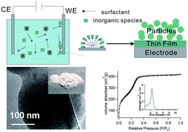
We report a facile electrochemistry assisted sol–gel approach for the preparation of mesoporous silica nanoparticles (MSNs) at the gram scale on common conductive substrates, such as the stainless steel surface. The formation of MSNs was triggered by electrochemical generation of hydroxide at the substrate/solution interface, which induced the self-assembly of surfactant micelles and meanwhile catalysed the polycondensation of silica precursors. The as-prepared MSNs were characterized in detail by several instrumental analysis techniques, revealing the obtained MSNs have a uniform pore size of about 2.4 nm and a high surface area of about 1164 m2 g−1. We believe that this approach can provide a simple, fast and low-cost way to produce MSNs and may be potentially useful for the large-scale industrial production.


Introduction

Since the discovery of the surfactant-templated approach for the synthesis of nanomaterials,1 it has been widely applied to prepare a large number of mesostructures with tailor-made properties.2–4 Due to the diverse morphologies of surfactant assemblies, mesoporous silica with different structures, dimensions and properties have been prepared by controlling the reaction conditions.5 Among them, mesoporous silica nanoparticles (MSNs) have been intensively investigated because of their excellent physical and chemical properties, such as high surface area, adjustable pore size, large pore volume and high biocompatibility.6,7 So far, MSNs have found a wide range of practical applications, for example in catalysis,8,9 chromatography,10,11 pollutant adsorption,12,13 cell imaging,14–16 drug delivery,17–22 bioanalysis23 and electroanalysis.24–26

MSNs can be synthesized by various methods, such as sol–gel reaction27 and microemulsion processing.28,29 The former one is relatively mature, with which the growth of MSNs can be controlled by carefully tailoring the reaction conditions. For example, the stöber or modified-stöber approach30,31 has been widely applied for preparation of monodispersed silica nanoparticles, using the base-catalysed hydrolysis of tetraalkyl silicates in a water-alcohol solution. Sol–gel synthesis is usually conducted in time-consuming bath operations,32 although many fast routes have been developed, such as using sodium silicate as the precursor,33 aerosol combined with sol–gel process,32 microwave irradiation34 and sonochemical synthesis.35

Electrochemistry, as a simple, facile and fast method, has been recently utilized to prepare highly ordered silica mesochannel film on various substrates.36,37 Formation of MSNs as a byproduct on the top surface of silica film has also been observed.37 However, the MSN formation process, as well as the physiochemical properties of formed MSNs were not studied in details. In this work, we investigated the effect of different factors, such as pH, current density, deposition time and surfactant concentration on the synthesis of MSNs. The structure and morphology of the MSNs were also characterized by various techniques in details. It was demonstrated that the electrochemically assisted sol–gel method can be employed for low-cost and large-scale synthesis of MSNs at room temperature.

Experimental section

Chemicals and materials

All chemicals and reagents were analytical grade or higher and used as received without further purification. Tetraethoxysilane (TEOS) and cetyltrimethylammonium bromide (CTAB) were purchased from Sigma. Anhydrous ethanol, hydrochloric acid (HCl) and sodium nitrate (NaNO3) were obtained from Sinopharm. Deionized water was used in all experiments. Gold electrodes (regular glass slides covered with 5 nm Cr and 100 nm Au) were purchased from Evaporated Metal Films (EMF) Inc. Prior to use, gold electrode was treated with the air plasma. Indium tin oxide (ITO) coated glasses (thickness of 100 nm, resistance of <15 Ω per square) were bought from Zhuhai Kaivo Electronic Components Co., Ltd. The ITO glasses or common stainless steel sheets were cleaned by sonication sequentially in acetone, ethanol and water, and then dried under a nitrogen stream.

Synthesis of MSNs

MSNs were prepared by electrochemistry assisted sol–gel method on various substrates, such as ITO conductive glass, gold coated glass and stainless steel sheet. Typically, a precursor mixture consisting of 100 mL ethanol, 100 mL aqueous solution of 0.1 M NaNO3 with pH adjusted by HCl to 2.6 unless otherwise specified, 68 mmol TEOS and 21.75 mmol CTAB was aged for 2.5 h under stirring. Then the three electrodes, involving stainless steel sheet (ITO or gold) as the working electrode, Ag/AgCl (saturated KCl) and stainless steel sheet with large area as reference and counter electrodes, were immersed into the precursor solution. A constant cathodic current density was applied for a certain time to deposit MSNs on surface of the substrate. Subsequently, the electrode was quickly removed from the solution and rinsed with large amount of water. Then the MSNs were scraped mechanically from the substrate and washed with water repeatedly. The powder was obtained through centrifugation and dried overnight at 80 °C. To remove the surfactants from mesochannels of MSNs, calcinations at 550 °C in air for 6 h was performed.

Instrumentation and characterization

Scanning electron microscopy (SEM) measurements were performed on a field-emission scanning electron microscope (FE-SEM, SU-70 or SU8010). High-resolution transmission electron microscopy (HR-TEM) images were obtained on a HT7700 transmission electron microscope (Hitachi, Japan) at an accelerating voltage of 200 kV. Nitrogen adsorption–desorption isotherms was carried out at 77 K using an auto-adsorption analyzer (micromeritics, TriStar II 3020 V1.03). The surface area calculations were based on Brunauer–Emmett–Teller (BET) model and pore size distribution was determined following the Barrett–Joyner–Halenda (BJH) method. X-ray diffraction (XRD) pattern was obtained on a Rigaku-D/Max-B automated powder X-ray diffractometer in a 2θ range of 1–10° (with a 2θ step of 0.02°) using Cu Kα radiation (λ = 1.5406 Å).

Results and discussion

As illustrated in Fig. 1, the formation of MSNs through electrochemistry assisted sol–gel method involves the following two steps. Firstly, the precursor solution containing TEOS, CTAB and NaNO3 was hydrolysed and aged at pH 2–4 for 2.5 h, in which three electrodes were then immersed to grow MSNs on the working electrode surface under the proper electrochemistry control. Upon applying an appropriate constant cathodic current density, the pH of electrolyte locally close to the electrode would be increased due to the consumption of H+ ions and the generation of OH through the electrochemical reduction of H+, H2O and NO3 in the vicinity of electrode-solution interface.36–40 The involved reactions are as follows (the standard redox potential is with respect to the standard hydrogen electrode):
2H+ + 2e → H2 E = 0.0 V

2H2O + 2e → H2 + 2OH E = −0.828 V

NO3 + H2O + 2e → NO2 + 2OH E = +0.01 V

image file: c5ra13482j-f1.tif
Fig. 1 Illustration of the electrochemistry assisted sol–gel method to produce MSNs.

These reduction reactions resulted in a sudden and sharp increase of the cathodic potential, as shown in Fig. 2a. The formed OH can induce the assembly of surfactants on the electrode surface, and meanwhile catalyse the formation of silica-surfactant nuclei and the growth of silica.41 These steps would lead to a slow increase of the potential due to the increase of surface impedance. In principle, OH generated at the electrode-solution interface spatially distributes over a thin layer with a thickness approximately given by image file: c5ra13482j-t1.tif (where D is the diffusion coefficient and equal to 4 × 10⁻⁵ cm2 s−1 for OH; t is the deposition time).42 It is about 16 μm when the deposition time is set to 2 ms. As shown in Fig. S1, the obtained mesoporous silica film (MSF) became thicker and thicker with increasing the deposition time, whereas the thickness of MSF increased slowly after 10 s. And the maximum thickness of MSFs is only about 330 nm in our experiments. In this case, OH can also diffuse beyond the MSF to induce the aggregation of silica-surfactant nuclei in the solution phase to generate MSNs (Fig. 2b) on the top of MSF. This mechanism is indeed consistent with the fast pH-changing method using pre-hydrolysis TEOS to prepare MSNs.43,44


image file: c5ra13482j-f2.tif
Fig. 2 (a) Electrochemical deposition curve and cross-section SEM image (b) of MSNs on the surface of ITO electrode (1 cm2) with a current density of 3 mA cm−2 for 15 s.

It is well known that the pH of precursor solution is a critical factor to affect the synthesis of MSNs. Hydrolysis of Si–OR bond in silanes can be promoted in both acid and base environments, whereas it is slow at neutral pH. The charge density of silica species is also dependent on the solution pH. The silica species are negatively charged when pH is above its isoelectric point (IEP = 2) and the charge density increases with increasing the pH. The negatively charged silicates can assemble with the positively charged surfactants via electrostatic interaction to form silica-surfactant nuclei. Hence, the pH of precursor solution was optimized in a range of 2–4. We found that the amount of MSNs obtained at pH = 2 (Fig. S2a) was much less than that at 2.6 and 4 (Fig. S2b and S2c), and MSNs can be readily synthesized in pH 2.6–4. Finally, pH = 2.6 was chosen as the optimal synthetic condition, which is consistent with the previous report on the silica film preparation.45

Apart from the solution pH, the amount of produced MSNs also relied on the electrode area, applied current density and time. The yield of MSNs increased apparently with increasing their values. For example, the amount of MSNs deposited on gold electrode at 30 mA cm−2 (Fig. S3) was larger than that at 3 mA cm−2 (Fig. S2b) obviously. In principle, MSNs can be prepared on various conductive substrates with this electrochemistry assisted sol–gel method. We tried three common electrode materials: indium tin oxide (ITO) coated glass, gold and stainless steel electrodes. If the applied current density is small and deposition time is short, all these three electrodes work well. However, when applying a higher current density or a longer deposition time, the ITO layer on glass will be destroyed due to the reduction of indium oxide. In order to prepare MSNs in a large-scale, the cheap and reusable stainless steel electrode with a big area (5 × 7 cm2) was used in our experiments. A current density of 3 mA cm−2 was applied for electro-deposition as an example. As shown in Fig. S4 and Fig. 3a, the yield of MSNs increased apparently with increasing the deposition time. With a simple treatment after deposition (described in Experimental section), powder of MSNs in the gram-scale was obtained (Fig. 3b). We believe that much more MSNs can be obtained if using a bigger size of electrode, a larger current density or a longer deposition time.


image file: c5ra13482j-f3.tif
Fig. 3 (a) Photograph of stainless steel sheet surface after MSNs deposition at 3 mA cm−2 for 30 min. (b) Photograph of the MSN powder. (c) The high-resolution TEM image of MSNs.

As-synthesized MSNs were characterized by several instrumental techniques. Fig. S5 showed that the size distribution of MSNs was narrow even for the deposition at large current densities. However, the size of MSNs was not uniform when increasing the deposition time, as depicted in Fig. S6. With increasing the deposition time, the produced particles were accumulated on the electrode surface in a layer by layer manner (Fig. 2b), resulting in the limited exposed surface of silanol groups on the existing particles and thus most of the additional silica source in solution reacts with each other rather than with the existing particles.46 As for the size of MSNs, it may be modulated by adding co-surfactant F127 (ref. 47) or other surface protecting agents, such as PEG,48 triethanolamine (TEA)49 and the amino acid L-lysine.50 Fig. 3c is the high-resolution TEM image of MSNs which clearly revealed that the obtained MSNs have uniform and ordered mesochannels.

The low-angle X-ray diffraction (XRD) pattern (Fig. 4) showed several diffraction peaks which can be exactly assigned to the (100), (110), (200) and (210) family planes of 2-D hexagonal meso-structure with the space group of p6m, similar to that of MCM-41.1 To characterize the porosity of MSNs, nitrogen sorption measurement which is an efficient tool for providing information about the pore system of various porous materials was conducted. Prior to this measurement, the CTAB surfactant was removed by calcinations in air at 550 °C for 6 h. The nitrogen adsorption–desorption isotherms of the MSNs was displayed in Fig. 5. It can be classified as a type IV curve with a distinct sharp capillary condensation step. The pore size distribution based on the adsorption branches, using the Barrett–Joyner–Halenda (BJH) method, reveals a uniform mesopore of about 2.4 nm in diameter (inset in Fig. 5). The BET surface area of MSNs is calculated from adsorption data to be 1164 m2 g−1, which is comparable with typical MCM-41 samples.51 All these characterizations indicate that the MSNs synthesized by the electrochemistry assisted sol–gel reaction are consistent with MCM-41 prepared by typical approach.1 Moreover, MSNs can also be prepared in a wide range of solution composition (Fig. S7 and S8).


image file: c5ra13482j-f4.tif
Fig. 4 Low-angle X-ray diffraction pattern of the synthesized MSN powder. The corresponding d-spacing: (100) = 3.81 nm; (110) = 2.17 nm; (200) = 1.92 nm and (210) = 1.42 nm.

image file: c5ra13482j-f5.tif
Fig. 5 Nitrogen adsorption–desorption isotherms at 77 K for MSNs after calcinations.

Conclusions

In summary, we reported a facile and simple electrochemistry assisted sol–gel method for the synthesis of mesoporous silica nanoparticles (MSNs) in the gram-scale. The prepared MSNs have a uniform mesochannel of 2.4 nm and a high surface area of about 1164 m2 g−1. This synthesis process does not involve any extra catalyst (basic compounds) and can finish in a pretty short time-scale. We believe it is an easy, fast and low-cost approach to produce MSNs in the large scale.

Acknowledgements

This work is supported by the Nature Science Foundation of China (21222504, 21335001), the Zhejiang Provincial Natural Science Foundation (R14B050003), the Program for New Century Excellent Talents in University and the Fundamental Research Funds for the Central Universities (2014XZZX003-04).

Notes and references

  1. C. Kresge, M. Leonowicz, W. Roth, J. Vartuli and J. Beck, Nature, 1992, 359, 710–712 CrossRef CAS PubMed.
  2. M. Kruk, Acc. Chem. Res., 2012, 45, 1678–1687 CrossRef CAS PubMed.
  3. Y. Deng, J. Wei, Z. Sun and D. Zhao, Chem. Soc. Rev., 2013, 42, 4054–4070 RSC.
  4. K. Egeblad, C. H. Christensen, M. Kustova and C. H. Christensen, Chem. Mater., 2007, 20, 946–960 CrossRef.
  5. S. H. Wu, C. Y. Mou and H. P. Lin, Chem. Soc. Rev., 2013, 42, 3862–3875 RSC.
  6. D. Y. Zhao, J. L. Feng, Q. S. Huo, N. Melosh, G. H. Fredrickson, B. F. Chmelka and G. D. Stucky, Science, 1998, 279, 548–552 CrossRef CAS.
  7. Y. Jiang, C. Lee, Q. Wang, Y. Deng and D. Fu, Microporous Mesoporous Mater., 2013, 181, 248–253 CrossRef CAS PubMed.
  8. A. Corma, Chem. Rev., 1997, 97, 2373–2420 CrossRef CAS PubMed.
  9. C. Li, Catal. Rev.: Sci. Eng., 2004, 46, 419–492 CAS.
  10. F. Hoffmann, M. Cornelius, J. Morell and M. Froba, J. Nanosci. Nanotechnol., 2006, 6, 265–288 CAS.
  11. H. M. Liu, Y. Guo, X. S. Wang, X. J. Liang, X. Liu and S. X. Jiang, RSC Adv., 2014, 4, 17541–17548 RSC.
  12. L. T. Gibson, Chem. Soc. Rev., 2014, 43, 5173–5182 RSC.
  13. L. T. Gibson, Chem. Soc. Rev., 2014, 43, 5163–5172 RSC.
  14. J. L. Vivero-Escoto, R. C. Huxford-Phillips and W. B. Lin, Chem. Soc. Rev., 2012, 41, 2673–2685 RSC.
  15. Y. S. Lin, S. H. Wu, Y. Hung, Y. H. Chou, C. Chang, M. L. Lin, C. P. Tsai and C. Y. Mou, Chem. Mater., 2006, 18, 5170–5172 CrossRef CAS.
  16. C. P. Tsai, Y. Hung, Y. H. Chou, D. M. Huang, J. K. Hsiao, C. Chang, Y. C. Chen and C. Y. Mou, Small, 2008, 4, 186–191 CrossRef CAS PubMed.
  17. M. Strømme, U. Brohede, R. Atluri and A. E. Garcia-Bennett, Wiley Interdiscip. Rev.: Nanomed. Nanobiotechnol., 2009, 1, 140–148 CrossRef PubMed.
  18. M. Sasidharan, H. Zenibana, M. Nandi, A. Bhaumik and K. Nakashima, Dalton Trans., 2013, 13381–13389 RSC.
  19. Z. Li, J. C. Barnes, A. Bosoy, J. F. Stoddart and J. I. Zink, Chem. Soc. Rev., 2012, 41, 2590–2605 RSC.
  20. M. Vallet-Regi, F. Balas and D. Arcos, Angew. Chem., Int. Ed., 2007, 46, 7548–7558 CrossRef CAS PubMed.
  21. J. K. Hsiao, C. P. Tsai, T. H. Chung, Y. Hung, M. Yao, H. M. Liu, C. Y. Mou, C. S. Yang, Y. C. Chen and D. M. Huang, Small, 2008, 4, 1445–1452 CrossRef CAS PubMed.
  22. S. Giri, B. G. Trewyn and V. S. Y. Lin, Nanomedicine, 2007, 2, 99–111 CrossRef CAS PubMed.
  23. R. J. Tian, H. Zhang, M. L. Ye, X. G. Jiang, L. H. Hu, X. Li, X. H. Bao and H. F. Zou, Angew. Chem., Int. Ed., 2007, 46, 962–965 CrossRef CAS PubMed.
  24. A. Walcarius, Chem. Soc. Rev., 2013, 42, 4098–4140 RSC.
  25. A. Walcarius, Electroanalysis, 2008, 20, 711–738 CrossRef CAS PubMed.
  26. A. Walcarius, Electroanalysis, 1998, 10, 1217–1235 CrossRef CAS.
  27. Y.-G. Lee, J.-H. Park, C. Oh, S.-G. Oh and Y. C. Kim, Langmuir, 2007, 23, 10875–10878 CrossRef CAS PubMed.
  28. C.-L. Chang and H. S. Fogler, Langmuir, 1997, 13, 3295–3307 CrossRef CAS.
  29. F. J. Arriagada and K. Osseoasare, Colloids Surf., 1992, 69, 105–115 CrossRef CAS.
  30. M. Grün, I. Lauer and K. K. Unger, Adv. Mater., 1997, 9, 254–257 CrossRef PubMed.
  31. W. Stober, A. Fink and E. Bohn, J. Colloid Interface Sci., 1968, 26, 62–69 CrossRef.
  32. C. Boissiere, D. Grosso, A. Chaumonnot, L. Nicole and C. Sanchez, Adv. Mater., 2011, 23, 599–623 CrossRef CAS PubMed.
  33. A. C. Voegtlin, A. Matijasic, J. Patarin, C. Sauerland, Y. Grillet and L. Huve, Microporous Mater., 1997, 10, 137–147 CrossRef CAS.
  34. C. G. Wu and T. Bein, Chem. Commun., 1996, 925–926 RSC.
  35. S. Vetrivel, C.-T. Chen and H.-M. Kao, New J. Chem., 2010, 34, 2109–2112 RSC.
  36. R. Shacham, D. Avnir and D. Mandler, Adv. Mater., 1999, 11, 384–388 CrossRef CAS.
  37. A. Walcarius, E. Sibottier, M. Etienne and J. Ghanbaja, Nat. Mater., 2007, 6, 602–608 CrossRef CAS PubMed.
  38. G. H. A. Therese and P. V. Kamath, Chem. Mater., 2000, 12, 1195–1204 CrossRef CAS.
  39. J. Joseph, H. Gomathi and G. P. Rao, Electrochim. Acta, 1991, 36, 1537–1541 CrossRef CAS.
  40. D. Aslanidis, J. Fransaer and J. P. Celis, J. Electrochem. Soc., 1997, 144, 2352–2357 CrossRef CAS PubMed.
  41. G. Herzog, E. Sibottier, M. Etienne and A. Walcarius, Faraday Discuss., 2013, 164, 259–273 RSC.
  42. A. J. Bard and L. R. Faulkner, Electrochemical Methods: Fundamentals and Applications, Wiley, New York, 1980 Search PubMed.
  43. C. E. Fowler, D. Khushalani, B. Lebeau and S. Mann, Adv. Mater., 2001, 13, 649–652 CrossRef CAS.
  44. H. P. Lin and C. P. Tsai, Chem. Lett., 2003, 32, 1092–1093 CrossRef CAS.
  45. W. Li, L. Ding, Q. Wang and B. Su, Analyst, 2014, 139, 3926–3931 RSC.
  46. T. Nakamura, M. Mizutani, H. Nozaki, N. Suzuki and K. Yano, J. Phys. Chem. C, 2007, 111, 1093–1100 CAS.
  47. K. Ikari, K. Suzuki and H. Imai, Langmuir, 2006, 22, 802–806 CrossRef CAS PubMed.
  48. Y. S. Lin and C. L. Haynes, Chem. Mater., 2009, 21, 3979–3986 CrossRef CAS.
  49. K. Moller, J. Kobler and T. Bein, Adv. Funct. Mater., 2007, 17, 605–612 CrossRef PubMed.
  50. T. Yokoi, Y. Sakamoto, O. Terasaki, Y. Kubota, T. Okubo and T. Tatsumi, J. Am. Chem. Soc., 2006, 128, 13664–13665 CrossRef CAS PubMed.
  51. V. Meynen, P. Cool and E. F. Vansant, Microporous Mesoporous Mater., 2009, 125, 170–223 CrossRef CAS PubMed.

Footnote

Electronic supplementary information (ESI) available. See DOI: 10.1039/c5ra13482j

This journal is © The Royal Society of Chemistry 2015
Click here to see how this site uses Cookies. View our privacy policy here.