DOI:
10.1039/C5RA13445E
(Paper)
RSC Adv., 2015,
5, 66420-66425
A polydiacetylene-based fluorescence assay for the measurement of lipid membrane affinity
Received
9th July 2015
, Accepted 17th July 2015
First published on 20th July 2015
Abstract
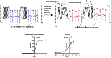
Polydiacetylene (PDA) is a promising membrane-screening tool because lipid constituents can be incorporated into the PDA framework to form lipid/PDA vesicles used as lipid bilayers. Previous reports have shown that the colorimetric signals of PDA could be utilized for the measurement of drug–lipid membrane interactions. In this study, the fluorescence signals of PDA vesicles were investigated for the measurement of lipid membrane affinity. Based on the fluorescence response of PDA vesicles (excitation wavelength 485 nm, emission wavelength 560 nm), the half maximal response concentration (EC50) was introduced for the evaluation of drug–membrane interactions. In order to validate this method, local anesthetics and flavonoids were selected as the reference compounds and their log(EC50) values correlated well with other lipid membrane affinity constants. Then the influence of buffer pH conditions and lipid constituents on the membrane affinity were investigated to show the wide application of this method using tetracaine hydrochloride as the reference compound. The particle size of vesicles before and after addition of tetracaine hydrochloride was determined to observe the extent of vesicle binding of the tested compound. The zeta potential results showed that the electrostatic interaction had less effect on the change of lipid membrane affinity at different pH value. Therefore, the hydrophobic interaction was assumed to play the most important role in the increase in lipid membrane affinity of tetracaine hydrochloride as the buffer pH value increased. The ratio of Chol in the lipid constituents affected the affinity of tetracaine hydrochloride less, but significantly weakened the sensitivity of PDA-based fluorescence signals. In summary, this work provides a simple, sensitive and reproducible PDA-based fluorescent method for the rapid measurement of lipid membrane affinity.
1. Introduction
Membrane affinity means the binding ability of a drug candidate to cell membranes and the lipid constituents has a major impact on it.1 Therefore, the measurement of lipid membrane affinity is an important early screening step during drug discovery. The n-octanol/water partition coefficient (Koct) is widely used to represent the lipid membrane affinity, but isotropic phase n-octanol is quite different from the structure of lipid bilayers. Compared with n-octanol, lipid vesicles almost exactly mimic real biological membranes. However, traditional methods for the determination of the lipid membrane partition coefficient, equilibrium dialysis and ultracentrifugation,2,3 are tedious and difficult to be used in the early screening step. Other existing methods also suffered from different problems, such as poor reproducibility (chromatographic techniques).4,5 Sensor techniques are widely used in the detection of biological interaction.6–9 For examining the membrane interactions, polydiacetylene (PDA) vesicles have several advantages over other platforms including molecular structure of the vesicle, ease of synthesis, inexpensive and commercially available monomers, etc.10–14
PDA undergoes blue-red color changes and fluorescence transitions due to the distortion of PDA conjugation plane, which may be induced by surface perturbations.15–19 When using as a membrane-screening tool, lipid constituents are incorporated into the PDA framework to form lipid/PDA vesicles. The interactions of target molecules with lipid/PDA vesicles change the fluidity of the lipid domain and induce distortion of the conjugation plane of PDA.20 Previous reports have shown that the colorimetric signals of PDA vesicles could be utilized for the measurement of drug–membrane interactions.10–12 We also have established a quantitative method for the measurement of lipid membrane affinity based on the PDA colorimetric signals in our previous report.21 The focus in this work was on the fluorescence properties of lipid/PDA vesicles, aiming to take advantage of the significant higher sensitivity of fluorescent signals.
In order to evaluate the applicability of the established PDA-based fluorescence method, local anesthetics and flavonoids were selected as the reference compounds. These drugs were the common-used target drugs for the evaluation of membrane affinity in previous studies.22,23 Local anesthetics are alkaline and prone to interact with the hydrophobic region of the lipid membrane.24 Flavonoids are weak acids and possess the protonophoric properties, which may be related with the ionisable hydroxyl groups.25 Application of the PDA colorimetric platform on the two types has been hampered by sensitivity problem. To resolve this problem, the PDA-based fluorescent signal was employed for the membrane affinity detection of local anesthetics and flavonoids. Moreover, the influence of pH value and lipid constituents on the membrane affinity was also investigated to show the wide application of this membrane screening method. Based on these studies, this paper presents a reproducible PDA-based fluorescence assay for the measurement of lipid membrane affinity.
2. Material and methods
2.1 Materials
Dimyristoylphosphatidylcholine (DMPC) and 10,12-tricosadiynoic acid were purchased from NOF (Tokyo, Japan) and Alfa-Aesar (Ward Hill, MA, USA), respectively. Cholesterol (Chol, 5AZZH-OL) was from TCI (Japan). Quercetin, luteolin, apigenin and daidzein were obtained from Yuanye Bio-Technology (Shanghai, China). Tetracaine hydrochloride, mepivacaine hydrochloride, lidocaine hydrochloride, propitocaine hydrochloride and bupivacaine hydrochloride were purchased from Jinan Chenghui-Shuangda Chemical (Jinan, China). All of the above-mentioned materials had purities greater than 98%. All other chemicals were of analytical grade and used as received.
2.2 Vesicles preparation
The preparation of PDA vesicles has been reported in our previous study.21 In brief, DMPC and diacetylene monomer 10,12-tricosadiynoic acid (2
:
3 molar ratio) were dissolved in a chloroform–ethanol solution (1
:
1) and dried together in vacuo, followed by addition of deionized water to produce a suspension. The suspension was heated to 70 °C and sonicated in an ultrasonic bath for 5 min. The vesicles solution was cooled to room temperature, kept at 4 °C overnight and then polymerized by irradiation at 254 nm for 5 min with stirring at room temperature to obtain the DMPC/PDA vesicles solution. The vesicles solution was stored at 4 °C before use. When used for the influence of Chol on the membrane affinity, the same procedure was performed as described above except the lipid added in the first step. For example, the molar ratio of Chol, DMPC and PDA was 0.2
:
0.8
:
1.5 in the 20% Chol liposome.
2.3 Preparation of buffer and sample solutions
Tris–HCl buffer solution, with a concentration of 150 mM and pH 6.3 to 9.0, was used for the investigation of pH influence. The stock solutions of local anesthetics and flavonoids were prepared by dissolving the compounds in the deionized water and DMSO, respectively. Different volumes of the stock solution were transferred to make a serious of standard solutions. Samples for the fluorescence (FL) studies were prepared by adding 0.2 mL of different standard solutions to 0.2 mL of PDA vesicles solution and 2 mL of Tris–HCl buffer solution. The sample solutions were kept at room temperature for 8–15 min before fluorescence assay. All other solutions were stored at 4 °C.
2.4 Fluorescence measurement and calculation of EC50
Fluorescence measurement of the sample solutions was performed on a 4600 FL spectrometer (Hitachi, Japan). The fluorescence emission intensity at 560 nm with an excitation wavelength of 485 nm was recorded. Spectral slits of emission and excitation wavelength were both 5 nm and the scan speed was 1200 nm min−1. FL was defined as FL = FL1 − FL0, where FL0 is the initial fluorescence response of vesicles before the addition of test compounds, and FL1 represents the final fluorescence response of vesicles after the addition of test compounds. EC50 was calculated by processing the FL response versus different concentrations with Graph-prism software (Version 5.01; GraphPad Software Inc., San Diego, CA) basing on the nonlinear regression.
2.5 Determination of zeta potential and size of vesicles
The vesicles size distribution and zeta potential values both before and after addition of tetracaine hydrochloride were measured using dynamic light scattering (Malvern Zetasizer 3000HSA, Malvern Instruments Ltd, United Kingdom). These measurements were performed at 25 °C using a suspension containing 0.1 mM DMPC in 150 mM Tris–HCl buffer solution with different pH value.
3. Results and discussion
3.1 The fluorescence spectra and calculation of the log(EC50) value
The fluorescence spectra of PDA/DMPC vesicles before and after addition of reference compounds were investigated (Fig. 1). It was found that the maximal fluorescence emission signal at the wavelength of 560 nm exhibited an obvious enhancement with a maximal excitation wavelength of 485 nm after addition of reference compounds. The intrinsic fluorescence of reference compounds had no obvious interference at this wavelength. Therefore, the fluorescence response of PDA/DMPC vesicles (excitation wavelength 485 nm, emission wavelength 560 nm) was selected as the signal for the further research. Previous reports used to evaluate the membrane affinity by the absolute response of the PDA, such as the first report utilizing PDA/lipid vesicles as membrane-screening tool.14 Actually, the absolute response of vesicles was also affected by the other properties of the tested compound besides the membrane affinity. It is necessary to use a reliable index to assess the membrane affinity.
 |
| Fig. 1 Fluorescence emission spectra of PDA with tetracaine hydrochloride (A), PDA with quercetin (B), PDA (C), quercetin (D) and tetracaine hydrochloride (E) at the excitation wavelength of 485 nm. Tetracaine hydrochloride (3 mM) and quercetin (5 mM). | |
We have established a simple quantitative model for the measurement of lipid membrane affinity using the index Kb based on the PDA colorimetric signals in our previous report.21 However, the Kb values of most tested compounds were negative based on the PDA fluorescence response using double-reciprocal plots. Therefore, the half maximal response concentration (EC50), a more general parameter for evaluating biological interaction,26,27 was introduced for the evaluation of drug–membrane interactions. As shown in Fig. 2, this index could be calculated according to the compound concentrations and fluorescence responses by the nonlinear regression. It is interesting that the FL response of tetracaine hydrochloride was significantly higher than luteolin, whereas the EC50 value of luteolin was lower than tetracaine hydrochloride at pH 7.6. This is an experimental proof that the absolute response change of PDA/lipid vesicles before and after addition of the tested compound cannot represent its lipid membrane affinity. Therefore, the log(EC50) value was selected for the evaluation of drug–membrane interactions in the next parts.
 |
| Fig. 2 Concentration-response curve and calculation of EC50 at pH 7.6. | |
3.2 Comparison of developed method with classical methods
To validate the performance of PDA-based fluorescence assay for the evaluation of lipid membrane interaction, the log(EC50) values of tested compounds were compared with other lipid membrane affinity constants. Local anesthetics and flavonoids were selected as the reference compounds because these drugs were the common-used target drugs for the evaluation of lipid membrane affinity in previous studies.22,23 The selected local anesthetics were alkalescent with pKa above 7.6 and the flavonoids were acidic compounds with pKa below 7.6.
For local anesthetics, the log(EC50) values of mepivacaine hydrochloride, lidocaine hydrochloride, propitocaine hydrochloride, bupivacaine hydrochloride and tetracaine hydrochloride were compared with the reported logarithm of the liposome/water partition coefficient (Km) measured by ultracentrifugation using DMPC liposome.28 As shown in Fig. 3A, the log(EC50) versus log(Km) of the five target compounds displayed a favorable linear correlation with R2 = 0.9829. When compared with the lipid membrane affinity constants measured by immobilized artificial membrane chromatography (IMAC) and micellar electrokinetic chromatography (MEKC), the favorable linear was good with R2 = 0.9864 and 0.9772, respectively (Fig. 3B and C). Furthermore, the correlation between log(EC50) and the potency of pharmacodynamic effect was also excellent with R2 = 0.9749 (Fig. 3D). It has been reported that the potency of local anesthetic drugs partly depend on the membrane affinity.29 Therefore, the good linear correlation illustrated that the developed PDA-based fluorescence method may be a certain prediction for anesthetic effect. For flavonoids, it has been reported that the lipid membrane partition coefficient (Kd) improved with the increase of the number of hydroxyl groups.30 In this study, daidzein (two OH groups), apigenin (three OH groups), luteolin (four OH groups) and quercetin (five OH groups) were selected for examining the influence of OH groups. As shown in Table 1, the affinity of flavonoids increased due to the improvement of the number of free hydroxyl groups. Although a limited number of flavonoids were tested due to the lack of classic data, the results illustrated that the developed method could be used for the research on the lipid membrane affinity of flavonoids.
 |
| Fig. 3 Comparison of log(EC50) in the fluorescence DMPC/PDA vesicle system, solute partitioning into DMPC liposome (Km), solute partitioning with IAMC and solute partitioning with MEKC, and potency of anesthetic drugs. (A) Correlation between log(EC50) and log(Km); (B) correlation between log(EC50) and log(KIAMC); (C) correlation between log(EC50) and log(KMEKC); (D) correlation between log(EC50) and potency of local anesthetic. Values were presented as mean ± SD. | |
Table 1 The log(EC50) values in the fluorescence vesicle system and comparison with the reported data
Compound |
log(EC50) (mean ± SD) |
Reference Kd |
Free hydroxyl groups |
Not studied in the previous reference. |
Daidzein |
0.2971 ± 0.03 |
a |
2 |
Apigenin |
0.1419 ± 0.02 |
a |
3 |
Luteolin |
−0.1461 ± 0.05 |
7.1 |
4 |
Quercetin |
−0.1841 ± 0.06 |
7.5 |
5 |
In conclusion, the log(EC50) value of FL response was demonstrated as an effective, accurate and reproducible index for the assessment of lipid membrane affinity.
3.3 Influence of buffer pH conditions
The buffer pH conditions had an important effect on the lipid membrane affinity of the polar compounds. Tetracaine hydrochloride was a common-used reference for the research on the factors affecting the membrane interaction due to its high affinity and good solubility.31 Therefore, the membrane affinity of tetracaine hydrochloride at different buffer pH conditions were compared to show the wide application of this method.
As show in Fig. 4, tetracaine hydrochloride was tested from pH 6.3 to pH 9.0 with 6 points to cover the normal physiological pH range. The size distribution of vesicles was determined both before and after addition of tetracaine hydrochloride in order to observe the possible changes of particle size (Fig. 5). It was found the particle size of vesicles became bigger after addition of the reference compound when the pH value was above 7.0. But the size change at pH 6.3 was quite little probably due to the low lipid membrane affinity of tetracaine hydrochloride at this pH value. Therefore, the changes in the size distribution might reflect the extent of vesicle binding of the tested compounds. To account for the electrostatic effects of the interaction, the zeta potential values of vesicles were detected before and after addition of tetracaine hydrochloride (Fig. 6). The zeta potential decreased with the increase of buffer pH value due to less Tris basic ions binding to the membrane interface. It was observed that the zeta potential increased as the addition of tetracaine hydrochloride at all pH value. Since the addition of the drug does not change the ionic strength and pH value of the solution significantly due to the high salt concentration of buffer, the binding of positively charged tetracaine hydrochloride with the lipid membrane interface should be responsible for the increase of zeta potential. Surprisingly, the change of zeta potential remained almost constant throughout pH range. The most probable explanation for the phenomenon was the electrostatic repulsion between positively charged drug molecules and the Tris basic ions binding to the membrane interface at low pH value, or limited positively charged drug molecules in the buffer at high pH value. The increase of lipid membrane affinity of tetracaine hydrochloride as the increase of buffer pH value might result from more neutral drug molecules formation in the buffer solution and binding to the hydrophobic region of membrane interface. Therefore, hydrophobic interaction between tetracaine hydrochloride and the lipid membrane should play the most important role in the changing of lipid membrane affinity at different pH value, while the electrostatic interaction had less effect on it.
 |
| Fig. 4 The log(EC50) values of tetracaine hydrochloride at different pH value. Values were presented as mean ± SD. | |
 |
| Fig. 5 Particle size of PDA with or without tetracaine hydrochloride and their differences. Values were presented as mean ± SD. | |
 |
| Fig. 6 Zeta potential of the test solution with or without tetracaine hydrochloride and their differences. Values were presented as mean ± SD. | |
In this part, we illustrated the application of polydiacetylene-based fluorescence method for the study with the buffer pH effect on the lipid membrane affinity of drug molecules. The zeta potential was detected to reflect the intrinsic mechanism of affinity change.
3.4 Influence of lipid constituents
Besides the phospholipid constituent, Chol was another important lipid constituent in the cell membrane.32–34 Thus, the effect of different lipid constituents (DMPC/Chol 100/0, DMPC/Chol 80/20 and DMPC/Chol 60/40) on the membrane affinity of tetracaine hydrochloride were compared using this PDA-based fluorescent method. As shown in Table 2, the ratio of Chol in the lipid constituents had less effect on the log(EC50) of the reference compound. The structural difference between Chol and phospholipid included the head region: the phospholipid with negatively charged head region and Chol with neutral head region. This result further indicated that hydrophobic interactions between tetracaine hydrochloride and lipid dominate the membrane energy in this case. However, the significant effect of Chol on the FL values could be found in Table 2. The FL response at the same concentration decreased significantly with the increase of the Chol ratio in the lipid constituents. This might result from the fact that Chol has less fluidity than DMPC at room temperature, leading to the less distortion of the conjugation plane of PDA. Therefore, it could be concluded that Chol had less effect on the interaction of target molecules with lipid/PDA vesicles, but weakened the sensitivity of this PDA-based fluorescent platform. This case also indicated that the log(EC50), rather than the FL value, represented the lipid membrane affinity because the PDA sensitivity would be affected by the lipid constituents.
Table 2 The fluorescence response and log(EC50) values of tetracaine hydrochloride for the influence of Chol
Chol (%) |
FL response (mean ± SD) |
log(EC50) (mean ± SD) |
0 |
500.18 ± 1.40 |
0.2429 ± 0.06 |
20 |
433.19 ± 0.76 |
0.2686 ± 0.03 |
40 |
204.17 ± 1.46 |
0.2275 ± 0.07 |
In summary, the influence of lipid constituents on the membrane affinity can be assessed under the established PDA-based fluorescent platform.
4. Conclusion
This study presents a PDA-based fluorescence assay for the measurement of lipid membrane affinity in drug discovery. As the fluorescence response might be affected by the other properties of test compounds besides membrane affinity or the lipid constituents in the lipid/PDA vesicles, the half maximal fluorescence response concentration (EC50) was introduced for the evaluation of membrane affinity. The log(EC50) values of local anesthetics and flavonoids correlated well with other lipid membrane affinity constants. The lipid membrane affinity of tetracaine hydrochloride increased as the increase of buffer pH value and the electrostatic interaction had less effect on this change. The ratio of Chol in the lipid constituents also less affected the affinity of tetracaine hydrochloride. Therefore, the hydrophobic interaction was assumed to play the most important role in the interaction between tetracaine hydrochloride and the lipid membrane. This work provides a simple, sensitive and reproducible PDA-based fluorescent method for the rapid measurement of lipid membrane affinity and showed its wide application to the study of affinity influencing factors.
Abbreviations
PDA | Polydiacetylene |
DMPC | Dimyristoylphosphatidylcholine |
FL | Fluorescence |
Chol | Cholesterol |
Koct | n-Octanol/water partition coefficient |
Km | Lipid membrane partition coefficient |
Acknowledgements
This work financially was supported by the National Natural Science Foundation of China (Grant No. 30901878, Grant No. 21275162, Grant No. 81373956 and Grant No. 81274064), the Open project Program of MOE Key Laboratory of Drug Quality Control and Pharmacovigilance (No. MKLDP2013MS03) and the Priority Academic Program Development of Jiangsu Higher Education Institutions.
References
- E. H. Kerns, J. Pharm. Sci., 2001, 90, 1838–1858 CrossRef CAS PubMed.
- M. Ikonen, L. Murtomaki and K. Kontturi, J. Electroanal. Chem., 2007, 602, 189–194 CrossRef CAS PubMed.
- C. Barzanti, R. Evans, J. Fouquet, L. Gouzin, N. M. Howarth, G. Kean, E. Levet, D. Wang, E. Wayemberg, A. A. Yeboah and A. Kraft, Tetrahedron Lett., 2007, 48, 3337–3341 CrossRef CAS PubMed.
- S. Ong, H.-L. Liu, X.-X. Qiu, G. Bhat and C. Pidgeon, Anal. Chem., 1995, 67, 755–762 CrossRef CAS.
- F. Beigi, I. Gottschalk, C. L. Hagglund, L. Haneskog, E. Brekkan, Y.-X. Zhang, T. Osterberg and P. Lundahl, Int. J. Pharm., 1998, 164, 129–137 CrossRef CAS.
- D. Lombardi, B. Cuenoud, H. Wunderli-Allenspach and S. D. Kramer, Anal. Biochem., 2009, 385, 215–223 CrossRef CAS PubMed.
- D. L. Melchior, J. Pharm. Sci., 2002, 91, 1075–1079 CrossRef CAS PubMed.
- E. Tait, S. P. Stanforth, S. Reed, J. P. Perry and J. R. Dean, RSC Adv., 2015, 5, 15494–15499 RSC.
- M. Sun, S. Wang, Q. Yang, X. Fei, Y. Li and Y. Li, RSC Adv., 2014, 4, 8295–8299 RSC.
- S. Rozner, S. Kolusheva, Z. Cohen, W. Dowhan, J. Eichler and R. Jelinek, Anal. Biochem., 2003, 319, 96–104 CrossRef CAS.
- S. Kolusheva, T. Shahal and R. Jelinek, J. Am. Chem. Soc., 2000, 122, 776–780 CrossRef CAS.
- S. Kolusheva, J. Friedman, I. Angel and R. Jelinek, Biochemistry, 2005, 44, 12077–12085 CrossRef CAS PubMed.
- D. Evrard, E. Touitou, S. Kolusheva, Y. Fishov and R. Jelinek, Pharm. Res., 2001, 18, 943–949 CrossRef CAS.
- S. Kolusheva, L. Boyer and R. Jelinek, Nat. Biotechnol., 2000, 18, 225–227 CrossRef CAS PubMed.
- X.-Q. Chen, G.-D. Zhou, X.-J. Peng and J.-Y. Yoon, Chem. Soc. Rev., 2012, 41, 4610–4630 RSC.
- M. Katz, I. Ben-Shlush, S. Kolusheva and R. Jelinek, Pharm. Res., 2006, 23, 580–588 CrossRef CAS PubMed.
- C. P. Oliveira, N. F. F. Soares, E. A. F. Fontes, T. V. Oliveira and A. M. M. Filho, Food Chem., 2012, 135, 1052–1056 CrossRef PubMed.
- T. Pattanatornchai, N. Charoenthai, S. Wacharasindhu, M. Sukwattanasinitt and R. Traiphol, J. Colloid Interface Sci., 2013, 391, 45–53 CrossRef CAS PubMed.
- R. Jelinek and M. Ritenberg, RSC Adv., 2013, 3, 21192–21201 RSC.
- R. Jelinek and S. Kolusheva, Biotechnol. Adv., 2001, 19, 109–118 CrossRef CAS.
- F. Zheng, Z. Wu and Y.-H. Chen, Anal. Biochem., 2012, 420, 171–176 CrossRef CAS PubMed.
- V. Abram, B. Berlec, A. Ota, M. Sentjurc, P. Blatnik and N. P. Ulrih, Food Chem., 2013, 139, 804–813 CrossRef CAS PubMed.
- J. Laine, J. Lokajova, J. Parshintsev, J. M. Holopainen and S. K. Wiedmer, Anal. Bioanal. Chem., 2010, 396, 2599–2607 CrossRef CAS PubMed.
- P. A. Zapata-Morin, F. J. Sierra-Valdez and J. C. Ruiz-Suarez, J. Mol. Graphics Modell., 2014, 53, 200–205 CrossRef CAS PubMed.
- N. P. Ulrih, A. Ota, M. Sentjurc, S. Kure and V. Abram, Food Chem., 2010, 121, 78–84 CrossRef PubMed.
- H. Yin and P. S. Casey, RSC Adv., 2014, 4, 26149–26157 RSC.
- T. van Oordt, G. B. Stevens, S. K. Vashist, R. Zengerle and F. von Stetten, RSC Adv., 2013, 3, 22046–22052 RSC.
- G. V. Betageri and J. A. Rogers, Int. J. Pharm., 1987, 36, 165–173 CrossRef CAS.
- L. Escuder-Gilabert, S. Sagrado, R. M. Villanueva-Camanas and M. J. Medina-Hernandez, Anal. Chem., 1998, 70, 28–34 CrossRef CAS PubMed.
- P. Tammela, L. Laitinen, A. Galkin, T. Wennberg, R. Heczko, H. Vuorela, J. P. Slotte and P. Vuorela, Arch. Biochem. Biophys., 2004, 425, 193–199 CrossRef CAS PubMed.
- M. Ikonen, L. Murtomäki and K. Kontturi, Colloids Surf., B, 2010, 78, 275–282 CrossRef CAS PubMed.
- D. Kurad, G. Jeschke and D. Marsh, Biophys. J., 2004, 86, 264–271 CrossRef CAS.
- J. A. Urbina, S. Pekerar, H.-B. Le, J. Patterson, B. Montez and E. Oldfield, Biochim. Biophys. Acta, 1995, 1238, 163–176 CrossRef.
- C. Lagerquist, F. Beigi, A. Karlen, H. Lennernas and P. Lundahl, J. Pharm. Pharmacol., 2001, 53, 1477–1487 CrossRef CAS.
Footnote |
† These two authors contributed equality to this article. |
|
This journal is © The Royal Society of Chemistry 2015 |
Click here to see how this site uses Cookies. View our privacy policy here.