Rohan D. Deshpande,
Gowda D. V.,
Naga Sravan Kumar Varma Vegesna*,
Rudra Vaghela* and
Kulkarni P. K.
Dept. of Pharmaceutics, JSS College of Pharmacy, JSS University, Mysuru, India-570015. E-mail: rudra52@gmail.com; vnskvarma@gmail.com; Tel: +91 9986101644
First published on 11th September 2015
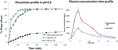
The aim of the present research was to improve the aqueous solubility and oral bioavailability of glibenclamide (GLB), a BCS class-II drug. A GLB nanosuspension (NS) was prepared using a liquid anti-solvent (LAS) precipitation technique and stabilized using HPMC K15M and lactose. Different in-process variables which directly affect the precipitated particle size have been thoroughly studied and optimized. The effect of a cryoprotective agent which could prevent agglomeration during lyophilisation was investigated. The optimal formulations of GD-H0.3d and GD-H0.4f exhibited a size range of 168.6 and 342.2 nm respectively and did not show any interaction when screened for incompatibility using FT-IR and DSC, but exhibited a decrease in crystallinity. The prepared GLB NPs exhibited superior aqueous solubility and dissolution when compared to pure GLB. The oral bioavailability of optimized formulations was found to exhibit 2.59, 1.67, 1.19, 2.50 and 2.40 folds of increment with respect to Cmax, Kel (h−1), t1/2, AUC0–24 h and AUC0–∞ for GD-0.3d in contrast to pure GLB.
Glibenclamide (GLB), 5-chloro-N-(4-[N-(cyclohexylcarbamoyl)sulfamoyl]phenethyl)-2-methoxybenzamide, also known as glyburide, is an anti-diabetic drug belonging to a class of sulphonylureas used for the management/control of type-II diabetes. It activates the β-cells of the pancreas and stimulates insulin release. However being a BCS class-II drug, the aqueous solubility is the rate limiting step which consequently hinders its oral absorption and its bioavailability.4–6
Nanonization aims at increasing the surface area to volume ratio of an individual particle via particle size reduction which subsequently improves the solubility as well as the dissolution of poorly water-soluble molecules.7 Strategies to nanonize GLB and to improve its aqueous solubility and bioavailability have been previously reported by Kumar et al., 2014,8 Guan et al., 2014 (ref. 9) and Salazar et al., 2013 (ref. 10) using a precipitation technique, supercritical fluid technology and combinative particle size reduction H 42 technologies. However, these technologies involve complicated procedures and inconvenient processes. Alternatively, the Liquid Anti-Solvent (LAS) precipitation technique is a simple bottom up process for control over particle properties such as size and morphology as well as crystallinity.11 Pointing out its advantages, the LAS technique is a more convenient process under ambient conditions without the need for any specialized equipment and is easily scalable.12
The underlying principle behind the LAS technique or any bottom-up technique involves crystallization or precipitation or solvent evaporation. The particles subjected to the LAS technique undergo supersaturation, nucleation and growth during the recrystallization process before precipitating out as nano- or micro-particles. However, the strict optimization of process parameters is a prerequisite for preparing optimized nanocrystals and to avoid undesirable agglomeration and uncontrolled crystal growth from the solvent system comprising of the drug.13,14 Thorat and Dalvi, 2012 (ref. 12) have highlighted the use of the LAS technique for recrystallizing poorly water soluble drugs with a controlled outcome of the particle size, size distribution and the stabilization of ultrafine particles. Shah et al., 2013 (ref. 15) previously prepared optimized GLB NPs using the LAS technique in order to improve their dissolution characteristics, and optimized the various significant parameters that could affect the response variables by using a Plackett–Burman screening design.
The aim of this research was to optimize and study the process parameters of the LAS technique that directly affect the physico-chemical properties of precipitated GLB NPs and further evaluate the aqueous solubility and oral bioavailability behaviour.
000 rpm) using a Polytron PT 1600E, Switzerland. The solvent to anti-solvent ratio used for the study was 5
:
5 to 1
:
9 ml/ml with an increment of 0.5 ml. The process time was kept constant at 5 min. The particle size of the GLB suspension was determined by photon correlation spectroscopy (PCS) using a Zeta sizer Nano ZS, Malvern Instruments, Malvern, UK. The optimal ratio that precipitated the minimal particle size was fixed and further considered for optimizing the drug concentration for the preparation of GLB NPs.Similarly, to select and study the effect of drug concentration on the precipitated particle size of GLB, a pre-fixed ratio of solvent to anti-solvent containing different amounts (10 to 60 mg ml−1) of GLB was used. The method of preparation and evaluation of the particle size of the GLB suspension was the same as mentioned above. The entire study was performed under ambient conditions. Details of the solvent
:
anti-solvent ratio (ml/ml) and drug concentration used for the study are further enclosed in the ESI.†
:
anti-solvent ratio and drug concentration that produced the minimal precipitated particle size were kept constant and to further understand the effect of stabilizer concentration on the precipitated particle size, HPMC K15M and lactose, in different concentrations, were selected for the stabilization of the GLB NSs. A prefixed amount of solvent containing GLB was added drop-wise to the anti-solvent containing HPMC K15M/lactose (0.1–0.6% w/v with an increment of 0.1% w/v) and homogenized at 10
000 rpm for 5 min under ambient temperature. The formed NS was centrifuged and re-suspended into fresh distilled water. This process was repeated twice before subjecting the prepared final NS to lyophilisation to obtain the GLB NPs. The GLB NS was re-suspended and evaluated for particle size. The formulation codes of the different prepared NS batches were coded as GD-H0.1to GD-H0.6 and GD-L0.1 to GD-L0.6, representing formulations stabilized using HPMC K15M and lactose respectively (details of the formulation codes are further enclosed in the ESI†).
Batches which reproduced the lowest particle size from each stabilizer used were subjected to lyophilisation studies. Mannitol in different concentrations (0–5% w/v, with an increment of 1% w/v) was used as a cryoprotective agent. The NSs were added to rubber stoppered vials and frozen using a deep freezer (Remi PVD-185 D, India) at −40 °C for 24 h, followed by freeze drying using a freeze dryer (Ilshin Laboratory Co. Ltd, Korea). The percentage yield of the freeze-dried GLB NPs was calculated gravimetrically (the formulation details are presented in the ESI†).
FT-IR spectral analysis was done by employing the KBr pellet press method and analysing the samples using an FT-IR spectrometer, Shimadzu, Model 8033. DSC was carried out by crimping the samples in aluminium pans and analysing them using a DSC Dupont9900. XRD analysis was performed using a Rigaku diffractometer coupled with copper as the anode material and a graphite monochromator and operated at 15 mA, 30 kV voltage. A scanning electron microscope (SEM) (Joel-LV-5600, USA) was used for surface morphology determination.
Initially, the rats were anaesthetized through intraperitoneal injection of urethane (1 g kg−1) and the jugular vein was cannulated to facilitate the collection of blood samples. The two pre-divided groups with six in each, were labelled as test and control. Oral administration of the optimized GLB NPs and pure drug (fixed dose of 5 mg kg−1 in water) to the test and control group was facilitated by a stomach sonde needle respectively. 0.3 ml blood samples were withdrawn, pre- and post-administration of the GLB NPs, from the jugular vein at pre-determined time intervals. Samples were collected in heparinised tubes, followed by centrifugation for 5 min at 10
000 rpm at 4 °C. The plasma was separated and stored at −50 °C for further analysis.
Plasma drug concentration was determined using a validated HPLC method. The drug was extracted from the plasma using 2 ml of methanol and vortexed for 5 min at 10
000 rpm. The supernatant was collected and was injected into a C18 column of HPLC using methanol
:
water in 80
:
20 v/v as the mobile phase. The pH was adjusted to 3.4 with orthophosphoric acid. The samples were injected into the column at a flow rate of 1 ml min−1 and quantified at 238 nm. Pharmacokinetic parameters such as maximum plasma concentration (Cmax), time to reach maximum plasma concentration (Tmax), half life (t1/2), absorption rate constant (Ka) and area under the curve (AUC0–t) were calculated from the plasma-concentration time profile.
Beck et al., 2010 (ref. 19) concluded that a higher polarity solvent results in increased particle size and vice versa. Considering this theory, DMSO exhibits superior solvent polarity when compared to acetone (7.2 against 5.1 respectively), making the latter a preferable candidate as a solvent system for solubilising GLB, and deionised water (DI) was taken as an anti-solvent.
:
anti-solvent. Different ratios of solvent
:
anti-solvent (ml/ml) were screened thoroughly to identify the appropriate ratio and to understand its influence on the precipitated particle size. The results obtained are depicted in Fig. 1. Pure GLB was found to be 14.3 ± 0.8 μm in size. During this study, it was observed that by decreasing the solvent to anti-solvent ratio from 5
:
5 to 3.5
:
6.5 ml/ml, the GLB mean particle size decreased subsequently from 4931.1 nm to 2815.5 nm (with a 1.751 fold decrease in particle size). However beyond 3.5
:
6.5 to 1
:
9 ml/ml, an increase in the drug mean particle size from 2815.5 nm to 5192.3 nm (with a 1.844 fold increase in particle size) was observed. Even though the particle size is in the micrometer range, a 5 fold decrease in the particle size was observed at the 3.5
:
6.5 ml/ml solvent
:
anti-solvent ratio when compared to the particle size of the pure drug. In contrast to other prepared ratios, the 3.5
:
6.5 ml/ml ratio offered a rapid reduction in drug concentration which led to a rapid drug precipitation upon addition of the solvent to the anti-solvent that ultimately resulted in the smallest particle size.20–22
Therefore based on the results, 3.5
:
6.5 ml/ml was selected and fixed as the optimal ratio for further studies. This study highlights the influence of the solvent/anti-solvent ratio on the resulting particle size of the precipitated drug.
:
6.5 ml/ml selected solvent
:
anti-solvent ratio constant. The results are shown in Fig. 2. Initially, a decrease in particle size from 2815.2 nm to 2276.4 nm was observed as the drug concentration increased from 10–40 mg ml−1, beyond which further increase in concentration from 40–60 mg ml−1 consequently increased the drug particle size from 2276.4 nm to 3089.3 nm. The effect of drug concentration has previously been investigated and reported by Park and Yeo, 2010 (ref. 23) for roxithromycin (ROX) and further reviewed by Abhijit and Sanjaykumar, 2013.20 According to the findings, drug concentration and the precipitated drug particle size are inversely proportional to one another. A decrease in the precipitated drug particle size is observed with a subsequent increase in drug concentration. This further relates the dependency of the nucleation rate on drug concentration in the prepared drug solution. Furthermore, the degree of supersaturation alters the nucleation rate, and it directly relates to the concentration of the drug in the solution. By extrapolating the findings on ROX by Park and Yeo, 2010,23 the increase in the particle size observed beyond 40 mg ml−1 of GLB in the drug solution might be due to the agglomeration of particles together during the process of precipitation, resulting in poor distribution of both size as well as shape. The observed phenomenon can be a consequence of the formation of several nuclei at the interface with respect to the influence on viscosity by drug concentration. Furthermore, the increased nuclei formation hinders diffusion from the solvent to anti-solvent which ultimately leads to particle aggregation.24–26 The solvent
:
anti-solvent ratio (3.5
:
6.5 ml/ml) and drug concentration (40 mg ml−1) were considered as optimized parameters and kept constant for the further course of the study.
The results obtained for the GLB NS prepared by individually employing HPMC K15M and lactose are presented in Fig. 3. From the results, the formulations GD-H0.3 and GD-L0.4 containing 0.3% w/v of HPMC K15M and 0.4% w/v of lactose, exhibited the smallest particle size of 136.1 ± 17.9 nm and 352.9 ± 20.9 nm respectively. At 0.3% w/v of HPMC K15M and 0.4% w/v of lactose, the interaction of the drug–polymer is superior when compared to the polymer–solvent affinity, subsequently resulting in the decreased precipitated particle size. The effect of the particle size in relation to the stabilizer can be explained by understanding the theory of the interaction parameter of the solvent–polymer–drug. As per the theory, the favourable interaction of drug molecules with the surroundings subsequently causes a quicker diffusion of stabilizer molecules towards the growing particle surface, which further inhibits the addition of drug molecules and controls the precipitated particle size.12
From the results, the formulations GD-H0.3 and GD-L0.4 were considered as optimized formulations depending on their particle size and were subjected to lyophilization to develop GLB NPs.
To conclude, GD-H0.3d and GD-L0.4f GLB NPs prepared using the 3.5
:
6.5 (ml/ml) solvent/anti-solvent ratio, comprising 40 mg ml−1 of GLB and 0.3% w/v of HPMC K15M and 0.4% w/v of lactose respectively, followed by lyophilisation using 0.3% w/v and 0.5% w/v of mannitol as a cryoprotectant were considered as optimized freeze-dried products with an average particle size of 136.1 ± 17.9 nm for GD-H0.3d and 342.2 ± 26.9 nm for GD-L0.4f. Even though the formulations GD-H0.3e and GD-H0.3f produced a desirable particle size upon redispersion, the redispersed particle size was larger than GD-H0.3d and therefore they were not used for further study. The percentage yield was found to be 71.8 ± 1.8% and 73.4 ± 2.3% for GD-H0.3d and GD-L0.4f after final gravimetric analysis with a drug content of 95.1 ± 0.8% and 92.6 ± 1.2% respectively.
O stretching and C–N stretching respectively. Similar observations with or without any modification in the characteristic peaks of GLB were seen in the NSs (GD-H0.3 and GD-L0.4) and the formulations GD-H0.3d and GD-L0.4f. The thermogram of GLB exhibits a sharp endothermic peak at 175.64 °C demonstrating its crystallinity. On the contrary, similar identical sharp peaks of GLB were found in GD-H0.3d and GD-L0.4f, reflecting the existence of a crystalline state. Hence FT-IR and DSC confirm the absence of any possible incompatibility.
![]() | ||
| Fig. 5 (a) FTIR spectra of (A) pure GLB, (B) GD-H0.3 and (C) GD-L0.4, (b) FTIR spectra of (A) pure GLB, (B) GD-H0.3d and (C) GD-L0.4f. | ||
The XRD data of GLB and the formulations are collated in Fig. 7. The characteristic sharp and intense peaks of GLB are found at 2θ angles of 10.84, 11.70, 18.92, 20.96, 23.06 and 27.26 degrees, indicating its crystalline nature. However, the formulations GD-H0.3d and GD-L0.4f exhibit peaks at similar positions to GLB but with a decreased intensity which represents the lower crystallinity/amorphous nature of the GLB NPs. During the preparation of the GLB NPs through the LAS technique, rapid crystallization retards the formation of compact crystalline structures, resulting in the formation of NPs with amorphous or decreased crystallinity.31
In order to understand the influence of the nanonization of pure GLB on in vitro drug release, the release profiles of pure GLB vs. GD-H0.3d, GD-L0.4f and GD-H0.3d vs. GD-L0.4f were compared by processing the obtained data with a paired t-test. The results indicated a statistically significant (P < 0.005) difference between pure GLB and GD-H0.3d and GD-L0.4f. However, not much significant (P > 0.005) difference was observed within the prepared GD-H0.3d and GD-L0.4f formulations. From the above results, GD-H0.3d was considered as a superior formulation compared to its counter-part GD-L0.4f due to its rapid dissolution rate. Hence, formulation GD-L0.4f was not used for the oral pharmacokinetics study.
| Parametersa | Cmax (ng ml−1) | Tmax (h) | t1/2 (h) | Kel (h−1) | AUC0→24 h (ng h ml−1) | AUC0→∞ (ng h ml−1) |
|---|---|---|---|---|---|---|
| a AUC0→∞ is the area under the curve from time 0 extrapolated to infinite time. | ||||||
| GLB | 9428.42 ± 897.8 | 4.0 ± 0.0 | 5.73 ± 0.42 | 0.1207 ± 0.0061 | 65 106.41 ± 60.3 |
71 680.24 ± 63.6 |
| GD-H0.3d | 24 451.14 ± 2170.5 |
4.0 ± 0.0 | 9.57 ± 0.23 | 0.1438 ± 0.0042 | 162 945.12 ± 241.5 |
172 383.64 ± 237.2 |
Footnote |
| † Electronic supplementary information (ESI) available. See DOI: 10.1039/c5ra12678a |
| This journal is © The Royal Society of Chemistry 2015 |