The quenching effect of chitosan crosslinking on ZnO nanoparticles photocatalytic activity

Anna Regiel-Futyraa, Małgorzata Kus-Liśkiewiczb, Szymon Wojtyłaa, Grażyna Stochela and Wojciech Macyk*a
aFaculty of Chemistry, Jagiellonian University in Kraków, Ingardena 3, 30-060 Kraków, Poland. E-mail: macyk@chemia.uj.edu.pl
bFaculty of Biotechnology, Biotechnology Centre for Applied and Fundamental Sciences, University of Rzeszów, Sokołowska 26, 36-100 Kolbuszowa, Poland

Received 30th June 2015 , Accepted 14th September 2015

First published on 14th September 2015


Abstract

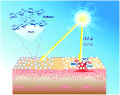
Zinc oxide (ZnO), the main component of several suntan lotions, generates highly oxidizing, cytotoxic and genotoxic reactive oxygen species (ROS) upon UV light absorption. In order to increase safety combined with ZnO use as a sunscreen, its photocatalytic activity should be efficiently quenched. In our studies commercial samples of zinc oxide nanoparticles were hybridized by ionotropic gelation with a natural biopolymer, chitosan (CS). The chemical crosslinking of the polymer in the presence of ZnO nanoparticles was performed. Significantly, in contrast to several CS–ZnO hybrid materials described in the literature, the obtained composites maintained the UV light absorption ability, while the photocatalytic activity towards chemical and biological substrates was totally quenched. Furthermore, a complete lack of photoelectrochemical response observed for the chitosan modified semiconductors confirmed the total inhibition of photoinduced interfacial electron transfer processes. Additionally, antibacterial activity against selected bacterial strains, Staphylococcus aureus and Escherichia coli was observed, although there was no cytotoxic effect against human keratinocytes. The nanocomposites resolve the problem of the risk associated with using semiconductor nanoparticles as ingredients of suntan lotions, cosmetics and dermatological formulations. The transparent polymeric coating allows the absorption of UV irradiation by ZnO particles and simultaneously blocks photogeneration of reactive radicals and oxygen species.


Introduction

Among the commercially available topical sunscreens two main classes based on their chemical nature can be distinguished – inorganic and organic ones. Nano- and microcrystalline semiconductors, such as titanium dioxide (TiO2) and zinc oxide (ZnO), belong to the inorganic sunscreens, constituting a specific UV radiation barrier. Their task is mainly to absorb UV radiation which is part of solar light, in particular UV-A (320–400 nm) and UV-B (290–320 nm).1 Both types of UV irradiation are cytotoxic to skin cells (keratinocytes and fibroblasts) and, moreover, damage the epidermis leading to skin cancer formation.2,3 Semiconductors are widely used in suntan lotions not only due to an excellent absorption of UV light, but also its low refractive index, chemical stability and relatively low toxicity.4,5 When exposed to UV irradiation, physical sunscreens do not decompose and thus are safer than chemical ones. Suntan lotions containing TiO2 or ZnO are nowadays usually transparent, non-adhesive and can be easily rubbed into the skin.6 It was further proposed that the ease of absorbing through the skin, without irritating it, provides the additional advantage.7,8 On the other hand, there are a lot of risks arising from TiO2/ZnO contact with the skin. The main concern is the toxicity of nanoparticles resulting mainly from their size, the ability to bypass the immune defence mechanisms, and the tendency to form complexes with proteins. However, the most important concern in the use of nano-oxides as the main components of the sunscreens is generation of reactive oxygen species (ROS). ROS are produced on the surface of the semiconductor in the photocatalytic process based on photogeneration of electron–hole pairs followed by the interfacial electron transfer (IFET) or energy transfer process.9 Through the IFET mainly hydroxyl radicals (OH˙), superoxide (O2˙) and hydrogen peroxide (H2O2) are being formed, while singlet oxygen (1O2) can be a product of either energy transfer process or the consecutive reduction and oxidation of O2.10,11 The created highly oxidizing hydroxyl radicals and singlet oxygen are known to be cytotoxic and mutagenic.10,12,13 They can lead to the oxidation of organic components of cosmetics and photodamages of the cells and their components – biological molecules, such as protein, lipids and DNA.14–16

In order to minimize the photocatalytic reactivity of TiO2 and ZnO, it is preferable to use deactivated forms of these semiconductors. Usually it is achieved through their surface chemical modifications or use of antioxidants formulated with the cosmetics, e.g. β-carotene, ascorbic acid, glutathione, etc.11 Another approach to TiO2/ZnO inactivation is their coating with SiO2 or Al2O3. Such core–shell nano- and microparticles demonstrate a reduced photocatalytic activity.14,16 An interesting and promising approach is the formation of a polymeric coatings with natural antioxidants designed to protect against photodamages.17 However, not every surface modification leads ultimately to a complete semiconductor photoactivity quenching.16 In this regard, the search of modern composite materials which constitute a barrier to UV radiation and simultaneously minimize toxic effects arising from photocatalytic reactions, remains a challenging research problem.

Another important threat coming from semiconductor nanoparticles presence in sunscreens is their size (<100 nm) allowing their easy penetration through the stratum corneum of the skin and, consequently, their penetration into the skin's deeper layers.8 The European Scientific Committee on Consumer Products analysed a number of studies in this field and highlighted a lack of sufficient data to assess the safety of nanoparticles application in topical lotions and their potential side effects.18 Several studies regarding the fate of nanoparticles administered topically to the skin demonstrated lymph nodes penetration, accumulation in macrophages, high capacity of nanoparticles internalization by endothelial cells, etc., which raises further concerns about potential inflammation and cytotoxic activity.19,20 While the debate over the safety of nanoparticles application continues, the need to minimize the side effects of NPs in sunscreen remains urgent.

Herein, we present a nanocomposite material based on zinc oxide nanoparticles, surface-modified with a biocompatible polymer, chitosan. ZnO–chitosan composites have been previously described in the literature as potent antibacterial and antifungal materials.4 Li et al. demonstrated that membranes of chitosan with embedded ZnO NPs were antimicrobial against Gram-(−) (Bacillus subtilis, Escherichia coli) and Gram-(+) (Staphylococcus aureus) bacterial strains, but there is no information on photoactivity of these composites.21 A similar bactericidal effect against Vibrio parahaemolyticus and Bacillus lechiniformis was demonstrated by Vaseeharan.22 In both cases materials retained UV light (<400 nm) absorption properties after chitosan incorporation. AbdElhady et al. reported the preparation of a UV-protective chitosan/ZnO-NPs/cotton cloth system.23 Also Farouk et al. presented antibacterial potential of cotton fibers modified with chitosan/ZnO-NPs sol.24 In contrast, Sanoop et al. presented photoactive ZnO/biopolymer nanocomposite coatings (including chitosan) used to modify a polyurethane/cotton foam.25 Other chitosan–ZnO hybrid materials were presented as highly efficient photocatalysts for methylene blue degradation under UV irradiation.26 Similarly, zinc oxide impregnated chitosan beads were found to be suitable for photocatalytic decolorization of Reactive Red 2 (RR) dye under UV and visible light irradiation.27

In contrast, the aim of this work was to minimize the photocatalytic activity of ZnO/CS materials induced by UV resulting in the formation of ROS. For our studies we have selected two commercially available ZnO nanoparticles samples with a particle size ≤50 nm (ZnO50) and ≤100 nm (ZnO100) and chitosan characterized by the average molecular weight. To obtain the ZnO nanoparticles surface modified with CS (CS@ZnO) an ionotropic gelation technique was applied. Presented materials arise as a step towards solving the problem of risks associated with using semiconductor nanoparticles as the active ingredients of sunscreen formulations described in the preceding paragraphs. Polymeric coating allows the absorption of UV irradiation and simultaneously provides an efficient photoprotection.

Experimental

Materials

Chitosan (obtained from chitin of shrimp shells; medium average molecular weight of ∼1278 ± 8 kDa; deacetylation degree 89 ± 2%), zinc oxide nanoparticles (ZnO; particle size <50 nm and <100 nm) and sodium tripolyphosphate (TPP) were purchased from Sigma-Aldrich and used as received. Aqueous solution of acetic acid (99.8% Sigma-Aldrich) was used as a solvent for chitosan. SDS, TEMED, dithiothreitol (DTT), Tris–HCl (VWR BDH Prolabo) and acrylamide/bisacrylamide gel (Sigma-Aldrich) were used for electrophoresis. Ammonium persulfate (APS) and Coomassie Brilliant Blue R-250 were supplied by Bio-Rad. Page Ruler Prestained Protein Ladder (Pierce, Thermo Scientific) was used as a size standard in protein electrophoresis (SDS-PAGE). Thiazolyl blue tetrazolium bromide (98%; 3-(4,5-dimethylthiazol-2-yl)-2,5-diphenyltetrazolium bromide) assay kit for cytotoxicity assessment was supplied by Sigma-Aldrich. DMSO and methanol were purchased from Chempur. Phosphate-Buffered Saline (PBS) without calcium and magnesium content, Dulbecco's Modified Eagle Medium (DMEM), with high glucose (4.5 g dm−3), with L-glutamine, with phenol red and antibiotic mix (penicillin, streptomycin, amphotericin) used in cell culturing were supplied by PAN BIOTECH Cell Culture Innovations. Materials for bacteria culturing were purchased from BIOMED (broth) and BIOCORP (agar).

Synthesis of CS@ZnO composites–polymer crosslinking

Two grades of commercially available zinc oxide nanoparticles, ZnO50 and ZnO100, were utilized. Chitosan modified ZnO-NPs were prepared by ionic-gelation technique with TPP as a cross-linking agent (Scheme 1). Briefly, ZnO was suspended in CS solution (1 g dm−3) and TPP solution was added in a drop wise manner upon stirring at room temperature. Weight ratio of components was 1[thin space (1/6-em)]:[thin space (1/6-em)]1[thin space (1/6-em)]:[thin space (1/6-em)]2 for CS[thin space (1/6-em)]:[thin space (1/6-em)]ZnO[thin space (1/6-em)]:[thin space (1/6-em)]TPP, respectively. Stirring (15 min) was followed by 30 minutes of sonication (PolSonic, 620 W). Samples were centrifuged (4000 rpm, 5 minutes) and the pellets were washed with distilled water twice. CS@ZnO were re-suspended in water directly before the further tests. Within the paper non-cross-linked composites of chitosan and zinc oxide are termed ZnO–CS, while the materials after gelation with TPP are termed CS@ZnO.
image file: c5ra12667c-s1.tif
Scheme 1 Modification procedure of zinc oxide nanoparticles.

Physicochemical characterization of ZnO and CS@ZnO

The hydrodynamic diameter and zeta potential (ζ) at pH = 5.5 of ZnO and CS@ZnO particles were determined via dynamic light scattering technique (DLS; Zetasizer Nano ZS with a 633 nm red laser, Malvern Instruments). UV-vis diffuse reflectance spectra of the bare and modified ZnO samples were collected using a UV-vis-NIR Shimadzu spectrophotometer UV-3600 equipped with the 15 cm dia. integrating sphere. Samples were ground in the agate mortar with BaSO4 (reference) with a weight ratio of ca. 1[thin space (1/6-em)]:[thin space (1/6-em)]30. FTIR spectra of materials were collected in the range of 4000–400 cm−1 (Perkin-Elmer Spectrum Two ATR). ZnO and CS@ZnO particles shape, morphology, chemical composition and purity was evaluated by scanning electron microscopy (SEM) using Tescan Vega3 LMU (LaB6 cathode) equipped with EDS spectrometer (Oxford Instruments, X-act, SDD 10 mm2). EDS spectra were collected from many points and elemental composition was subsequently averaged. Crystal structures of investigated materials were confirmed by X-ray diffraction (XRD) using Rigaku MiniFlex 600 diffractometer (Cu Kα radiation obtained at 40 kV, 15 mA, scan speed 1 deg per min, scanning rate 0.05 deg per step, with 2θ ranging from 10 to 90 deg). Photoelectrochemical measurements were carried out using Autolab PGSTAT302N potentiostat and XBO150 xenon lamp equipped with a monochromator and a shutter (Instytut Fotonowy). Measurements have been done in the typical three-electrode setup using platinum wire and Ag/AgCl as a counter and reference electrodes respectively. The working electrode was prepared by casting the tested material onto ITO-covered (indium tin oxide) foil. The electrodes were placed in a cuvette filled with 0.1 mol dm−3 argon-saturated KNO3 solution (pH = 6.1) as an electrolyte (oxygen free conditions). Irradiation was done in the range of 330–450 nm (steps of 10 nm). The working electrodes were irradiated from the backside (through the ITO-foil) in order to eliminate the influence of the thickness of the material layer. Photocurrents were measured in the potential range of 0.1–1 V vs. Ag/AgCl.

Photocatalytic degradation of Azure B

Photocatalytic activity was tested by monitoring the progress of Azure B (AB) photodegradation. All tests of photocatalytic activity were done under identical conditions. A 150 W xenon lamp (XBO-150, with an emission spectra covering UV-A and UV-B range), equipped with a CuSO4 solution filter (0.1 mol dm−3), cutting off the NIR irradiation (780–2500 nm) and the 325–500 nm band-pass filter was used for irradiation. ZnO and CS@ZnO were suspended in aqueous AB solution (0.25 mmol dm−3) and sonicated for 5 minutes (20 cm−3, 1.0, 1.5, 2.0 g dm−3). During 30 minutes of irradiation suspensions were stirred magnetically and saturated with air. Samples were collected each 5 minutes, filtered through the Millipore filter (0.2 μm) and subjected to UV-vis analysis at Hewlett Packard HP8453 spectrophotometer. The progress of AB degradation was monitored by the absorbance changes at 610 nm. Tests were performed in triplicate (three independent trials).

Photocatalytic activity towards α-terpinene

Photocatalytic activity was also tested by monitoring the process of α-terpinene oxidation. The suspensions of ZnO materials (1 g dm−3) in methanolic α-terpinene solution (10−3 mol dm−3) were irradiated for 3 hours in the quartz cuvette using a XBO 150 xenon lamp (Instytut Fotonowy) as a light source. Substrate concentration changes were determined with HPLC analysis (Perkin-Elmer, Flexar system) equipped with UV-vis detector and C18 column (Perkin-Elmer, Spheri 5, ODS 5 μm, 250 × 4.6 mm, cat. no. 0712-0019). In the developed HPLC method the mobile phase was a mixture of methanol and water with a ratio of 60[thin space (1/6-em)]:[thin space (1/6-em)]40 (v/v), (flow rate 1 cm3 min−1). Detection of the α-terpinene oxidation product, ascaridole, was carried out at λ = 226 nm. A standard sample of ascaridole (PhytoLab) was used as a reference.

Biological model molecule (bovine serum albumin – BSA) photodegradation studies

Photodegradation of model biological molecule, Bovine Serum Albumin (BSA), was studied prior to the assessment and comparison of the safety of ZnO and CS@ZnO in contact with biomolecules. The suspensions of ZnO materials (1 g dm−3) in BSA solution (0.4 mg cm−3) were irradiated for 30 min in the quartz cuvette using XBO 150 xenon lamp (Instytut Fotonowy) equipped with a CuSO4 solution filter (0.1 mol dm−3) cutting off the NIR irradiation and the 320 nm cut-off filter. Solutions were saturated with air during irradiation. Samples were collected after each 10 minutes and subjected to the protein degradation analysis by a gel electrophoresis technique (Bio-Rad, PowerPacTM Basic). Changes of BSA concentration were determined by densitometric analysis of SDS-PAGE result. After the electrophoresis each gel was stained with Coomassie Brilliant Blue R-250. The imaging of the stained gels was performed with GelDoc-It™ Imaging System (UVP). Images were further processed with a computer software. The strands of BSA were integrated and compared to the control, which was a BSA initial concentration. Additional controls were also performed – BSA solution irradiated in the absence of both ZnO and CS@ZnO in order to exclude a direct protein photodegradation. Tests were performed in triplicate.

Bacterial culture conditions and colony-forming capability tests

Two bacterial strains, representing Gram-(+) Staphylococcus aureus ATCC 25923 and Gram-(−) Escherichia coli PCM 2209, were selected for antibacterial activity assessment. Strains were maintained in enriched Tryptone Soy Broth (TSB, BIOMED) and kept at 4 °C. Bacterial cells were inoculated into 10 cm−3 of TSB and incubated in aerobic conditions at 37 °C for 18–24 h to give a final concentration of ∼109 CFU mL−1 (CFU – Colony Forming Unit, stationary phase). Enriched agar (BIOCORP) was used for seeding plates preparation. Phosphate buffered saline (PBS) was employed for serial dilutions (1[thin space (1/6-em)]:[thin space (1/6-em)]1.2[thin space (1/6-em)]:[thin space (1/6-em)]7.2[thin space (1/6-em)]:[thin space (1/6-em)]40[thin space (1/6-em)]:[thin space (1/6-em)]5000 weight ratio of KCl, KH2PO4, Na2HPO4, NaCl and distilled water, respectively). Bacterial cultivation was carried out in a bacteriological incubator (Thermo Scientific, MaxQ 6000). All assays were carried out in a laminar flow hood (Thermo Scientific, MSC Advantage). Bacterial growth inhibition was tested by adding 24 h inoculum to ZnO or CS@ZnO suspensions in 1[thin space (1/6-em)]:[thin space (1/6-em)]9 v/v ratio (bacteria cells concentration of ∼109 CFU mL−1, ZnO or CS@ZnO final concentration of 0.45 mg cm−3). Samples were incubated in a shaker incubator at 37 °C for 4, 8 and 24 h (under diffused day light). After each incubation time serial dilutions were performed and seeded on agar plates. Bacteria colonies were counted and the numbers of CFU mL−1 were calculated after 24 h of plates incubation at 37 °C.

Cell culture conditions and cytotoxicity studies

To determine the potential cytotoxic activity of ZnO and CS@ZnO dispersions, human immortal keratinocytes cell line was chosen (HaCaT). Cells were maintained in high-glucose Dulbecco's Modified Eagle's Medium (DMEM) with phenol red, with 1% of antibiotics (streptomycin, penicillin, amphotericin) and 10% of Fetal Bovine Serum (FBS). Cells were cultured at 37 °C in 5% CO2 saturated air. Culture media were replaced every 2 days. Cells were passaged at least once a week. Cells were seeded in 96-well flat bottom plates at a density of 1 × 10⁴ cells per well with 200 μL of medium (24 h, 37 °C, 5% CO2 atmosphere). After 24 h of culturing the medium was aspirated out, and cells were washed with phosphate-buffered saline (PBS). Each well was treated with 0.25 mg cm−3 ZnO or CS@ZnO suspended in DMEM with 10% FBS and incubated for 24 h (37 °C, 5% CO2 atmosphere). Cell viability was determined by the MTT assay.28 Briefly, each well was washed with PBS, treated with 200 μL of the MTT solution (0.5 mg cm−3 in DMEM without FBS) and incubated at 37 °C for 4 h. Insoluble purple formazan crystals were subsequently dissolved in DMSO[thin space (1/6-em)]:[thin space (1/6-em)]CH3OH (1[thin space (1/6-em)]:[thin space (1/6-em)]1). The absorbance at 565 nm was measured in a microplate reader (Infinite 200 M PRO NanoQuant, Tecan). Results are presented as surviving cells fraction, [absorbance570 nm of treated cells/absorbance570 nm of untreated cells] × 100%, and expressed as mean values ± standard deviation. The results are the average values of three independent experiments. Any potential interference from nanoparticles was evaluated and ruled out during the assay.

Results and discussion

Materials characterization

Nanocrystalline ZnO suspensions and chitosan-modified ZnO colloidal suspensions were slightly white and translucent. The hydrodynamic diameter and surface charge at pH = 5.5 of ZnO and CS@ZnO particles were determined via dynamic light scattering technique. The results are listed in Table 1.
Table 1 Results of dynamic light scattering measurements
Sample Size/nm PdI Zeta potential (ζ)/mV
ZnO50 167 ± 10 0.27 ± 0.02 36.4 ± 0.2
CS@ZnO50 233 ± 8 0.4 ± 0.01 26.6 ± 0.8
ZnO100 288 ± 12 0.23 ± 0.01 32.9 ± 0.5
CS@ZnO100 342 ± 14 0.17 ± 0.01 20.9 ± 0.9


The increase of particles size can be observed for the materials with chitosan modification which proves the polymeric layer formation. The polydispersity index (PdI) values indicate a narrow size distribution of particles. Surface charge or zeta potential relationship with the stability of ZnO in aqueous media was previously described in the literature.29,30 ZnO point of zero charge occurs usually at pH ∼ 9 (pHPZC).31,32 Since chitosan possesses numerous amino groups (determined by the deacetylation degree), the acid dissociation constant (pKa) is ca. 6.5. Below that pH amino groups become protonated and thus the surface charge of CS@ZnO modified nanoparticles reaches positive values. The zeta potential at pH = 5.5 of unmodified ZnO NPs, in comparison to CS@ZnO, was distinctly higher. Still, the ζ value around 20–30 mV evidences the incipient stability of modified NPs in the solution.

The electronic absorption spectra presented as Kubelka–Munk function are shown in Fig. 1. Importantly, surface modification of ZnO nanoparticles with chitosan did not block the UV light absorption of materials.


image file: c5ra12667c-f1.tif
Fig. 1 Normalized diffuse reflectance spectra of photocatalysts before and after chitosan modification (transformed to Kubelka–Munk function).

The infrared absorption spectra of materials are shown in Fig. 2. In order to prove the presence of chitosan on the surface of ZnO nanoparticles the infrared spectra were collected for the unmodified materials (ZnO), cross-linked chitosan, materials with chitosan without cross-linking (ZnO–CS) and for composites CS@ZnO. The absorption peak ∼3290 cm−1 for the bare polymer is attributed to stretching vibrations within the amino (–NH2) and hydroxyl (–OH) groups. For cross-linked materials a significant intensity enhancement, broadening and shift to lower wavenumber, indicates a strong interaction of chitosan functional groups with ZnO surface.33 For CS@ZnO50 (Fig. 2A) and CS@ZnO100 (Fig. 2B) the maximum occurs at ∼3276 cm−1 and ∼3259 cm−1, respectively. In addition, changes in the position and intensity of the amide I and amide II bands located at 1450, 1550 and 1650 cm−1 also confirm the interaction of ZnO with –OH and –NH2 groups.23 Absorption peaks at 1647 and 1078 cm−1, characteristic for chitosan, are ascribed to bending –NH2 and stretching C–O group vibrations. They are not noticeable for CS@ZnO, due to the binding of amide groups to ZnO.34 A comparison of the spectra recorded for materials without cross-linked polymer to those with the cross-linked chitosan, confirms the absence of chitosan on ZnO surface in the former case. Moreover, it points at the significance of the polymer crosslinking on the ZnO nanoparticles coverage with chitosan.


image file: c5ra12667c-f2.tif
Fig. 2 FTIR spectra of materials based on ZnO50 (A) and ZnO100 (B).

To determine the crystal structure and phase composition of zinc oxide nanoparticles XRD measurements were performed (data not shown). X-ray diffraction patterns of ZnO50 and ZnO100 confirmed the most common and stable structure of polycrystalline wurtzite, and the hexagonal crystal structure of zinc oxide nanoparticles (typical diffraction peaks were assigned to (100), (002), (101), (102), (110), (103), (112) and (202) planes, based on the database (JCPDS 36-1451)).35

Scanning electron microscopy (SEM) pictures present the morphology of obtained materials (Fig. 3). The unmodified zinc oxide 50 (Fig. 3A) and 100 (Fig. 3D) are composed of fine particles (size ≤ 100 nm). Although incubation of ZnO50 and ZnO100 in chitosan solution showed no significant effect on the morphology of the polymer samples; they remain in the form of small particles forming bigger aggregates (Fig. 3B and E). In contrast, materials modified with cross-linked chitosan show significant changes in morphology (Fig. 3C and F). The polymer covers aggregates of ZnO, observed as a rounded and smooth shapes.


image file: c5ra12667c-f3.tif
Fig. 3 Scanning electron microscopy (SEM) images with EDS composition analysis of ZnO50 (A), ZnO50-CS (B), CS@ZnO (C), ZnO100 (D), ZnO100-CS (E) and CS@ZnO100 (F).

The photocurrent dependence on the incident light wavelength at a constant potential (200 mV vs. Ag/AgCl) is presented in Fig. 4. Under these conditions anodic photocurrents can be observed for both ZnO50 and ZnO100 materials.36,37 When light of energy higher than the band gap energy is absorbed by the material, the electrons are excited to the conduction band of semiconductor. Photogenerated charges can undergo an interfacial electron transfer observed as the photocurrent. The anodic photocurrent results in the electron transfer from the semiconductor to the electrode and from the electron donor (e.g. water) to the semiconductor. ZnO–CS materials generate much lower photocurrents than bare ZnO, while the photoactivity of CS@ZnO appears almost completely quenched. These results, coherent with photocatalytic activity of tested samples, point at an efficient barrier for the electron transfer constituted by the cross-linked chitosan.


image file: c5ra12667c-f4.tif
Fig. 4 Photocurrents recorded for ZnO50-based (A) and ZnO100-based (B) materials. The measurements were carried out in aqueous 0.1 M KNO3 electrolyte in an inert atmosphere of argon. The changes in measured current, associated with the opening and closing of the shutter, are the measure of photocurrent.

Photocatalytic degradation of Azure B

Azure B (AB; thiazine dye) can be degraded by hydroxyl radicals, photogenerated holes in the valence band of a semiconductor (e.g. nanocrystalline TiO2) or oxidized by singlet oxygen.38 Therefore degradation of this dye occurs in the presence of several reactive oxygen species. Since photogenerated ROS may be responsible for oxidation of organic components of suntan lotions and photodamages of skin or DNA, it is crucial to determine whether chitosan modified materials generate these reactive species. Fig. 5 shows the results of AB photodegradation tests in the presence of various concentrations of ZnO (A and C) and CS@ZnO (B and D) samples. The decrease of Azure-B absorbance was observed within irradiation, indicating degradation of the dye. In the timescale of experiment (30 min) the most efficient AB degradation (∼80%) under applied irradiation conditions occurred in the presence of ZnO100 even at the lowest concentration level of 1.0 g dm−3 (Fig. 5C). For 1.5 and 2.0 g dm−3 ZnO100 AB was totally degraded within 30 minutes. Also ZnO50 appeared to act as an efficient photocatalyst, although AB degradation at the highest material concentration reached ∼70%. In contrast to unmodified ZnO, AB was photostable during irradiation in the presence of CS@ZnO50 (Fig. 5B) and CS@ZnO100 (Fig. 5D) even at the highest concentrations of composites. The test of Azure B photodegradation confirmed the lack of photoactivity of CS@ZnO materials resulting from the chitosan crosslinking on the surface of ZnO nanoparticles.
image file: c5ra12667c-f5.tif
Fig. 5 Photodegradation of Azure B in the presence of ZnO50 (A), CS@ZnO50 (B), ZnO100 (C) and CS@ZnO100 (D) at different levels of concentration (denoted in graphs). The results are shown as the relative AB concentration (A/A0).

Photocatalytic activity towards α-terpinene

Singlet oxygen generation was followed by monitoring the progress of α-terpinene oxidation to ascaridole.39 The study showed no formation of ascaridole during irradiation (data not shown). Obtained results prove that neither bare nor modified ZnO enables the singlet oxygen generation at the detection level (0.05 μM) of the applied method.

BSA photodegradation

The photocatalytic activity of ZnO and CS@ZnO was tested towards biological model molecule, Bovine Serum Albumin. The results are shown in Fig. 6. Similarly to Azure B photodegradation process, ZnO50 (Fig. 6A) and ZnO100 (Fig. 6B) samples appeared to be effective in BSA degradation under applied irradiation conditions (∼50% of degradation). Again, photocatalysts samples modified with cross-linked chitosan were not photoactive towards the tested protein. These results are parallel to the photocatalytic test and photocurrent measurements results. Stability of BSA under irradiation in the presence of CS@ZnO materials confirms the absence of ROS and inhibition of any energy and electron transfer processes.
image file: c5ra12667c-f6.tif
Fig. 6 BSA photodegradation curves for ZnO50 (A) and ZnO100 (B) based materials.

All presented photoactivity studies confirm a complete quenching of photocatalytic activity of ZnO modified with cross-linked chitosan.

Antibacterial activity

The antibacterial activity of ZnO and chitosan modified ZnO nanoparticles was tested by bacteria incubation in materials suspensions against Gram-(−) Escherichia coli PCM 2209 and Gram-(+) Staphylococcus aureus ATCC 25923. The colony-forming capability test was performed. The results, presented as CFU mL−1 in Fig. 7, indicate for both of the tested strains the enhancement of the antibacterial activity of materials after chitosan modification. However, E. coli appeared to be totally resistant against bare ZnO nanoparticles (Fig. 7A and B) and more resistant than S. aureus for CS@ZnO upon 24 h incubation time (Fig. 7C and D). The reduction of bacterial growth reached ∼5[thin space (1/6-em)]log. In the case of S. aureus strain, both ZnO50 and ZnO100 nanoparticles caused a 6[thin space (1/6-em)]log reduction of the bacteria number within 24 hours. Interestingly, for CS@ZnO50 and CS@ZnO100 a total bacterial growth reduction was achieved after 8 hours of incubation. The results are in agreement with Farouk et al. who demonstrated an enhanced antibacterial activity of ZnO–chitosan composite for textile finish against E. coli and M. luteus.24 Also AbdElhady et al. presented antibacterial activity of chitosan/ZnO composite against E. coli and S. aureus.23 Bacterial growth reduction by a CS/ZnO composite was also demonstrated against other Gram-(−) and Gram-(+) bacterial strains.22 The antibacterial potential of ZnO nanoparticles strongly depends on their size, concentration and intensity of visible and UV light. The smaller the NPs the higher bactericidal effect was obtained by N. Jones et al.40 Moreover, the UV light photoactivated samples caused a stronger bacterial growth reduction.40 It is important to note that the toxicity of nanoparticles depends not only on their size and concentration but also on the method of application on the tested organism. When ZnO quantum dots in the form of powder, film and gel were tested, the bactericidal properties against three bacterial pathogens were ZnO nanoparticles form dependent. In contrast to the quantum dots bound in the polystyrene films, ZnO powder and gel showed a significant antibacterial activity. The sustained bactericidal effect of zinc oxide after coating with chitosan seems to be important for its applications.41 Our results confirmed the antibacterial potential of ZnO nanoparticles with dimensions lower than 100 nm and demonstrated the dependence between the bacterial strain and growth reduction rate. The antimicrobial mode of chitosan and its derivatives is well described in the literature.42–44 We presented the enhanced antibacterial activity of chitosan modified ZnO which suggest the synergic mode of action of both components. The reduction of bacterial growth for CS@ZnO50 and CS@ZnO100 reached ∼5[thin space (1/6-em)]log for E. coli and total reduction for S. aureus. These results are consistent with those presented by Luo, who showed that application of ZnO with antibiotic against bacteria improved the antimicrobial activity of both applied agents.45
image file: c5ra12667c-f7.tif
Fig. 7 Colony-forming capability tests presented as CFU mL−1 for Gram-(−) Escherichia coli PCM 2209 (A and B) and Gram-(+) Staphylococcus aureus ATCC 25923 (C and D). (Mean value ± standard deviation; n = 3).

Cytotoxicity assay

Several papers demonstrate ZnO antibacterial activity based mainly ROS generation and on free radicals photogeneration.46–48 However, the cytotoxic effect towards human cells is not fully examined. Size dependent cytotoxicity against leukemia cancer cells was demonstrated by Guo et al.49 In other studies the cytotoxicity of 60 nm ZnO NPs was evaluated on normal and tumor cells, especially glioma cells. No toxicity on normal human astrocytes was demonstrated at mmol L−1 concentration level.50 Herein, we present results of cytotoxicity assay on human keratinocytes cell line of ZnO and CS@ZnO materials (Fig. 8). The final concentration of materials was 0.25 mg cm−3. Only for ZnO50 a slight cytotoxic effect was observed (a cell viability was reduced to ∼85%). It stays in agreement with the literature, where the size dependent toxicity of ZnO was proved. Chitosan modified ZnO nanoparticles, both CS@ZnO50 and CS@ZnO100, did not cause cytotoxicity on human keratinocytes at the tested concentration level. Coating nanoparticles approaches seem to be reasonable to minimize toxicity or block photocatalytic activity. Ramasamy reported that after exposure of human dermal fibroblast cells to surface modified zinc oxide nanoparticles a decreased cytotoxicity was observed. Furthermore, comparing two different silica layer thicknesses, the material with a thicker coating was much less toxic compared to that with a thinner layer, as well as to bare ZnO-NPs.51 Presented results demonstrate a huge potential of CS@ZnO materials application in cosmetics and biomedical field.
image file: c5ra12667c-f8.tif
Fig. 8 Cellular viability after incubation with ZnO and CS@ZnO for HaCaT cell line. Data were expressed as the mean ± standard error (n = 9).

Conclusions

Zinc oxide, one of the main components of suntan lotions, after photoactivation by UV light produces reactive oxygen species, which are known to be highly cytotoxic and genotoxic. In order to minimize the photoactivity of ZnO materials, it is preferable to use deactivated forms of this semiconductor. Although several singlet oxygen and radical scavengers are used as additives, no significant ROS related risk decrease was observed.13 The aim of this work was to solve the problem of photoactivity of one of the inorganic sunscreens, ZnO nanoparticles. We presented a novel nanocomposite material based on zinc oxide nanoparticles, surface-modified with a biocompatible chitosan by ionotropic gelation technique. The preserved UV light absorption abilities of synthesized composites were proven. Cross-linked chitosan quenched completely the photocatalytic activity of ZnO nanoparticles. As a result materials do not generate reactive oxygen species, in particular singlet oxygen or hydroxyl radicals. Furthermore, a complete photocurrent quenching for cross-linked chitosan modified zinc oxide was confirmed, which approves the lack of photoinduced energy or electron transfer processes. In contrast to other chitosan–ZnO hybrid materials presented in the literature, cross-linked chitosan coating allows a simultaneous absorption of UV light by the entrapped ZnO particles and efficient photoprotection. Moreover, composites exhibit an enhanced antibacterial activity against Staphylococcus aureus and Escherichia coli in comparison to unmodified ZnO. Simultaneous lack of cytotoxic effect against human keratinocytes makes the described CS@ZnO materials a perfect candidate for active and safe component of suntan lotions. Presented materials resolve the problem of risks associated with the use of ZnO as the active ingredient of suntan lotions, cosmetics and dermatologic formulations.

Acknowledgements

This work has been supported by the Ministry of Science and Higher Education, Poland, within the IDEAS PLUS project, grant No. IdP2012000362. A part of this work was carried out at the equipment financed by the European Development Fund in the framework of the Polish Innovation Economy Operational Program (contract no. POIG.02.01.00-12-023/08). The authors are grateful to Dr Olexandr Korchynskyi and Prof. Mykhailo Gonchar for enabling experiments on HaCaT cell line.

Notes and references

  1. M. Buchalska, G. Kras, M. Oszajca, W. Łasocha and W. Macyk, J. Photochem. Photobiol., A, 2010, 213, 158 CrossRef CAS PubMed.
  2. M. Pidoux and R. M. Tyrrell, Cancer Res., 1987, 47, 1825 Search PubMed.
  3. F. P. Gasparro, Environ. Health Perspect., 2000, 108, 71 CrossRef CAS.
  4. M. Mirhosseini and F. B. Firouzabadi, Int. J. Dairy Technol., 2013, 66, 291 CrossRef CAS PubMed.
  5. A. C. Dodd, A. J. McKinley, M. Saunders and T. Tsuzuki, J. Nanopart. Res., 2006, 8, 43 CrossRef CAS.
  6. A. Kołodziejczak-Radzimska and T. Jesionowski, Materials, 2014, 7, 2833 CrossRef PubMed.
  7. A. B. G. Lansdown and A. Taylor, Int. J. Cosmet. Sci., 1997, 19, 167 CrossRef CAS PubMed.
  8. S. E. Cross, B. Innes, M. S. Roberts, T. Tsuzuki, T. A. Robertson and P. McCormick, Skin Pharmacol. Physiol., 2007, 20, 148 CrossRef CAS PubMed.
  9. K. Szaciłowski, W. Macyk, A. Drzewiecka-Matuszek, M. Brindell and G. Stochel, Chem. Rev., 2005, 105, 2647 CrossRef PubMed.
  10. A. Jańczyk, E. Krakowska, G. Stochel and W. Macyk, J. Am. Chem. Soc., 2006, 128, 15574 CrossRef PubMed.
  11. A. Jańczyk, A. Wolnicka-Głubisz, K. Urbanska, G. Stochel and W. Macyk, J. Photochem. Photobiol., B, 2008, 92, 54 CrossRef PubMed.
  12. A. Jańczyk, A. Wolnicka-Głubisz, K. Urbanska, H. Kisch, G. Stochel and W. Macyk, Free Radical Biol. Med., 2008, 44, 1120 CrossRef PubMed.
  13. N. Serpone, D. Dondi and A. Albini, Inorg. Chim. Acta, 2007, 360, 794 CrossRef CAS PubMed.
  14. M. D. Newman, M. Stotland and J. I. Ellis, J. Am. Acad. Dermatol., 2009, 61, 685 CrossRef CAS PubMed.
  15. P. J. McHugh and J. Knowland, Photochem. Photobiol., 1997, 66, 276 CrossRef CAS PubMed.
  16. R. Dunford, A. Salinaro, L. Cai, N. Serpone, S. Horikoshi, H. Hidaka and J. Knowland, FEBS Lett., 1997, 418, 87 CrossRef CAS.
  17. W. A. Lee, N. Pernodet, B. Li, C. H. Lin, E. Hatchwell and M. H. Rafailovich, Chem. Commun., 2007, 4815,  10.1039/b709449c.
  18. G. Oberdorster, A. Maynard, K. Donaldson, V. Castranova, J. Fitzpatrick, K. Ausman, J. Carter, B. Karn, W. Kreyling, D. Lai, S. Olin, N. Monteiro-Riviere, D. Warheit and H. Yang, Part. Fibre Toxicol., 2005, 2, 8 CrossRef PubMed.
  19. S. Kim, Y. T. Lim, E. G. Soltesz, A. M. de Grand, J. Lee, A. Nakayama, J. A. Parker, T. Mihaljevic, R. G. Laurence, D. M. Dor, L. H. Cohn, M. G. Bawendi and J. V. Frangioni, Nat. Biotechnol., 2004, 22, 93 CrossRef CAS PubMed.
  20. K. Peters, R. Unger, C. J. Kirkpatrick, A. Gatti and E. Monari, J. Mater. Sci.: Mater. Med., 2004, 15, 321 CrossRef CAS.
  21. L.-H. Li, J.-C. Deng, H.-R. Deng, Z.-L. Liu and L. Xin, Carbohydr. Res., 2010, 345, 994 CrossRef CAS PubMed.
  22. B. Vaseeharan, J. Sivakamavalli and R. Thaya, J. Compos. Mater., 2013, 49, 177 CrossRef PubMed.
  23. M. M. AbdElhady, Int. J. Carbohydr. Chem., 2012, 2012, 6 Search PubMed.
  24. A. Farouk, S. Moussa, M. Ulbricht and T. Textor, Int. J. Carbohydr. Chem., 2012, 2012, 8 Search PubMed.
  25. P. K. Sanoop, K. V. Mahesh, K. M. Nampoothiri, R. V. Mangalaraja and S. Ananthakumar, J. Appl. Polym. Sci., 2012, 126, E233 CrossRef PubMed.
  26. Y. Haldorai and J.-J. Shim, J. Appl. Polym. Sci., 2013, 20, 365 CAS.
  27. M. H. Farzana and S. Meenakshi, Int. J. Biol. Macromol., 2015, 72, 900 CrossRef CAS PubMed.
  28. T. L. Riss, R. A. Moravec, A. L. Niles, et al., ed. L. Minor, G. S. Sittampalam, N. P. Coussens, H. Nelson, et al., Assay Guidance Manual [Internet], Eli Lilly & Company and the National Center for Advancing Translational Sciences, Bethesda (MD), 2004, p. 2004 Search PubMed.
  29. K.-M. Kim, T.-H. Kim, H.-M. Kim, H.-J. Kim, G.-H. Gwak, S.-M. Paek and J.-M. Oh, J. Toxicol. Environ. Health Sci., 2012, 4, 121 CrossRef.
  30. S.-W. Bian, I. A. Mudunkotuwa, T. Rupasinghe and V. H. Grassian, Langmuir, 2011, 27, 6059 CrossRef CAS PubMed.
  31. S. S. Alias, A. B. Ismail and A. A. Mohamad, J. Alloys Compd., 2010, 499, 231 CrossRef CAS PubMed.
  32. F. Mohd Omar, H. Abdul Aziz and S. Stoll, Sci. Total Environ., 2014, 468–469, 195 CrossRef CAS PubMed.
  33. R. Salehi, M. Arami, N. M. Mahmoodi, H. Bahrami and S. Khorramfar, Colloids Surf., B, 2010, 80, 86 CrossRef CAS PubMed.
  34. P. Bhadra, M. K. Mitra, G. C. Das, R. Dey and S. Mukherjee, Mater. Sci. Eng., C, 2011, 31, 929 CrossRef CAS PubMed.
  35. L. Kumari and W. Z. Li, Cryst. Res. Technol., 2010, 45, 311 CrossRef CAS PubMed.
  36. E. M. P. Steinmiller and K.-S. Choi, Proc. Natl. Acad. Sci. U. S. A., 2009, 106, 20633 CrossRef CAS PubMed.
  37. K. Szaciłowski, in Infochemistry, John Wiley & Sons, Ltd, 2012, p. 165,  DOI:10.1002/9780470710883.ch6.
  38. H. Gnaser, M. R. Savina, W. F. Calaway, C. E. Tripa, I. V. Veryovkin and M. J. Pellin, Int. J. Mass Spectrom., 2005, 245, 61 CrossRef CAS PubMed.
  39. M. Matsushita, T. H. Tran, A. Y. Nosaka and Y. Nosaka, Catal. Today, 2007, 120, 240 CrossRef CAS PubMed.
  40. N. Jones, B. Ray, K. T. Ranjit and A. C. Manna, FEMS Microbiol. Lett., 2008, 279, 71 CrossRef CAS PubMed.
  41. T. Jin, D. Sun, J. Y. Su, H. Zhang and H. J. Sue, Int. J. Food Sci., 2009, 74, M46 CrossRef CAS PubMed.
  42. M. Kong, X. G. Chen, K. Xing and H. J. Park, Int. J. Food Microbiol., 2010, 144, 51 CrossRef CAS PubMed.
  43. D. Raafat and H. G. Sahl, Microb. Biotechnol., 2009, 2, 186 CrossRef CAS PubMed.
  44. E. I. Rabea, M. E. T. Badawy, C. V. Stevens, G. Smagghe and W. Steurbaut, Biomacromolecules, 2003, 4, 1457 CrossRef CAS PubMed.
  45. Z. Luo, Q. Wu, J. Xue and Y. Ding, Biomed. Nanotechnol., 2013, 9, 69 CrossRef CAS PubMed.
  46. J. Sawai, S. Shoji, H. Igarashi, A. Hashimoto, T. Kokugan, M. Shimizu and H. Kojima, J. Ferment. Bioeng., 1998, 86, 521 CrossRef CAS.
  47. Y. Xie, Y. He, P. L. Irwin, T. Jin and X. Shi, Appl. Environ. Microbiol., 2011, 77, 2325 CrossRef CAS PubMed.
  48. G. Applerot, A. Lipovsky, R. Dror, N. Perkas, Y. Nitzan, R. Lubart and A. Gedanken, Adv. Funct. Mater., 2009, 19, 842 CrossRef CAS PubMed.
  49. D. Guo, C. Wu, H. Jiang, Q. Li, X. Wang and B. Chen, J. Photochem. Photobiol., B, 2008, 93, 119 CrossRef CAS PubMed.
  50. S. Ostrovsky, G. Kazimirsky, A. Gedanken and C. Brodie, Nano Res., 2009, 2, 882 CrossRef CAS.
  51. M. Ramasamy, M. Das, S. S. A. An and D. K. Yi, Int. J. Nanomed., 2014, 9, 3707 CAS.

This journal is © The Royal Society of Chemistry 2015
Click here to see how this site uses Cookies. View our privacy policy here.