Ortensia Ilaria Parisi†
ab,
Catia Morelli†a,
Luca Scrivanoa,
Maria Stefania Sinicropia,
Maria Grazia Cesarioa,
Sebastiano Candamanoc,
Francesco Puoci‡
*a and
Diego Sisci‡a
aDepartment of Pharmacy, Health and Nutritional Sciences, University of Calabria, 87036 Rende (CS), Italy. E-mail: francesco.puoci@unical.it; Tel: +39 0984 493151, +39 0984 496211
bDepartment of Informatics, Modeling, Electronics and Systems Engineering, University of Calabria, 87036 Rende (CS), Italy
cDepartment of Environmental and Chemical Engineering, University of Calabria, 87036 Rende (CS), Italy
First published on 9th July 2015
The aim of the present work was the preparation of a magnetic hydrogel for guided release of sunitinib malate (SUM), an anticancer drug. Precipitation polymerization method has been employed in order to synthesize the hydrogel, using methacrylic acid (MAA), ethylene glycol dimethacrylate (EGDMA), glycidyl methacrylate (GMA) and 2,2′-azobisisobutyronitrile (AIBN) as monomer, cross-linker, pro-hydrophilic monomer and initiator, respectively. To confer magnetic responsiveness, particles of magnetite have been encapsulated during the polymerization process, followed by epoxide ring opening on the nanospheres' surface, in order to obtain a restricted access material (RAM) with low protein adsorption ability and improved biocompatibility. The successful introduction of magnetite has been confirmed through FT-IR and TG analyses, while protein adsorbing tests have been conducted to verify the RAM features. Furthermore, swelling properties have been evaluated before and after epoxide ring opening. Finally, in vitro tests have been performed to evaluate the release profile and cytotoxic effect on ARO, WRO, HeLa and MCF-7 cell lines.
This proves its wide application in many kinds of tumoral diseases, due to its unspecific and unselective activity towards human tissues. For this reason, concentrating efforts in the study and development of new materials able to confer selectivity towards cancerous tissues to antineoplastic compounds is important. Cancer therapy is indeed always accompanied with adverse and undesirable systemic toxic effects. Furthermore, anticancer drugs suffer of low solubility in aqueous solution, narrow therapeutic index and limited penetration into cytoplasmic area.
The purpose of the present study was the development of a novel magnetic nanospherical hydrogel, in order to achieve a guidable and biocompatible system for targeted SUM release. Hydrogels are known to be, by definition, polymeric materials organized in hydrophilic networks capable of a great retention of water. Hydrophilic groups, such as –OH, –CONH–, –CONH2 and –SO3H, are responsible of their high affinity for aqueous and biological media. The water absorbing capability confers to the polymer a hydration degree up to 90%, without dissolution of the material.9 Hydrogels are extensively used as delivery systems for chemotherapeutic agents in cancer therapy,10,11 often able of a stimuli-triggered release.12,13 However, today, guidable devices towards the site of interest are of great importance, aiming to non-systemic effects and limiting cytotoxicity to cancer cells. For this reason, encapsulation of magnetite in hydrogel network is a good strategy to obtain a magnetic guidance.14
Polymers are advantageous for hydrogel synthesis, thanks to the possibility of manipulation of their physico-chemical properties. It is indeed possible to achieve micro- or nano-sized polymeric hydrogel, modifying the polymerization conditions.
Microgels and nanogels have been already reported as delivery vehicles in cancer therapy.15,16 In particular nanogels are interesting nanoscale networks that provide several advantages inherent to their nanometric size.
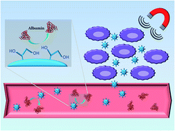
Magnetically responsive micro- and nanogels have been previously prepared.17,18 However, polymeric materials can strongly adsorb on their surface biological components, such as lipids and proteins.19 Different kinds of proteins compete in vivo with each other to surfaces causing the so-called Vroman effect.20 These undesired interactions, occurring in biological fluids, are responsible of decreased biocompatibility and bioavailability, causing a reduction of therapeutic efficacy and possible thrombotic episodes.21,22 To avoid the interference of these biological substances many strategies could be carried out.23,24 Therefore, the purpose of this work was also the synthesis of a restricted access material (RAM) to prevent these non-specific interactions. RAMs are supports designed to limit the access into inner core only to small molecules, excluding biological macromolecules, thanks to a chemical barrier created on polymeric surface.25 In order to achieve the RAM system, glycidyl methacrylate has been used for the insertion of hydrophilic moieties on polymers outer surface, responsible of the reduced adsorption of proteins.26
Therefore, the purpose of the present work was the development of a hybrid material in which multiple characteristics co-exist in the same system. The magnetic responsiveness of nanogel is, indeed, combined with the restricted accessibility.
Thus in this paper the synthesis of magnetic nanospherical hydrogel as RAM for guided release of sunitinib malate is reported and the nanospheres obtained are characterized. Also in vitro toxicity of several cellular lines has been evaluated.
In the polymerization environment monomers (methacrylic acid, MAA), cross-linkers (ethylene glycol dimethacrylate, EGDMA), pro-hydrophilic monomers (glycidyl methacrylate, GMA) and initiator (2,2′-azobisisobutyronitrile, AIBN) are present, dissolved in toluene/acetonitrile mixture. The magnetite particles (Fe3O4) are, instead, dispersed in this system. The first step of the process is the formation of radical oligomers by action of radical species originated from initiator molecules by UV photo-triggering (λ = 360 nm). Oligomers crosslink simultaneously surrounding the magnetite particles, serving as seeds for nanospheres formation. After the nucleation phase, the second step is represented by microgels growth that produces uniformly sized spheres (Fig. 2). In this second phase of the reaction, the number of particles are constant, while size is increasing,27,28 continuously capturing oligomers and preventing any further nucleation.
The grown beads are, in the end, separated from the continuous phase by enthalpic or entropic precipitation, if repulsive interactions between polymer and solvent occur or positive interactions are avoided, respectively.28
The ideal MAA:
GMA
:
EGDMA ratio for the formation of nanospheres has been found to be 8
:
8
:
10, resulting in spherical nanoparticles with a mean diameter of 170 nm, in the dry-collapsed form (Table 1), as confirmed by scanning electron microscope image (Fig. 1). In addition, the dimensional data obtained from the analysis performed with the stereomicroscope agree with the SEM image information about the narrow size distribution (Table 1).
Magnetic nanospheres | Mean diameter | Polydispersity index |
---|---|---|
Dry MNs (collapsed) | 170 ± 11 nm | 0.256 |
Hydrated MNs (swollen) | 287 ± 15 nm | 0.270 |
Moreover, dimensional evaluation of hydrated MNs has been performed with dynamic light scattering, putting a small amount of MNs in a phosphate buffer saline solution at pH 7.4. In this case the mean diameter was 287 nm (Fig. 3). Data, collected in Table 1, show two different size for the two different states of MNs. This could be easily ascribed to the volume of water within the MNs in the swollen state, absent, instead, in the dry-collapsed form. In both cases polydispersity index is <0.3 and mean diameter is below 300 nm.
The importance of obtaining monodisperse spherical nanoparticles for anticancer drugs delivery is validated by the concept of the enhanced permeability and retention (EPR) effect. EPR effect is described as the capability of cancer tissue of being selectively targeted by nanosized particles that are then retained in the site. The explanation of this effect rely on increased vascularization, defective permeability of endothelium and absence of lymphatic drainage in cancer tissue.30
![]() | ||
Fig. 4 FT-IR spectra; (A) magnetite; (B) nanospheres synthesized in absence of magnetite; (C) nanospheres synthesized in presence of magnetite. |
Thermogravimetric analyses (TGA) have been carried out at the range of 30 to 800 °C to determine the amount of magnetite encapsulated in MNs (Fig. 5). In case of magnetite weight loss around 3.5% was due to the physically adsorbed water molecules on the magnetic particles surface (Fig. 5 – black line).
The TGA curve of MNs showed a weight loss of 92.3% associated with the complete thermal decomposition of the polymeric material (Fig. 5 – red line). In fact, the TGA curve of non magnetic nanospheres showed a complete decomposition at 800 °C (Fig. 5 – blue line). The residual weight at 800 °C in the TGA curve of MNs could be easily ascribed to the presence of magnetite. The remaining weight at 800 °C was about 7.7% of the amount of MNs used for the experiment i.e. the percentage of magnetite encapsulated in MNs.
As seen in Fig. 6, MNs have been found to be responsive to the magnetic field. The lower response, compared to magnetite, is supposed to be caused by the shielding of the Fe3O4 particles by the surrounding polymeric matrix.
The confirmed magnetic property of Fe3O4-encapsulated beads, together with the passive targeting of nanospheres based on EPR effect, is the other characteristic exploitable for selective drug delivery. It is, indeed, possible to guide the magnetic nanospheres, applying an appropriate magnetic field, towards a specific target, i.e. cancer site.
Data showed a reduced adsorption of BSA after GMA epoxide ring opening on the surface of the nanospheres, from 21.9 ± 1.0% to 3.0 ± 0.8% after the reaction. The same experiment has been conducted using polymeric nanospheres as control, synthesized in same condition of MNs but in absence of GMA. Also the control showed a similar behaviour to MNs with unreacted epoxide rings (see Table 2). These results demonstrated the importance of hydrophilic groups in decreasing albumin adsorption, thus increasing biocompatibility.
Magnetic hydrogel | Water uptake (%) | BSA adsorption (%) |
---|---|---|
Control (MNs without GMA) | 368.6 ± 1.1 | 16.2 ± 0.9 |
MNs before epoxide ring opening | 343.1 ± 0.9 | 21.9 ± 1.0 |
MNs after epoxide ring opening | 477.2 ± 1.1 | 3.0 ± 0.8 |
The hydrophilic moieties are also fundamental in order to confer swelling ability to polymeric matrix in aqueous solutions. For the evaluation of swelling properties, small amounts of nanospheres have been immersed in a biological-like solution (a phosphate buffer saline solution at pH 7.4, PBS) and the water uptake was measured. As expected, the nanomaterial showed a water uptake of 343.1 ± 0.9% before the opening of epoxide ring and 477.2 ± 1.1% after the reaction. Furthermore, compared to the control, MNs showed also a higher water content, due to the higher number of hydrophilic groups. Data confirm a good swelling behaviour and epoxide opening improved hydrophilic property thanks to the new –OH groups present on the surface. All data are collected in Table 2.
Furthermore in vitro release studies were performed by immersing aliquots of MNs/SUM in PBS pH 7.4 at 37 °C, simulating the physiological environment. The release profile of SUM from nanospherical hydrogels is shown in Fig. 7.
In order to understand the mechanism involved in the in vitro release studies, drug release kinetics and mechanism of drug release from MNs have been investigated using zero order, first order and Korsmeyer–Peppas models.
The release data have been analyzed with zero order and first order kinetics.
Zero order kinetic eqn (1) explains the drug dissolution from various drug delivery systems, as well as from polymeric systems.
Qt = Q0 + K0t | (1) |
On the other hand, first order kinetic describes drug release dependent on concentration. The release rate is indeed proportional to the amount of drug still present within the system. A first order kinetic is expressed by eqn (2).
![]() | (2) |
Data obtained from SUM release profile study fit better first order kinetic than zero order kinetic. This could be stated on the basis of the higher R2 value (see Table 3).
Kinetic order | R2 value |
---|---|
Zero order kinetic | 0.9105 |
First order kinetic | 0.9978 |
In order to understand the drug release mechanism, the Korsmeyer–Peppas model was used. The Korsmeyer–Peppas eqn (3) can be applied up to 60% of the total amount of drug released
![]() | (3) |
According to Ritger–Peppas considerations, for swellable spherical systems the diffusion exponent n could assume value n ≤ 0.43, indicating a Fickian diffusion of the drug through the polymer. For value of n in the range of 0.43 < n < 0.85 the mechanism is non-Fickian or anomalous diffusion, in which polymer relaxation and drug diffusion rates are comparable. When n ≥ 0.85 the mechanism is case II diffusion, where diffusion is rapid and relaxation is slow.33
For SUM release from MNs, the value obtained is n = 0.63 clearly indicating that the mechanism involved is non-Fickian diffusion and both relaxation of polymer and drug diffusion participate in SUM release.
MNs/SUM was also tested in HeLa (human cervix tumor) and MCF-7 (human breast cancer) cell lines, which have been reported to be sensible to SUT treatment.34,35 Compared to controls, MCF-7 cells showed up to 30% growth retardation after 3 days of MNs/SUM treatment (Fig. 8D), while HeLa cells reached almost the 60% growth inhibition on the same day (Fig. 8C). As for ARO cells, the high proliferative rate of HeLa and MCF-7 cells might be responsible of the lower efficacy of MNs/SUM in suppressing the growth of these cell lines compared to WRO cells.
Also the controlled release of SUM from hydrogel can explain the difference when compared to the free drug. Exposition of cells to free SUM is indeed responsible for the rapid cell death. In fact, it is still able to induce cell death in these two cell lines, as observed in thyroid tumor cells.
It is worth to underline that, in all cell lines tested, polymers (MNs) alone did not show any significant effect at all time-points if compared to controls (Fig. 8). These results confirm previously published data reporting non detectable hydrogels toxicity on cell systems.36
MAA was purified before use by distillation under reduced pressure and AIBN was purified by recrystallization from methanol.
All solvents were reagent grade or HPLC-grade and purchased from Carlo Erba reagents (Milan, Italy).
Human anaplastic ARO and follicular WRO thyroid tumor cells (gifts from F. Arturi and A. Belfiore, University of Magna Grecia, Catanzaro, Italy), were maintained in Dulbecco's Modified Eagle's Medium (DMEM) with GlutaMAX™, containing 10% fetal bovine serum (FBS; Invitrogen). HeLa cervical adenocarcinoma cells and MCF-7 breast cancer cells were purchased from Interlab Cell Line Collection, ICLC, Genoa, Italy, and grown in modified Eagle's medium (MEM, Sigma-Aldrich, Milan, Italy) plus 10% fetal bovine serum (FBS) or Dulbecco's modified Eagle's/Ham's F-12 medium (1:
1) (DMEM-F12) plus 5% FBS, respectively. All culture media were supplemented with 100 IU mL−l penicillin, 100 mg mL−l streptomycin, and 0.2 mM L-glutamine (all from Life Technologies, Monza, Italy). Cells were maintained as monolayer culture in a humidified incubator at 5% CO2 and 37 °C.
IR spectra were recorded as KBr pellets on a Jasco FT-IR 4200.
The scanning electron microscopy (SEM) micrographs were obtained with a Jeol JSMT 300 A; the samples were made conductive by gold layer deposition on samples surfaces in a vacuum chamber. Particles size distribution range was calculated employing an image processing and analysis system, a Leica DMRB endowed with a LEICA Wild 3D stereomicroscope.
Particles size and distribution were determined by Dynamic Light Scattering (DLS) analyses using a 90 Plus particle size analyzer (Brookhaven Instruments Corporation, New York, NY USA), at 25.0 ± 0.1 °C by measuring the autocorrelation function at 90°. The laser was operating at 658 nm. The distribution size was directly obtained from the instrument fitting data by the inverse “Laplace transformation” method. The Polydispersity index (P.I.) was used as a measure of the width of size distribution. P.I. less than 0.3 indicates a homogenous population. Each sample was measured three times and the results are expressed as mean ± standard deviation.
The thermoanalytical measurements were performed on an automatic TG instrument (Netsch STA 490) under nitrogen flow of 40 cm3 min−1 in the range of 30–800 °C at a rate of 10 °C min−1.
Magnetization of the samples was measured as function of the applied magnetic field (H) with a 9600 VSM (LDJ, USA) superconducting quantum interference device (SQUID) magnetometer. The magnetic hysteresis loop graphs were obtained by changing H between +20000 and −20
000 Oe, at 25 °C.
In 100 mL of water 0.01 mol of FeCl2·4H2O and 0.02 mol of FeCl3·6H2O were firstly dissolved together. The mixture was stirred vigorously until the complete dissolution. Afterwards the solution was purged with nitrogen gas and the temperature was increased to 80 °C. 40 mL of a NaOH solution (2 M) were then added into it. After 1 h a black solid appeared, confirming the completion of reaction. The precipitate magnetite was collected using an external magnetic field and washed several times, with water first and then ethanol, and finally dried under vacuum.
Two different polymeric materials as controls have been prepared using same conditions, but in absence of magnetite or in absence of GMA.
Three different sets of three experiments each were performed. In the first set 150 mg of MNs before epoxide ring opening were packed into 6.0 mL polypropylene SPE columns. The columns were attached with a stop cock and a reservoir at the bottom end and the top end, respectively. The columns were preconditioned before use by successive washing steps with water, HCl (0.07 M), water, methanol/water (50:
50, v/v), water, and finally 25 mM phosphate buffer (pH 7.4). The adsorption test was performed by loading the cartridges with 2.0 mL of the prepared BSA standard solution. The second set was prepared in same conditions, except for MNs used after epoxide ring opening. In the third set, control with no GMA was used.
The amount of adsorbed protein after loading step was calculated by UV-Vis spectrophotometer at λmax 290 nm.
![]() | (4) |
The experiments were also performed on nanospheres synthesized in the same reaction conditions, but in the absence of GMA.
Experiment was carried out in triplicate.
The amount of SUM not loaded, contained in the excess of solvent, was measured by UV-Vis absorption at their maximum wavelength (λmax = 430 nm).38 Thus, the amount of SUM loaded was calculated by difference from the total amount.
The drug loading content (DLC) and the drug loading efficiency (DLE) were obtained using eqn (5) and (6), respectively,
![]() | (5) |
![]() | (6) |
Analyses were performed with a UV-Vis spectrophotometer and the amounts of released SUM were calculated from the equation of the calibration curve of SUM, previously determined.
Experiments were repeated three times.
The encapsulation of magnetite in polymeric matrix contributes to targeted release of SUM, due to the magnetic responsiveness showed.
The efficacy of MNs as carriers for SUM has been proven on several cancer cell lines showing a good behaviour, especially in thyroid tumor cells, and opening to new possible applications of such antineoplastic drug.
Footnotes |
† Ortensia Ilaria Parisi and Catia Morelli have contributed equally to the manuscript. |
‡ Joint senior author. |
This journal is © The Royal Society of Chemistry 2015 |