Dianming Wang‡
a,
Ziran Da‡b,
Bohan Zhanga,
Mark Antonin Isbellab,
Yuanchen Donga,
Xu Zhoua,
Huajie Liuc,
Jerry Yong Yew Heng*b and
Zhongqiang Yang*a
aKey Laboratory of Organic Optoelectronics & Molecular Engineering of the Ministry of Education, Department of Chemistry, Tsinghua University, Beijing 100084, P. R. China. E-mail: zyang@tsinghua.edu.cn
bSurfaces and Particle Engineering Laboratory (SPEL), Department of Chemical Engineering, Imperial College London, South Kensington Campus, London SW7 2AZ, UK. E-mail: jerry.heng@imperial.ac.uk
cLaboratory of Physical Biology, Shanghai Institute of Applied Physics, Chinese Academy of Sciences, Shanghai 201800, China
First published on 29th June 2015
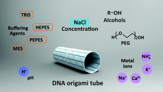
This work offers a methodology for screening compatible buffer conditions for both DNA origami and protein crystallisation and studied how protein crystallisation buffer conditions notably cations, buffering agents, precipitants, and pH, influenced the stability of tubular DNA origami.
DNA origami with a tubular structure, 100 nm in length, 22 nm in diameter and 2 nm in wall thickness, was prepared following established procedure. The advantages of using this tubular structure are that the preparation is quick and straightforward. The DNA origami was prepared in assembly buffer: 12.5 mM Mg(OAc)2, 20 mM CH3COOH, 40 mM Tris, 2 mM EDTA, pH 8.0. Atomic force microscopy (AFM) was employed to characterise the integrity of the tubular structure, verifying that the DNA origami was achieved as designed (Fig. 1).
The shot gun approach refers to DNA origami either assembled in protein crystallisation buffer, or assembled in DNA assembly buffer first and followed by buffer exchange. Table 1 lists crystallisation buffers for four model proteins. It turned out that these solutions were not compatible in the assembly of DNA, and no complete origami was observed, Fig. S2.†
Protein | Buffer solution (solvent water) | Precipitant solution (solvent buffer) |
---|---|---|
Lysozyme | 0.1 M sodium acetate pH 4.5 | 1.1 M NaCl |
Thaumatin | 0.05 M PIPES pH 6.8 | 0.34 M Na–K tartrate |
Human serum albumin | 0.05 M monopotassium phosphate pH 5.15 | 5% (w/v) PEG 4k |
Catalase | 0.1 M Tris pH 8.4 | 5% (w/v) PEG 4k; 5% (v/v) 2-methyl-1,3-propanediol (MPD) |
Several studies report that DNA origami can remain structurally intact in cell lysate.8 This is indicative that once the DNA origami forms the structure is stable. Therefore, we employed buffer exchanges by three methods, (1) one-off: an abrupt buffer exchange by adding 400 μL protein crystallisation into 40 μL, 1 nM DNA origami solution, removing the buffer by a 100000 molecular weight cut-off, centrifuging at 3000g for 5 min. The exchange was repeated 3 times; (2) gradient buffer exchange: in order to minimise the damage to the DNA origami structures, we used five kinds of gradient buffers with crystallisation/DNA origami buffer (v/v) ratios at 1
:
7, 1
:
3, 1
:
2, 3
:
1, up to pure crystallisation buffer respectively, with the removal of the DNA buffer identical to the one-off method; (3) gradual buffer exchange by overnight dialysis: we added 40 μL, 1 nM DNA origami solution to a dialysis bag (Mw cut-off 12
000), placing it into 500 mL of protein crystallisation and stirred it overnight. All three methods ensured that the DNA assembly buffer was fully replaced with protein crystallisation. After changing the buffer, the solution was left at room temperature for at least 2 h followed by AFM characterisation. The protein crystallisation buffer for catalase stabilised the DNA origami significantly better than the other three, with the structure remaining stable after the one-off buffer exchange for at least 24 h, Fig. 2. The gradient buffer exchange method yielded the same outcome for catalase, Fig. S3;† however, even the mildest condition for the exchange buffer via dialysis did not result in intact DNA origami structure with few DNA origami observed, maybe due to the DNA sticking to the dialysis tube,9 Fig. S3.† Taking all of these results into account, we concluded that protein crystallisation may not be ideal or compatible conditions during the assembly of DNA origami probably due to an adding-up effect. Whereas, by exchanging the original DNA assembly buffer with protein crystallisation via either one-off or gradient buffer exchange, the DNA origami structure was retained for catalase. For the rest of the studies the most straightforward one-off method was employed.
The common variations of the four components in protein crystallisation buffer conditions are summarised as follows: cations are Na+, K+, Ca2+, NH4+ at concentrations of 0.1–0.2 M; Tris, HEPES, PEPES and MES buffers are most often used; precipitating agents mainly fall into three categories: (1) salts (high concentration, e.g., 3–5 M), (2) alcohols, (3) polymers; the pH of the buffer usually ranges from 4 to 10. Hence, the impact of salts, buffering agents, precipitants (alcohol, polymer and salt at high concentration) and pH were examined individually. In each experiment, the DNA assembly buffer was employed as the baseline recipe, apart from the factor of interest, all other aspects were maintained the same.
First, the impact of salt on origami was examined. It was noted that monovalent ions were not good for adhering DNA origami structures onto mica.10 Therefore, due to imaging purposes the buffer was exchanged with DNA assembly buffer right before AFM characterisation. Fig. S4† showed that when the 12.5 mM Mg2+ was replaced with 200 mM Na+, the buffer exchange did not influence the formation of DNA origami. However, in the 200 mM Ca2+ solution, tubular DNA origami could not form so was not a suitable condition for DNA hybridisation. When the 12.5 mM Mg2+ was replaced with 200 mM K+ or 200 mM NH4+ cations, large rectangles and wreckages were observed attributed to the loop tension and electrostatic repulsion effects.
Next, we sought to study the stability of DNA origami in different buffering agents, such as Tris, HEPES, PEPES and MES. The concentrations were all set at 100 mM. The AFM images in Fig. 1 and S5† showed that DNA origami formed in all four buffers. Though in Fig. S5c,† a few unusually long tubular structures were spotted, these structures were dimers or trimmers due to the blunt-end stacking, which does not influence the stability of DNA origami structure. Hence, the tubular structures were still deemed intact. From this observation, it was concluded that the buffering agents in protein crystallisation buffer were amiable for the DNA origami.
Ethanol, 2-methyl-1,3-propanediol (MPD), and glycerol were the selected alcohols and added at a concentration of 10% (v/v). Fig. S6† showed that the presence of alcohol did not interfere with the tubular structure of DNA origami. Buffer mixture contained precipitants PEG at different molecular weights of 2000, 4000, and 8000 g mol−1 were prepared at a concentration of 30% (w/v). The AFM images in Fig. S7† revealed that PEG was mild and did not affect the formation of DNA origami. According to previous studies, PEG has been used as a crowding agent to assist the formation of G-quadruplex11 and double crossover,12 which explains why the DNA origami could still form in the presence of PEG. High concentrations of NaCl are widely used as precipitants in protein crystallisation. Results shown in Fig. S8† highlighted that there were no DNA wreckages or aggregates at 2 M or 3 M NaCl, However, there was no DNA origami formed in the presence of NaCl solution at a high concentration of 4 M, likely due to the large amounts of Na+ strongly promoting aggregation of DNA origami.
We made a final observation on the influence of pH which is a key factor in origami assembly. The buffer at a pH range of 4–10, was prepared (adjusted by HCl and NaOH), and it was observed that except for pH 4, DNA origami remained stable and intact at pH 5–10, Fig. S9.† The results of the variation of these four factors in protein crystallisation are summarised in Table 2.
Cation | Na+ | K+ | Ca2+ | NH4+ |
✓ | ✗ | ✗ | ✗ | |
Buffering agent | Tris | HEPES | PEPES | MES |
✓ | ✓ | ✓ | ✓ |
Precipitant | Alcohol | ||
Ethanol | MPD | Glycerol | |
✓ | ✓ | ✓ | |
Salt | |||
2 M NaCl | 3 M NaCl | 4 M NaCl | |
✓ | ✓ | ✗ | |
Polymer | |||
PEG2000 | PEG4000 | PEG8000 | |
✓ | ✓ | ✓ |
pH | 4 | 5 | 6 | 7 | 8 | 9 | 10 |
✗ | ✓ | ✓ | ✓ | ✓ | ✓ | ✓ |
The results of this entire study can be summarised as follows: (i) the condition for origami assembly was crucial, whereas the stability range of already assembled origami was broader; (ii) sudden exchange of buffer did not disrupt the tubular structure of the DNA origami, hence, abrupt exchange using cut-off was more straightforward and recommended for future work; (iii) the systematic studies indicated that most variations, from the baseline condition, made to match individual crystallisation buffer components retained origami structure. It was found that Na+ (0.2–3 M), common buffer agents, Tris, HEPES, PEPES and MES buffer, 10% (v/v) alcohol/30% (w/v) PEG precipitants, and a wide pH range (5–10) in the buffer, would not disrupt the structure of tubular DNA origami. However, K+, Ca2+, NH4+ at a concentration of 200 mM and Na+ at a concentration of 4 M, may break the tubular DNA origami structure, though the mechanisms are unknown; (iv) we successfully stabilised the tubular DNA origami structures in catalase protein buffer; however, the other three non-extreme conditions chosen in this study failed to stabilise it. One may conclude that the factors of concern have an adding-up effect and should be analysed more thoroughly not simply as individual components. Further work should be done to investigate the exact mechanism of the adding-up effect. In addition, attempts on making more robust origami structure may also be made to off-set these challenges.
Footnotes |
† Electronic supplementary information (ESI) available: Experimental details including DNA sequences, protein and typical crystallisation conditions. See DOI: 10.1039/c5ra12159k |
‡ The authors contributed equally to this work. |
This journal is © The Royal Society of Chemistry 2015 |