
H2S bubbles-assisted synthesis of hollow Cu2−xSeyS1−y/reduced graphene oxide nanocomposites with tunable compositions and localized surface plasmon resonance†
Wen Long
Li‡
a,
Hong Yan
Zou‡
b,
Jing
Lan
b,
Qiang
Wang
b,
Yuan Fang
Li
a and
Cheng Zhi
Huang
*ab
aKey Laboratory of Luminescent and Real-Time Analytical Chemistry (Southwest University), Ministry of Education, College of Chemistry and Chemical Engineering, Southwest University, Chongqing 400715, China. E-mail: chengzhi@swu.edu.cn; Fax: +86-23-68367257; Tel: +86-23-68254659
bChongqing Key Laboratory of Biomedical Analysis (Southwest University), Chongqing Science & Technology Commission, College of Pharmaceutical Science, Southwest University, Chongqing 400715, China
First published on 19th October 2015
Abstract
Hollow nanostructures have been diversely applied in many fields and efforts have been made to develop their various synthesis methodologies. Herein, homogeneous hollow Cu2−xSeyS1−y/reduced graphene oxide (rGO) nanocomposites were prepared for the first time via a facile and green aqueous chemical approach using H2S gas bubble templates as soft templates at room temperature. It is found that 2D hexagonal Cu2−xSeyS1−y nanosheets assemble around the gas–liquid interface of H2S bubbles to form hollow nanospheres on the rGO sheets, wherein rGO sheets as molecular templates play a critical role in maintaining the structural integrity of single-crystal hollow Cu2−xSeyS1−y nanospheres. Increasing the S content in the precursors, i.e., increasing the amount of H2S bubbles, both the diameter and chalcogen composition (S/Se molar ratio) of hollow Cu2−xSeyS1−y/rGO nanocomposites gradually increase. The near-infrared (NIR) localized surface plasmon resonance (LSPR) in Cu2−xSeyS1−y/rGO nanocomposites, arising from the free carriers (holes) in Cu2−xSeyS1−y, gets red shifts with increasing the S content, due to the higher carrier mass and assembly degree of Cu2−xSeyS1−y.
Introduction
Hollow nanostructures have received considerable attention recently because of their diverse potential applications in chemical catalysis,1,2 H2 evolution,3,4 gas sensing,5,6 drug delivery7–9 and biomedical therapy.7,8 Various methodologies have been developed to prepare hollow nanostructures, which involve templates such as polymer spheres,4 silica spheres,10 emulsion droplets,11 polymer/surfactant micelles,12 and gas bubbles,3,13,14 as well as chemical processes based on the Kirkendall effect,5,15 or Ostwald ripening.1 Among these preparation methods, gas bubble template seems more simple, effective and applicable. It is not necessary for gas bubble method to integrally remove the template cores, which has the advantages of impurity-free procedure, short reaction time and low temperature.13,14 In addition, one-step procedure obviously stands out for generating hollow nanostructures in gas bubble methodology.Copper chalcogenides (i.e., Cu2−xSe and Cu2−xS), besides their excitonic properties similar to semiconductor nanocrystals (NCs) that have been employed with highly efficiency in solar cells due to their tailorable bandgap,16–19 possess a near-infrared (NIR) localized surface plasmon resonance (LSPR), which arises from free carriers (holes) in the valence band that undergo collective oscillation.19–27 Theoretically, the free carriers in copper chalcogenides, opposite to the free electrons in metals, must be provided by cationic vacancies or extrinsic dopants.20,22,25,27 Recent investigations have showed that these new plasmonic nanomaterials have potential for use in photoacoustic imaging,28,29 photothermal therapy,7,30–33 plasmonic optoelectronic devices,17,34 and chemical sensing.35–37
Ternary Cu–Se–S semiconductor NCs are of great significance for not only inheriting the perfect optoelectronic and biochemical properties from their parent binary NCs (Cu2−xSe and Cu2−xS), but also possessing unique properties from the chalcogen composition variation.38,39 For example, Cu2−xSe and Cu2−xS NCs reveal that the NIR LSPR can be well tuned by adjusting the concentration of copper vacancies, and show enhanced absorption with increasing copper vacancies in NCs.22,23,27 Furthermore, the NIR LSPR can be finely tuned by controlling the ratio of chalcogen in ternary Cu2−xSeyS1−y NCs.40,41 Therefore, controllably synthesizing ternary Cu2−xSeyS1−y NCs and theoretically understanding the influence of anion composition variation on their physical and chemical properties are of great interest.
Commonly, the nucleation temperature of Cu2−xSe NCs is higher than that of Cu2−xS NCs,41 so the reactivity of Se and S precursor will not be balanced at the same temperature, which make it hard to synthesize the desired ternary Cu2−xSeyS1−y NCs with homogeneous structure rather than separate binary NCs. Up to now, few works concerning the synthesis of ternary Cu2−xSeyS1−y NCs have been reported, wherein high temperature was typically adopted.38–44
In this article, we firstly report a facile and green one-pot aqueous chemical approach for controllably prepare homogeneous hollow Cu2−xSeyS1−y/rGO nanocomposites on the basis of H2S gas bubble template mechanism at room temperature. Ascorbic acid (AA), a relatively safe and green reducing agent, has been used in many solution-based synthesis of inorganic nanomaterials, including Cu2−xSe NCs.15,45 H2S gas bubbles, produced by Na2S and AA in the synthesis, induce the 2D Cu2−xSeyS1−y nanosheets assembled around the gas–liquid interface of H2S bubbles to form the hollow nanospheres on the rGO sheets, wherein rGO sheets as molecular templates play a critical role in maintaining the structural integrity of homogeneous hollow Cu2−xSeyS1−y nanospheres. Owing to the different amount of H2S bubbles, morphology evolution and composition variation of Cu2−xSeyS1−y/rGO nanocomposites are obtained by adjusting the S/Se molar ratios in the precursors. The NIR LSPR in Cu2−xSeyS1−y/rGO nanocomposites, arising from the free carriers (holes) in Cu2−xSeyS1−y NCs, continuously red-shifts with increasing the S/Se atomic ratio, due to the higher carrier mass and assembly degree in Cu2−xSeyS1−y NCs.20
Experimental section
Chemicals and materials
Graphene oxide (GO) was purchased from XF NANO INC (Nanjing, China) and dissolved in deionized water via sonication for several hours to obtaining the GO aqueous solution. Ascorbic acid (AA) was obtained from Dingguo Changsheng Biotechnology Co., Ltd (Beijing, China). Polystyrene sulfonate (PSS, MW = 70 kDa) was purchased from Alfa Aesar Co. Ltd (MA, USA). SeO2 was supplied by Aladdin Chemistry Co., Ltd (Shanghai, China) and stored in a desiccator at room temperature. CuSO4·5H2O and Na2S·9H2O were analytical reagent grade. All chemicals were used as received without further purification. Milli-Q purified water (18.2 MΩ cm−1) was used throughout the experiments.Apparatus
Transmission electron microscopy (TEM), high resolution transmission electron microscopy (HRTEM), and selected area electron diffraction (SAED) measurements were obtained from a Tecnai G2 F20 S-TWIN microscopy (FEI, USA). Scanning electron microscopy (SEM) and energy-dispersive X-ray spectroscopy (EDX) were performed with an S-4800 scanning electron microscope (Hitachi, Japan). X-ray diffraction (XRD) analyses were carried out on an XRD-7000 with Cu Kα radiation source (Shimadzu, Japan). Diffraction patterns were collected in the range of 2θ = 5–80° under ambient conditions at a scanning rate of 2° min−1. X-ray photoelectron spectroscopy (XPS) analysis was conducted by an ESCALAB 250 X-ray photoelectron spectrometer (Thermo Scientific, USA). Fourier transform infrared (FT-IR) spectrum was measured by an FTIR-8400S spectrophotometer (Shimadzu, Japan). Nitrogen adsorption–desorption isotherms were measured on a Quantachrome Instrument (AS1-MP-9, USA) at 77 K. Absorption spectra were recorded by a UV-3600 UV-vis-NIR spectrophotometer (Shimadzu, Japan).Preparation of Cu2−xSeyS1−y/rGO nanocomposites
Cu2−xSeyS1−y/rGO nanocomposites were synthesized in reference to our previous report with small modifications.46 In a typical synthesis procedure, GO aqueous solution (10 mL, 0.1 mg mL−1) was at first magnetically stirred to form a homogeneous pale-yellow solution in a water bath at 30 °C. Then, 100 μL SeO2 (0.02 mmol) and 200 μL AA (0.08 mmol) were added into the above solution, followed by adding 100 μL Na2S (0.06 mmol). After continuous stirring for 10 min, 100 μL CuSO4 (0.04 mmol) and 500 μL AA (0.2 mmol) were added, and the resulted mixture was sustained stirring for 10 h. The products were purified through a 10 kDa dialysis membrane for 12 h with 6 changes of distilled water, and then centrifuged for 10 min at 8000 rpm and washed thrice with distilled water. The final products were redispersed in water and stored in a 4 °C refrigerator for characterization and further use. By fixing the Se reactant and regulating the S reactant, different molar ratios of S/Se (0![[thin space (1/6-em)]](https://www.rsc.org/images/entities/char_2009.gif) :1, 1:1, 2:1, 3:1, 4:1, and 5:1) in the precursors were used to control the composition of Cu2−xSeyS1−y/rGO nanocomposites.
:1, 1:1, 2:1, 3:1, 4:1, and 5:1) in the precursors were used to control the composition of Cu2−xSeyS1−y/rGO nanocomposites.
Results and discussion
Characterization of hollow Cu2−xSeyS1−y/rGO nanocomposites
The morphologies of the samples were confirmed by transmission electron microscopy (TEM) and scanning electron microscopy (SEM) measurements. GO sheets were 2D lamellar with clear wrinkles and folding on the smooth surface (Fig. S1†). Obvious evolution in the morphology of Cu2−xSeyS1−y/rGO nanocomposites was observed with increasing the reactant S/Se ratio (Fig. 1). Without addition of S precursor, spherical Cu2−xSe nanoparticles (NPs), with an average diameter of 50 nm, could be uniformly grown on the surface of rGO sheets without apparent aggregation (Fig. 1A). When an S/Se molar ratio of 1:1 was adopted, individual spherical Cu2−xSeyS1−y NPs, with an average diameter of 30 nm, were also uniformly grown on the rGO sheets without apparent aggregation (Fig. 1B). As the S/Se molar ratio increased to 2:1, both quasi-spherical Cu2−xSeyS1−y NPs (∼40 nm) and hollow nanospheres (∼65 nm) were anchored on the rGO sheets in Cu2−xSeyS1−y/rGO nanocomposites (Fig. 1C). Interestingly, some 2D Cu2−xSeyS1−y nanosheets appeared on the rGO sheets. Increasing the S/Se molar ratio (3:1), hollow Cu2−xSeyS1−y nanospheres were basically formed on the rGO sheets with a large size increase, about 140 nm (Fig. 1D). Notably, some Cu2−xSeyS1−y nanosheets were inlaid in the surface of hollow nanospheres, while some of the Cu2−xSeyS1−y nanospheres surfaces were cracked. The hollow-structured Cu2−xSeyS1−y nanospheres were likely the assemblies of Cu2−xSeyS1−y nanosheets (Fig. S2†). Continuing to increase the S/Se molar ratio (4:1), the surfaces of hollow Cu2−xSeyS1−y nanospheres (∼160 nm) became smoother, while Cu2−xSeyS1−y nanosheets decreased (Fig. 1E). Hollow Cu2−xSeyS1−y nanospheres were entirely formed on the rGO sheets, with an average diameter of 190 nm and shell thickness of 20 nm, as the S/Se molar ratio increased to 5:1 (Fig. 1F).
 | ||
| Fig. 1 TEM images of Cu2−xSeyS1−y/rGO nanocomposites prepared with various reactant S/Se molar ratios. (A) 0:1; (B) 1:1; (C) 2:1; (D) 3:1; (E) 4:1; (F) 5:1. Obvious evolution in the morphology of Cu2−xSeyS1−y/rGO nanocomposites were observed as increasing the S/Se molar ratio in the reactants, and 2D Cu2−xSeyS1−y nanosheets assembled into the large hollow nanospheres on the rGO sheets. | ||
As shown in Fig. 2, the interplanar distance of Cu2−xSe NPs was measured to be 0.22 nm (Fig. 2A), corresponding to the (220) plane of cubic Cu2−xSe.46 The high-resolution TEM (HRTEM) images demonstrated all the Cu2−xSeyS1−y NCs with a lattice plane distance of 0.33 nm (Fig. 2B–F), corresponding to the (101) plane of hexagonal Cu2−xSeyS1−y.39 Furthermore, the HRTEM results indicated that these Cu2−xSeyS1−y NCs were single crystals.
 | ||
| Fig. 2 HRTEM image of Cu2−xSeyS1−y/rGO nanocomposites prepared with various reactant S/Se molar ratios. (A) 0:1; (B) 1:1; (C) 2:1; (D) 3:1; (E) 4:1; (F) 5:1. | ||
Few literatures have involved ternary Cu2−xSySe1−y single-crystal assemblies and their conversion mechanism. Generally, monodispersed NPs self-assemble into a 2D superlattice and 2D nanosheets self-assemble into a 3D stacked nanoplate.38,39 Herein, since the synthesis condition was surfactant-free, the high surface tension of Cu2−xSeyS1−y nanosheets should be released to reach a stable state. H2S gas bubbles, generated from Na2S and AA, provide numerous gas–liquid interfaces inside the solution phase (Fig. S3†). As a gas bubble-template mechanism, the in situ generated Cu2−xSeyS1−y nanosheets tended to move into the H2S gas bubbles interface region.10,13 When the concentration of Cu2−xSeyS1−y nanosheets in this region was sufficiently high, the Cu2−xSeyS1−y nanosheets would interact with each other to form hollow Cu2−xSeyS1−y spheres with the bubbles as soft templates. Thus, the as-obtained hollow Cu2−xSeyS1−y nanospheres were generated via the assembly of 2D Cu2−xSeyS1−y nanosheets around the gas–liquid interface of H2S gas bubbles rather than through the Ostwald ripening process.3,11,12
The final chemical compositions of Cu2−xSeyS1−y/rGO nanocomposites prepared with different S/Se molar ratios in the precursors were measured by energy-dispersive X-ray (EDX) spectroscopy (Fig. S4†). When the molar ratio of S/Se increased from 1:1 to 5:1, the S content dramatically increased from 0.25 to 0.74, and the Se content dramatically decreased from 0.75 to 0.26. No regular tendency could be found in copper vacancy concentration.41 The mean Cu/Se/S atomic ratios were summarized in Table 1. So, the elemental compositions of Cu2−xSeyS1−y/rGO nanocomposites could be easily tuned by adjusting the molar ratio of S/Se in the reactants.
| S/Se molar ratios in reactants | EDX analysis (mean atomic percent) | Compositions of Cu2−xSeyS1−y/rGO nanocomposites | ||
|---|---|---|---|---|
| Cu | Se | S | ||
| 0:1 |
12.90 | 10.51 | — | Cu1.23Se/rGO |
| 1:1 |
36.45 | 23.36 | 7.62 | Cu1.18Se0.75S0.25/rGO |
| 2:1 |
43.42 | 20.11 | 9.03 | Cu1.49Se0.69S0.31/rGO |
| 3:1 |
44.40 | 12.67 | 16.07 | Cu1.54Se0.44S0.56/rGO |
| 4:1 |
38.61 | 8.86 | 20.62 | Cu1.31Se0.30S0.70/rGO |
| 5:1 |
48.60 | 7.94 | 22.88 | Cu1.58Se0.26S0.74/rGO |
Powder X-ray diffraction (XRD) patterns showed that all of these Cu2−xSeyS1−y/rGO nanocomposites exhibited a hexagonal phase of Cu2−xSeyS1−y NCs,41 while the characteristic peak at 11.3° corresponding to GO (002) had almost disappeared (Fig. 3), indicating the formation of homogeneous Cu2−xSeyS1−y NCs and the reduction of GO.47,48 The absences of the diffractions peaks of layered rGO might come from the regular stacks of rGO were exfoliated,49,50 and the anchored Cu2−xSeyS1−y nanospheres might also prevent the restacking of the exfoliated rGO sheets.48,51 Remarkably, the diffraction peaks slightly shifted to higher 2θ degrees with increasing S content, reflecting a decrease in the lattice constant.40,41 The continuous shift of the diffraction peaks toward higher 2θ could be more clearly seen in the expanded view of the (110) peak (shown in Fig. 3B). The decrease in lattice parameter was accountable, because the Se ions in the lattice were replaced by the smaller S ions.39,43
 | ||
| Fig. 3 (A) XRD patterns of Cu2−xSeyS1−y/rGO nanocomposites synthesized with various reactant S/Se ratios (from bottom to top) of 1:1 (black curve), 2:1 (red curve), 3:1 (blue curve), 4:1 (magenta curve) and 5:1 (green curve), respectively. (B) An expanded view of the (110) peak, showing the shift to higher diffraction angles (lower lattice constant) with increasing S content. | ||
The Brunauer–Emmett–Teller (BET) surface area and the porosity of Cu2−xSeyS1−y/rGO nanocomposites (a reactant S/Se molar ratio of 3:1, typically) were evaluated after degassing at 200 °C. The nitrogen adsorption–desorption isotherms of Cu2−xSeyS1−y/rGO nanocomposites belonged to the typical type IV isotherm (Fig. 4A), revealing a disordered mesoporous structure.48,52 It was calculated that the BET surface area of Cu2−xSeyS1−y/rGO nanocomposites was 8.8143 m2 g−1 with Barrett–Joyner–Halenda (BJH) desorption pore diameter of 13.8913 nm (Fig. 4B), respectively.
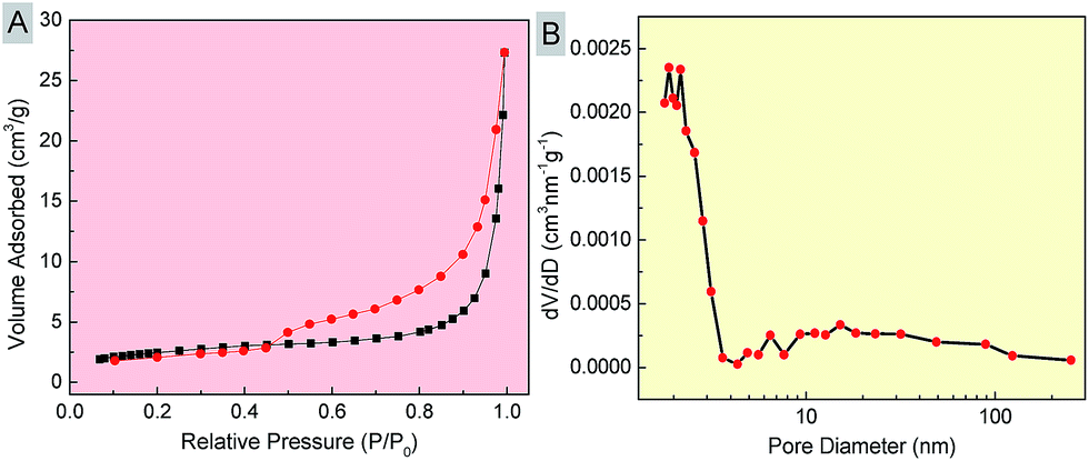 | ||
| Fig. 4 Characterization of the as-synthesized hollow Cu2−xSeyS1−y/rGO nanocomposites (a reactant S/Se molar ratio of 3:1). (A) N2 adsorption–desorption isotherms and (B) the corresponding pore size distribution of Cu2−xSeyS1−y/rGO nanocomposites. | ||
X-ray photoelectron spectra (XPS) analysis was performed to investigate the chemical composition and valence state of Cu2−xSeyS1−y/rGO nanocomposites (a reactant S/Se molar ratio of 3:1, typically, Fig. 5). The obvious peaks of C and O in the survey spectrum of GO could be clearly detected (Fig. S5†). In comparison with GO, the survey spectrum of Cu2−xSeyS1−y/rGO nanocomposites clearly showed the peaks of Cu 2p, Se 3d, S 2p, C 1s and O 1s (Fig. 5A). The C 1s and O 1s peaks mainly originated from rGO. Two predominant peaks at 932.5 and 952.4 eV were associated with the binding energies of Cu 2p3/2 and Cu 2p1/2 for Cu+,42 respectively (Fig. 5B), and no Cu2+ satellite peaks were detected. The binding energy for Se 3d was 55.2 eV (Fig. 5C), close to the value of lattice Se2−.42 The S 2p region showed the binding energies of S 2p3/2 and S 2p1/2 at 162.1 and 163.4 eV. The Se 2p3/2 and Se 2p1/2 peaks, with binding energies of 161.2 and 167.2 eV, respectively, were overlapped with S 2p peaks (Fig. 5D).42 From the C 1s XPS spectrum of Cu2−xSeyS1−y/rGO nanocomposites (Fig. 5E), the peaks of 284.8, 286.8 and 288.7 eV were assigned to C–C, C–O and C![[double bond, length as m-dash]](https://www.rsc.org/images/entities/char_e001.gif) O, respectively.53 Compared with the C 1s spectrum of GO (Fig. 5F), the peak intensities of oxygen functional groups greatly decreased in Cu2−xSeyS1−y/rGO nanocomposites, confirming that GO had been reduced to rGO (also seen in the FTIR spectrum of Cu2−xSeyS1−y/rGO nanocomposites, Fig. S6†).48,53,54
O, respectively.53 Compared with the C 1s spectrum of GO (Fig. 5F), the peak intensities of oxygen functional groups greatly decreased in Cu2−xSeyS1−y/rGO nanocomposites, confirming that GO had been reduced to rGO (also seen in the FTIR spectrum of Cu2−xSeyS1−y/rGO nanocomposites, Fig. S6†).48,53,54
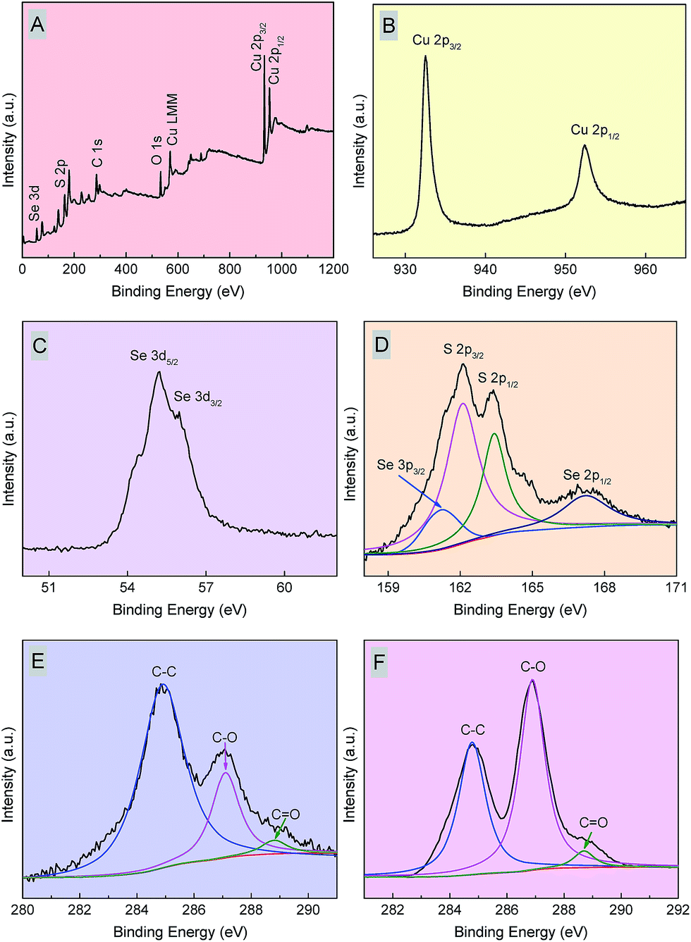 | ||
| Fig. 5 Typical XPS spectra of the as-synthesized hollow Cu2−xSeyS1−y/rGO nanocomposites (a reactant S/Se molar ratio of 3:1): (A) survey spectra, (B) Cu 2p spectrum, (C) Se 3d spectrum, (D) Se 3p and S 2p spectra. C 1s spectra of (E) Cu2−xSeyS1−y/rGO nanocomposites and (F) GO. | ||
The formation mechanism of hollow Cu2−xSeyS1−y/rGO nanocomposites
It was found that the hollow-structured Cu2−xSeyS1−y/rGO nanocomposites were the result of 2D Cu2−xSeyS1−y nanosheets assembled around the gas–liquid interface of H2S gas bubbles on the rGO sheets, and rGO sheets was critical for maintaining the structural integrity of hollow Cu2−xSeyS1−y nanospheres. When absence of rGO and using PSS as surfactants, the morphological differences of individual Cu2−xSeyS1−y NCs also occurred as the reactant S/Se molar ratio increased, but the hollow nanospheres were damaged: the 2D Cu2−xSeyS1−y nanosheets did not tend to self-assemble into a larger nanospheres (Fig. S7†). Increasing the S/Se molar ratio (3:1), the products were the aggregation of spherical NPs and 2D nanosheets. The larger quasi-spherical Cu2−xSeyS1−y alloy NPs were synthesized with a reactant S/Se molar ratio of 4:1, and ∼100 nm 2D Cu2−xSeyS1−y nanosheets were obtained with a reactant S/Se molar ratio of 5:1. EDX analysis revealed the presence of Cu, Se and S in Cu2−xSeyS1−y alloy NPs, but the Se/S elemental molar ratios were absent of regularly tunability (Fig. S8 and Table S1†).
With the aim of synthesis hollow Cu2−xSeyS1−y nanospheres, H2S gas bubbles, produced by Na2S and AA, played the main role, which acted as appropriate soft templates. These H2S gas bubbles created numerous gas–liquid interfaces inside the solution phase.13 In gas bubble-template mechanism, the gas–liquid interfaces in the solution could serve as the nucleation or agglomeration centers for Cu2−xSeyS1−y nanosheets.10,12 Especially, in our aqueous solution, since no coating of surfactants on Cu2−xSeyS1−y nanosheets, they had a tendency to aggregate together to release their high surface energy (Scheme 1, step a).14 The presence of rGO sheets provided flexible supports, while lots of H2S gas bubbles generated in the synthesis supplied aggregation centres, which could enable this agglomeration process to proceed in a controllable way (Scheme 1, step b).13 Driven by the minimization of interfacial energy, small Cu2−xSeyS1−y nanosheets could aggregate around the gas–liquid interface of H2S gas bubbles (Scheme 1, step b).10 Finally, well-defined hollow Cu2−xSeyS1−y nanospheres with compacted shells were formed on the rGO sheets (Scheme 1, step c).
 | ||
| Scheme 1 Schematic illustration of the formation of hollow Cu2−xSeyS1−y/rGO nanocomposites. 2D Cu2−xSeyS1−y nanosheets were capable of assembling around the gas–liquid interface of H2S gas bubbles generated from Na2S and AA to form hollow-structured nanospheres on the rGO sheets. | ||
In comparison, when PSS was used as capping agent, the growth dimension of Cu2−xSeyS1−y NCs was restricted and the high surface energy was decreased, which would diminish the attractive force between the Cu2−xSeyS1−y NCs. So, the agglomeration process would not happened immediately, resulted in the failure formation of hollow Cu2−xSeyS1−y nanospheres.
Tunable NIR LSPR in Cu2−xSeyS1−y/rGO nanocomposites
Since rGO exhibited negligible absorption above 400 nm,46 the NIR absorption of Cu2−xSeyS1−y/rGO nanocomposites was attributed to Cu2−xSeyS1−y NCs. Arisen from the high density of free carriers (holes) in Cu2−xSeyS1−y NCs, strong NIR LSPR absorptions were observed in these Cu2−xSeyS1−y/rGO nanocomposites.20,41 Consistent with the previous studies, the S content played an important role in determining the LSPR peak position in Cu2−xSeyS1−y/rGO nanocomposites.38,41 Actually, the NIR LSPR continuously red-shifted with increasing S content regardless of the change of copper vacancies (Fig. 6).41 The red shift of LSPR with increasing S content could be attributed to the higher holes effective mass in Cu2−xSeyS1−y NCs. As seen in the Drude model:20higher carrier mass produces a lower bulk plasmon frequency and, in turn, longer LSPR wavelength (red shift).20 (Where εr is the real part of the dielectric function, ωp is the bulk plasmon frequency, γ is the damping frequency, Nh is the density of free carriers, e is the charge of the electron, ε0 is the vacuum permittivity and mh is the effective mass of the free carriers.)

 | ||
| Fig. 6 LSPR absorption of Cu2−xSeyS1−y/rGO nanocomposites synthesized with different S/Se molar ratios: black curve, 0:1; red curve, 1:1; blue curve, 2:1; magenta curve, 3:1; green curve, 4:1; violet curve, 5:1 (the intensity of the LSPR peak was normalized). | ||
Meanwhile, the LSPR can be tuned toward the red part of the absorption spectra upon assembly, such as supercrystals and stacked nanoplates.27,38,55 Hollow Cu2−xSeyS1−y/rGO nanocomposites were the result of 2D Cu2−xSeyS1−y nanosheets assembled on the rGO sheets, so the NIR LSPR of Cu2−xSeyS1−y/rGO nanocomposites red-shifted more as increasing the degree of assembly in Cu2−xSeyS1−y nanospheres (increasing the S content).
Conclusions
In summary, homogeneous hollow Cu2−xSeyS1−y/rGO nano-composites, with tunable Se/S ratios and NIR LSPR, have been controllably synthesized by self-assembly coupled with H2S gas bubble template through a facile and green one-pot aqueous chemical approach at room temperature, without the use of any surfactants. The reactions proceed through an environmentally benign reducing reagent, ascorbic acid (AA). 2D hexagonal Cu2−xSeyS1−y nanosheets assemble around the gas–liquid interface of H2S gas bubbles to form the hollow-structured nanospheres on the rGO sheets. Hollow Cu2−xSeyS1−y/rGO nanocomposites grow to a larger diameter with increasing the S content in the precursors, i.e., increasing the amount of H2S bubbles. The compositions of the hexagonal Cu2−xSeyS1−y/rGO nanocomposites can be easily tuned by adjusting the reactant molar ratio of S/Se, as ascertained from EDX analysis. rGO sheets as molecular templates play a critical role in maintaining the structural integrity of homogeneous hollow Cu2−xSeyS1−y nanospheres. A gas bubble template mechanism is proposed to explain the morphology and crystal phase formation in the synthesis. Cu2−xSeyS1−y/rGO nanocomposites exhibit strong NIR LSPR due to the presence of copper vacancies in Cu2−xSeyS1−y NCs, which create the free carriers (holes). Owing to the higher free carriers mass and assembly degree in Cu2−xSeyS1−y NCs, a continuous red-shift of LSPR is revealed with increasing the S content in Cu2−xSeyS1−y/rGO nanocomposites. Due to the large specific surface area and prominent LSPR properties, the as-synthesized hollow Cu2−xSeyS1−y/rGO nanocomposites could be further applied in biomedicine, chemical sensing, optoelectronic devices, catalysis, and lithium-ion batteries.Acknowledgements
This work was financially supported by the National Natural Science Foundation of China (NSFC, no. 21375109).Notes and references
- W.-S. Wang, L. Zhen, C.-Y. Xu, W.-Z. Shao and Z.-L. Chen, CrystEngComm, 2013, 15, 8014–8021 RSC.
- Q. W. Shu, C. M. Li, P. F. Gao, M. X. Gao and C. Z. Huang, RSC Adv., 2015, 5, 17458–17465 RSC.
- Z.-J. Li, X.-B. Fan, X.-B. Li, J.-X. Li, C. Ye, J.-J. Wang, S. Yu, C.-B. Li, Y.-J. Gao, Q.-Y. Meng, C.-H. Tung and L.-Z. Wu, J. Am. Chem. Soc., 2014, 136, 8261–8268 CrossRef CAS PubMed.
- J. Huang, R. Ma, Y. Ebina, K. Fukuda, K. Takada and T. Sasaki, Chem. Mater., 2010, 22, 2582–2587 CrossRef CAS.
- G. Xiao, Y. Zeng, Y. Jiang, J. Ning, W. Zheng, B. Liu, X. Chen, G. Zou and B. Zou, Small, 2013, 9, 793–799 CrossRef CAS PubMed.
- Z. Wang, F. Peng, Y. Wu, L. Yang, F. Zhang and J. Huang, CrystEngComm, 2012, 14, 3528–3533 RSC.
- K. Dong, Z. Liu, Z. Li, J. Ren and X. Qu, Adv. Mater., 2013, 25, 4452–4458 CrossRef CAS PubMed.
- S. Ramadan, L. Guo, Y. Li, B. Yan and W. Lu, Small, 2012, 8, 3143–3150 CrossRef CAS PubMed.
- J. Wang, P. P. Gao, X. X. Yang, T. T. Wang, J. Wang and C. Z. Huang, J. Mater. Chem. B, 2014, 2, 4379–4386 RSC.
- J. Liu, F. Liu, K. Gao, J. Wu and D. Xue, J. Mater. Chem., 2009, 19, 6073–6084 RSC.
- J. Hu, M. Chen, X. Fang and L. Wu, Chem. Soc. Rev., 2011, 40, 5472–5491 RSC.
- X. W. Lou, L. A. Archer and Z. Yang, Adv. Mater., 2008, 20, 3987–4019 CrossRef CAS PubMed.
- M. Mozafari, F. Moztarzadeh, A. M. Seifalian and L. Tayebi, J. Lumin., 2013, 133, 188–193 CrossRef CAS PubMed.
- J. Liu and D. Xue, J. Cryst. Growth, 2009, 311, 500–503 CrossRef CAS PubMed.
- J. Xu, Y.-B. Tang, W. Zhang, C.-S. Lee, Z. Yang and S.-T. Lee, Cryst. Growth Des., 2009, 9, 4524–4528 CAS.
- S. C. Riha, D. C. Johnson and A. L. Prieto, J. Am. Chem. Soc., 2011, 133, 1383–1390 CrossRef CAS PubMed.
- X. Liu, X. Wang, B. Zhou, W.-C. Law, A. N. Cartwright and M. T. Swihart, Adv. Funct. Mater., 2013, 23, 1256–1264 CrossRef CAS PubMed.
- H. Zhou, W.-C. Hsu, H.-S. Duan, B. Bob, W. Yang, T.-B. Song, C.-J. Hsu and Y. Yang, Energy Environ. Sci., 2013, 6, 2822–2838 CAS.
- Y. Zhao and C. Burda, Energy Environ. Sci., 2012, 5, 5564–5576 CAS.
- X. Liu and M. T. Swihart, Chem. Soc. Rev., 2014, 43, 3908–3920 RSC.
- A. Comin and L. Manna, Chem. Soc. Rev., 2014, 43, 3957–3975 RSC.
- J. M. Luther, P. K. Jain, T. Ewers and A. P. Alivisatos, Nat. Mater., 2011, 10, 361–366 CrossRef CAS PubMed.
- D. Dorfs, T. Härtling, K. Miszta, N. C. Bigall, M. R. Kim, A. Genovese, A. Falqui, M. Povia and L. Manna, J. Am. Chem. Soc., 2011, 133, 11175–11180 CrossRef CAS PubMed.
- F. Scotognella, G. Della Valle, A. R. Srimath Kandada, D. Dorfs, M. Zavelani-Rossi, M. Conforti, K. Miszta, A. Comin, K. Korobchevskaya, G. Lanzani, L. Manna and F. Tassone, Nano Lett., 2011, 11, 4711–4717 CrossRef CAS PubMed.
- Y. Zhao, H. Pan, Y. Lou, X. Qiu, J. Zhu and C. Burda, J. Am. Chem. Soc., 2009, 131, 4253–4261 CrossRef CAS PubMed.
- I. Kriegel, J. Rodríguez-Fernández, A. Wisnet, H. Zhang, C. Waurisch, A. Eychmüller, A. Dubavik, A. O. Govorov and J. Feldmann, ACS Nano, 2013, 7, 4367–4377 CrossRef CAS PubMed.
- I. Kriegel, C. Jiang, J. Rodríguez-Fernández, R. D. Schaller, D. V. Talapin, E. da Como and J. Feldmann, J. Am. Chem. Soc., 2012, 134, 1583–1590 CrossRef CAS PubMed.
- G. Ku, M. Zhou, S. Song, Q. Huang, J. Hazle and C. Li, ACS Nano, 2012, 6, 7489–7496 CrossRef CAS PubMed.
- X. Liu, W.-C. Law, M. Jeon, X. Wang, M. Liu, C. Kim, P. N. Prasad and M. T. Swihart, Adv. Healthcare Mater., 2013, 2, 952–957 CrossRef CAS PubMed.
- W. Li, R. Zamani, P. Rivera Gil, B. Pelaz, M. Ibáñez, D. Cadavid, A. Shavel, R. A. Alvarez-Puebla, W. J. Parak, J. Arbiol and A. Cabot, J. Am. Chem. Soc., 2013, 135, 7098–7101 CrossRef CAS PubMed.
- C. M. Hessel, V. P. Pattani, M. Rasch, M. G. Panthani, B. Koo, J. W. Tunnell and B. A. Korgel, Nano Lett., 2011, 11, 2560–2566 CrossRef CAS PubMed.
- Q. Tian, F. Jiang, R. Zou, Q. Liu, Z. Chen, M. Zhu, S. Yang, J. Wang, J. Wang and J. Hu, ACS Nano, 2011, 5, 9761–9771 CrossRef CAS PubMed.
- Q. Tian, M. Tang, Y. Sun, R. Zou, Z. Chen, M. Zhu, S. Yang, J. Wang, J. Wang and J. Hu, Adv. Mater., 2011, 23, 3542–3547 CrossRef CAS PubMed.
- S. Deka, A. Genovese, Y. Zhang, K. Miszta, G. Bertoni, R. Krahne, C. Giannini and L. Manna, J. Am. Chem. Soc., 2010, 132, 8912–8914 CrossRef CAS PubMed.
- J. Xu, W. Zhang, Z. Yang, S. Ding, C. Zeng, L. Chen, Q. Wang and S. Yang, Adv. Funct. Mater., 2009, 19, 1759–1766 CrossRef CAS PubMed.
- S. Q. Lie, H. Y. Zou, Y. Chang and C. Z. Huang, RSC Adv., 2014, 4, 55094–55099 RSC.
- H. Y. Zou, P. F. Gao, M. X. Gao and C. Z. Huang, Analyst, 2015, 140, 4121–4129 RSC.
- J. Xu, X. Yang, Q. Yang, W. Zhang and C.-S. Lee, ACS Appl. Mater. Interfaces, 2014, 6, 16352–16359 CAS.
- J.-J. Wang, D.-J. Xue, Y.-G. Guo, J.-S. Hu and L.-J. Wan, J. Am. Chem. Soc., 2011, 133, 18558–18561 CrossRef CAS PubMed.
- E. Dilena, D. Dorfs, C. George, K. Miszta, M. Povia, A. Genovese, A. Casu, M. Prato and L. Manna, J. Mater. Chem., 2012, 22, 13023–13031 RSC.
- X. Liu, X. Wang and M. T. Swihart, Chem. Mater., 2013, 25, 4402–4408 CrossRef CAS.
- P. L. Saldanha, R. Brescia, M. Prato, H. Li, M. Povia, L. Manna and V. Lesnyak, Chem. Mater., 2014, 26, 1442–1449 CrossRef CAS.
- J. Xu, Y. B. Tang, X. Chen, C. Y. Luan, W. F. Zhang, J. A. Zapien, W. J. Zhang, H. L. Kwong, X. M. Meng, S. T. Lee and C. S. Lee, Adv. Funct. Mater., 2010, 20, 4190–4195 CrossRef CAS PubMed.
- K. Miszta, R. Brescia, M. Prato, G. Bertoni, S. Marras, Y. Xie, S. Ghosh, M. R. Kim and L. Manna, J. Am. Chem. Soc., 2014, 136, 9061–9069 CrossRef CAS PubMed.
- S. Q. Lie, D. M. Wang, M. X. Gao and C. Z. Huang, Nanoscale, 2014, 6, 10289–10296 RSC.
- W. L. Li, S. Q. Lie, Y. Q. Du, X. Y. Wan, T. T. Wang, J. Wang and C. Z. Huang, J. Mater. Chem. B, 2014, 2, 7027–7033 RSC.
- J. Bai and X. Jiang, Anal. Chem., 2013, 85, 8095–8101 CrossRef CAS PubMed.
- G. Nie, L. Zhang, X. Lu, X. Bian, W. Sun and C. Wang, Dalton Trans., 2013, 42, 14006–14013 RSC.
- C. Xu, X. Wang and J. Zhu, J. Phys. Chem. C, 2008, 112, 19841–19845 CAS.
- D. Cai and M. Song, J. Mater. Chem., 2007, 17, 3678–3680 RSC.
- G. Xie, J. Cheng, Y. Li, P. Xi, F. Chen, H. Liu, F. Hou, Y. Shi, L. Huang, Z. Xu, D. Bai and Z. Zeng, J. Mater. Chem., 2012, 22, 9308–9314 RSC.
- J. Zhang, L. Fan, Y. Zhu, Y. Xu, J. Liang, D. Wei and Y. Qian, Nanoscale, 2014, 6, 12952–12957 RSC.
- L. F. Zhang and C. Y. Zhang, Nanoscale, 2014, 6, 1782–1789 RSC.
- X. M. Zhao, S. W. Zhou, L. P. Jiang, W. H. Hou, Q. M. Shen and J. J. Zhu, Chem.–Eur. J., 2012, 18, 4974–4981 CrossRef CAS PubMed.
- I. Kriegel, J. Rodríguez-Fernández, E. D. Como, A. A. Lutich, J. M. Szeifert and J. Feldmann, Chem. Mater., 2011, 23, 1830–1834 CrossRef CAS.
Footnotes |
| † Electronic supplementary information (ESI) available: TEM image of GO sheets. EDX spectra of Cu2−xSeyS1−y/rGO nanocomposites, FTIR spectra of GO and Cu2−xSeyS1−y/rGO nanocomposites, SEM images and EDX analysis of Cu2−xSeyS1−y@PSS NPs. See DOI: 10.1039/c5ra12019e |
| ‡ These people contributed equally to this work. |
| This journal is © The Royal Society of Chemistry 2015 |