A. P. Subramanian
,
S. K. Jaganathan
* and
Eko Supriyanto
IJN-UTM Cardiovascular Engineering Centre, Faculty of Biosciences and Medical Engineering, Universiti Teknologi Malaysia, Johor Bahru 81310, Malaysia. E-mail: jaganathaniitkgp@gmail.com; Fax: +60 7-5558553; Tel: +60 7-5558548
First published on 11th August 2015
Cancer is the second leading cause of mortality around the globe, despite the various advancements in science. The success of cancer treatment lies with early diagnosis and effective therapy making them inseparable. In recent years, there has been an extraordinary development in the field of nanomedicine with the development of new nanoparticles for the diagnosis and treatment of cancer. These studies are generally focused on creating novel nanocomposites for combating cancer. This review will serve as a one-stop arrangement for collating and providing future perspectives about the various nanocomposite based in vitro and in vivo investigations for cancer diagnosis and treatment. From our study, it is revealed that nanocomposite based diagnosis engrosses nanosensors for detection and conjugation of biomarkers, quantum dots, radiolabeling, delivery of contrast agent for better imaging of cancer development. In cancer therapeutics, nanocomposites hold enormous potential in maximizing the benefits of both targeted chemotherapy and photodynamic therapy. This review will encourage the need for in-depth molecular level examinations in relation to the cytotoxicity and bio-distribution of the developed nanocomposite should be evaluated in the clinical setting for better understanding. These supplementary studies on nanooncology would help in personalizing cancer theranostics making nanooncology a future trend.
![]() Eko Supriyanto | Dr Eko Supriyanto is the director of IJN-UTM cardiovascular center, Universiti Teknologi Malaysia. His area of interests includes biomaterials for cancer diagnosis. |
Cancer claims to be the second cause of death worldwide. The cancer ultimately results from uncontrollable growth of cells that may spread to other parts of the body. According to the World Health Organization (WHO), about 1658
370 new cancer cases are expected to be diagnosed in 2015.4 Physicians around the globe strongly believe that early detection of cancer can greatly improve the odds of successful treatment and survival. Detection of cancer is not only limited planning of treatment procedures. Various visualization techniques are used to follow the course of cancer treatment, to monitor the metastasis formation and proper remission of the developed cancer.5 Thus, visualization and therapeutics of cancer seem to have an interrelationship among one another for an extended instance and seems inseparable. This paper links the recently developed nano-based diagnosis and treatment methods to the killer disease cancer.
Imaging modalities like X-rays, CT, MRI and PET wisely utilizes a range of contrast agents for better diagnosis.6 Biocompatible nanodevices made of biomaterials usually deliver these agents. This adaptation allows very high resolution and an accurate mapping of lesions facilitating the surgeons for planning the surgical removal of the tumor. More advanced nanodevices have features to detect, evaluate, treat and report to the clinical doctor automatically.7 Cancer treatments are generally categorized as surgery, radiotherapy, chemotherapy, immunotherapy, hormone therapy, or gene therapy. Of these the chemotherapy and radiotherapy are conventional. The chemotherapists and radiotherapists still face the biggest challenge of side effects after the treatment. These conventional cancer therapies have side effects such as nausea, insomnia, delirium, vomiting, fatigue and severe hair loss. To overcome these shortcomings, several researchers and scientists focus on nanotherapeutics.8 This nanotherapeutics mainly deals with the targeted drug delivery by the nanocarriers, which are mostly made of nanocomposites.
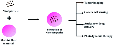
Nanocomposite is a multiphase solid material where one of the phases is in nano-scale that is recently used for this purpose. Due to their quantum-scale sizes, they exhibit different properties from that of conventional microcomposites.9 They have good biocompatibility and biodegradability apart from being thermodynamically stable within biological systems.10 The nanocomposites are tailored, based upon the specific application. The nanodevices are synthesized using nanocomposites by the researchers around the globe. Most of their applications are related to in vivo visualization and therapy with anticancer drugs11 in the field of oncology. This review emphasis only on the various in vivo and in vitro investigations of nanocomposite materials in diagnosis and therapy employed for various cancers thereby promoting them in the clinical usage (Fig. 1).
Martirosyan et al. produced a nanocomposite made of poly(methyl methacrylate) with single walled carbon nanotube to promote the interstitial brachytherapy, a treatment for prostate cancer. The treatment is usually monitored by magnetic resonance imaging. The developed nanocomposite was designed to promote the monitoring. The contrast agents (choline derivate- and gadolinium-based) are introduced inside the nanocomposite using high pressure stainless steel syringe. The polyglycolic acid was used as a biodegradable encapsulation. The in vitro testing of the nanocomposites showed visualization under 1.5 T, revealing the biocompatibility and radiation resistance.14
A novel nanocomposite gel was made of gold nanoparticle and chitosan gel coated glassy carbon electrode (GCE) for immobilization and electrochemical study of K562 leukemia cells. This cell-based sensor was based on the measurement of electron-transfer resistance with [Fe(CN)6]3−/4− as a redox probe. Higher amount of K562 cells was immobilized to the electrode with the increasing concentration of cells to the immersed GCE. The density of the cells adhered to the film increased with respect to the incubation time, which was evidenced by the morphology of the distinguishable filopodia, a good indicator of cell adhesion to material surfaces and cell viability.15 A nanocomposite platform for detection of chronic myelogenous leukemia (CML) was prepared and analyzed in clinical samples. The nanocomposite was developed by deposition of nanostructured composite of chitosan (CS)–cadmium-telluride quantum dots (CdTe-QDs) onto indium-tin-oxide coated glass substrate. After which the amine terminated probe DNA has been covalently immobilized onto CS–CdTe/ITO electrode using glutaraldehyde as a cross linker. Four cDNA samples were used to test the specificity of pDNA/CS–CdTe/ITO bioelectrode. The change in the peak current during the voltametric analysis revealed that the fabricated nucleic acid sensor had excellent scope for detection of CML in clinical patient samples.16
Shen et al. developed an electrochemical biosensing system for detecting leukemia cells based on TiO2/CNT nanocomposites modified electrodes. Titanium isopropoxide and multiwalled CNT were dispersed in a ratio of 70:
30 (w/w) to form the nanocomposite. Electrochemical studies were carried out to test the ability of the developed nanocomposite with respect to detection. The peak current obtained was 10-fold higher than that without the cancer cells. The electrochemical response of the probe on the K562 cells film is apparently stronger than that in the K562 cells film, accompanying with a 30 mV negative shift of the peak potential.17 A novel impedance cell sensor based on the polystyrene/polyaniline/gold nanocomposite was prepared for the detection of HL-60 leukemia cells. The nanocomposite was prepared by assembling the gold nanoparticles on the surface of polystyrene (PS) and polyaniline (PANI) core–shell nanocomposite. The electrochemical sensor was suspended in the HL-60 leukemia cells for 2 h. The immobilized cells exhibited irreversible voltammetric response and increased the electron transfer resistance with a good correlation to the logarithmic value of concentration ranging from 1.6 × 10³ to 1.6 × 10⁸ cells per mL with a limit of detection of 7.3 × 10² cells per mL at 10σ.18
Bai et al. developed a copper monosulfide nanoparticle-decorated reduced graphene oxide-based electrochemical biosensor for the reliable detection of H2O2. The high levels of H2O2 are closely associated with cancer and progressive neurodegenerative diseases. The levels of extracellular H2O2 were detected from the HeLa cell culture. This method was used to detect the H2O2 in the human cervical cancer cell lines HeLa in phosphate-buffered saline (pH 7.4) containing 25 mM glucose at the applied potential of −0.27 V versus Ag/AgCl. An increased cathodic current was observed after treatment of the HeLa cells with 0.4 mg mL−1 CdTe QDs. Thus, promoting CuS nanoparticle-decorated reduced graphene oxide-based electrochemical biosensor for cervical cancer detection.19
An aptamer biosensor for breast cancer cell detection was developed on the ultrasensitive electrochemical basis. This aptamer is a probe that can recognize and bind to the MCF-7 cells. The aptamer was made up of porous graphene oxide/Au composites and porous polytetrafluoroethylene alloy. The mucin protein 1 was selected as the tumor marker for MCF-7 breast cancer cell detection. The aptamer had an analytical performance ranging from 100 to 5.0 × 10⁷ cells per mL with a limitation of mL−1, which was reproducible.20 An integrated nanocomposite loaded with 5 nm gold nanoparticle conjugated with poly[9,9-bis((6′-N,N,N-trimethylammonium)hexyl)fluorenyldivinylene-alt-4,7-(2,1,3-benzothiadiazole)dibromide] (PFVBT) polymer was used for dual-modal targeted cellular imaging of MCF-7 cells. The PLGA–PEG 2000–folate was used for encapsulation of the nanocomposite. The eccentrically loaded gold nano particle helped to maintain the fluorescence. The cellular uptake of the nanocomposite was improved due to the presence of folic acid groups. The nanocomposites were then suspended in both 3T3 fibroblast cells as well as the MCF-7 breast cancer cells. The fluorescence intensities from 3T3 cells upon treatment with folic acid functionalized nanocomposites were not significantly higher than the intensities from MCF-7 cells. This was related to the presence of folic acid receptors in the cancer cells.21
A label-free electrochemical bioanalyte immunosensor was developed for simultaneous detection of lung cancer biomarkers such as anti-MAGE A2 and anti-MAGE A11. These antibodies belong to the melanoma associated gene (MAGE) that recognizes the MAGA antigens responsible for tumor progression present in the cancer cells. This was done using carbon nanotubes–chitosan (CNT–CHI) composite. They used single walled CNT and the electrodes were fabricated by drop casting method onto graphite surface. The differential pulse voltammeter (DPV) measurements displayed that both (MAGE A2/CNT–CHI/graphite and MAGE A11/CNT–CHI/graphite) immunoelectrodes had successful detection of analytes anti-MAGE A2 and anti-MAGE A11 from 5 fg mL−1 to 50 ng mL−1. These CNT-graphite electrodes were independently able to distinguish the anti-MAGE A2 and anti-MAGE A11 independently in a single experimental run, when exposed to a mixture of various analyte concentrations in different combinations irrespective of the presence of other analyte present in the same vessel.22 Zhuo et al., developed a more susceptible method for detection of progastrin releasing-peptide (ProGRP) tumor marker with small cell lung cancer (SCLC), which may indicate an early tendency of cancer metastasis. Pro-GRP is a member of the bombesin family of peptides is produced by the SCLC cells to have mitogenic activity. The Au/TiO2 nanocomposites (nano-Au/TiO2) was prepared by attaching the Au nanoparticles to the TiO2 nanoparticles using the linkage reagent 3-aminopropyltriethoxysilane. After this, the glucose oxidase (GOD) and ferrocene labeled secondary antibodies (Fc-Ab2) were used to bind Au/TiO2 nanocomposites to provide amplified signals. Apart from this, the nano-Au functionalized graphene sheets (GS) were used as biosensor platform to increase the surface area and the electronic transmission rate to capture a large amount of primary antibodies (Ab1). The clinical analysis was done in eleven serum samples from lung cancer patients and healthy volunteers, in order to evaluate their performance. It was found that for the proposed immunosensor, the current is linear with the concentration of ProGRP being within a concentration range from 10.0 to 500 pg mL−1 with a limit of detection down to 3.0 pg mL−1 (S/N = 3).23
A combined magnetic enrichment and optical detection strategy for lung cancer cells detection was developed by Ma et al. The bifunctional nanocomposites (BNPs) with quantum dots had excellent fluorescence with ability to conjugate with monoclonal anti-CEA antibodies, which allows specific reorganization of SPCA-1 human lung adenocarcinoma cells in short time. This antibody helps in the identification of the carcinoembryonic (CEA) antigen present in certain types of cancer cells. The BNPs was developed by modification of silica coated superparamagnetic nanoparticle (SiO2/γ-Fe2O3) with N-(2-aminoethyl)-3-aminopropyltrimethoxysilane and conjugated with anti-CEA antibodies. The benign and malignant pleural effusions of 10 patients was used to examined the ability of the BPNs in relation to detection. The immunonanoparticles had 70% sensitivity and 100% specificity in the pleural effusion samples when examined within 1 hour.24 Zhao et al. developed a localized surface plasmon resonance (LSPR) biosensor made of silver nanoparticles array to detect the cervical cancer using the squamous cell carcinoma antigen (SCCa), as a tumor biomarker. The LSPR helps in overcoming disadvantages and provides good sensitivity and selectivity. The anti-SCCa antibodies were functionalized using 11-mercaptoundecanoic acid. The biosensors were incubated in different concentrations of standard SCCa solution and LSPR values increased stepwise with increasing SCCa concentrations. Thus, the custom-built LSPR system is available for quantitative analysis of SCCa level in human serum with advantages in terms of a rapid test time, label-free and dilution-free process.25
In another study, a label-free surface plasmon resonance cytosensor for breast cancer cell detection was produced by the nano-conjugation of monodisperse magneticnanoparticle and folic acid. The biosensor was based on surface plasmon resonance (SPR) spectroscopy and had a magnetic nanoparticle combined with MUC1 aptamer and folic acid. The detection ability of this nanosystem was as low as 500 MCF-7 breast cancer cells per mL. It was concluded that the MUC1 aptamer and folic acid improved the sensitivity and selectivity of SPR biosensors.26
Chen et al. developed a generic nanosensor for targeting-free cancer cell screening. The visual screening of cancer cells from normal cells was done by glucose sensitivity of the cancer cells. The Ag/Au nanoshells were glucose oxidase-modified to act as a plasmonic diagnostic tool. The nanosenors were capable of distinguishing the malignant human cervical cancer cell line (HeLa) and the nonmalignant mouse embryonic fibroblast cell line (L929). The cells were found to adhere to the nanosensor with an increase in the period of incubation.27
An in vivo evaluation of nanodevices made of porous silicon nanoparticles encapsulated with solid lipid nanoparticles (THCPSi-SLNCs) in the mice model was recorded. The 18F radiolabeling was incorporated for proper imaging of the breast cancer cells found. Tumor uptake of 18F loaded THCPSi-SLNCs was found to be higher when compared to the 18F loaded THCPSi nanoparticles in the subsequent 7 weeks after tumor inoculation. Both the nanodevices were found to be cleared quickly from the circulation and to accumulate in the liver and spleen. Further material characterization of the nanodevice is needed to promote them.28
Tian et al. synthesized nanoamplifiers from gadolinium and gold nanocomposites for magnetic resonance imaging of the colon cancer in both in vivo and in vitro conditions. Electron transfer between water and gadolinium-doped nanoparticle, which is apparent in the presence of gold, explained the enhancement of signal sensitivity. The tumor-targeted nanoamplifiers were injected into mouse models of colon cancer liver metastasis. It was inferred that the nanoamplifiers enhanced the MRI and optical imaging with considerable contrast improvement despite the gold nanoparticles departure. The amplification was more noticed during the invasive targeted imaging.29
The various principals involved in the in vitro and in vivo experimentation of nanocomposite based cancer diagnosis are tabulated in Table 1. From the table, nanocomposite based cancer diagnosis includes the gold nanoparticles, TiO2 nanoparticles, chitosan, CNT and graphene. The TiO2 nanoparticle possesses redox selectivity, photocatalyst and tunable magnetic property, whereas the chitosan is said to be more biodegradable and biocompatible.10 The gold nanoparticles have unique optical, electronic and molecular-recognition properties, which makes them promising a nanomaterial.9,71 Whilst, the carbon allotropes have their extraordinary strength, thermal conductivity, electrical properties and specifically graphene are transparent helping the nanocomposite to be flexible.73 It is evident that the carbon allotropes such as CNT and graphene are equally employed as the gold nanoparticles. Every nanomaterials have their own pros and cons. It is difficult to weigh up a particular nanomaterial to be perfect. Regardless of a nanocomposite's exclusive properties, they may yield different ability depending upon the application. Hence, it becomes more complicated to find a versatile nanocomposite by comparing their property for all applications. The various characteristics of cancer such as stages, type, site it develops varies from person to person, making it more difficult to find a particular nanocomposite as best.
Nanocomposite | Principle involved | Reference |
---|---|---|
In vitro | ||
Poly(methyl methacrylate), single walled carbon nanotube, polyglycolic acid encapsulation | The choline derivate- and gadolinium-based contrast agents enhanced the MRI visualization | 14 |
Gold nanoparticle & chitosan | The electron-transfer resistance was measured with different concentration of K562 leukemia cells | 15 |
Chitosan, cadmium-telluride & quantum dots | Amine terminated probe DNA was conjugated for detection of chronic myelogenous leukemia (CML) | 16 |
Titanium isopropoxide and multiwalled carbon nanotube | Electrochemical studies was conducted in K562 cells with nanocomposite | 17 |
Gold nanoparticles, polystyrene (PS) & polyaniline (PANI) core–shell nanocomposite | Measurement of the electron transfer resistance in the HL-60 leukemia cells with nanocomposite suspension | 18 |
Copper monosulfide nanoparticle & graphene oxide | Levels of extracellular H2O2 were detected from the HeLa cell culture | 19 |
Porous graphene oxide/Au composites & polytetrafluoroethylene alloy | The mucin protein 1, tumor marker showed amplification and selectivity of MCF-7 cells | 20 |
Gold nanoparticle, [9,9-bis((6′-N,N,N-trimethylammonium)hexyl)fluorenyldivinylene-alt-4,7-(2,1,3-benzothiadiazole) dibromide] (PFVBT) polymer, folate | The folate enhanced the selectivity while gold nanoparticle helped in maintain the fluorescence | 21 |
Carbon nanotube, chitosan & graphite | The electrodes contained the lung cancer biomarkers such as anti-MAGE A2 and anti-MAGE A11 | 22 |
Au nanoparticles & TiO2 nanoparticles | Glucose oxidase (GOD) and ferrocene labelled secondary antibodies (Fc-Ab2) were used to provide amplified signals | 23 |
Silica coated superparamagnetic nanoparticle & N-(2-aminoethyl)-3-aminopropyltrimethoxysilane containing quantum dots | The conjugated anti-CEA antibodies recognized the antigens in the SPCA-1 cells and emitted fluorescence | 24 |
Surface plasmon resonance (LSPR) biosensor made of silver nanoparticles array | The anti-SCCa antibodies were functionalized to the biosensors to help in the quantitative analysis of cancer | 25 |
Monodispersed magnetic nanoparticles | Magnetic nanoparticle combined with MUC1 aptamer and folic acid to improve the selectivity | 26 |
Glucose oxidase-modified Ag/Au nanoshells | Glucose responsive ability of the cancer cells were used for detection | 27 |
![]() |
||
In vivo | ||
Porous silicon nanoparticles & solid lipid nanoparticles | 18F radiolabeling was incorporated for imaging of the breast cancer cells in mice model | 28 |
Gadolinium & gold nanoparticles | The electron transfer between water and gadolinium-doped nanoparticle enhanced the MRI images | 29 |
It is also inferred that more concentration is given to the precise properties of a cancer cell than an improvement to conventional visualization techniques leading to the development of immunosensors and nanoaptamers. As there are more trails have been conducted using clinical samples with these nanocomposites, it is high time that steps are taken to introduce these types of diagnosis in human trials. The experimentation of nanocomposite with contrast agent and radiolabeling has been, presently initiated in in vivo condition. In depth studies regarding the effects of these immunosensors, contrast agents, radiolabels in normal renal cells and other allergic reactions must be studied. These examinations may be carried out in diverse animal models promoting the personalized diagnosis of cancer.
The synthesis, characterization and in vitro studies doxorubicin-loaded magnetic nanocomposites was done. The co-polymer prepared from N-isopropylacrylamide (NIPAAm) and methacrylic acid (MAA) via radical polymerization was loaded with doxorubicin. As the N-isopropylacrylamide (NIPAAm) and methacrylic acid (MAA) polymers are temperature sensitive, the release of doxorubicin was influenced by temperature changes. About 0.05% of doxorubicin was released at 37 °C and 2.5% of doxorubicin was released at 40 °C, with 40 min of exposure. The doxorubicin-loaded PNIPAAm–MAA-grafted magnetic nanoparticles had time-dependent effect on the A549 lung cancer cells. The IC50 of doxorubicin-loaded PNIPAAm–MAA-grafted magnetic nanoparticles was found to be 0.16 to 0.20mg mL−1.36 A recent study on the controlled release of doxorubicin from electrospun PEO/chitosan/graphene oxide nanocomposite (PEO/CS/GO) nanofibrous for the treatment of A549 cells in in vitro conditions. The release rate of doxorubicin from PEO/CS/GO/doxorubicin nanofibrous was very slow in both neutral and acidic conditions compared with GO/doxorubicin. It was found that the hydrophobic force including π–π stacking, hydrogen bonding, electrostatic interaction between GO and doxorubicin and diffusion of doxorubicin from pores of nanofibers were responsible for slower release of doxorubicin from PEO/CS/GO/doxorubicin nanofibrous. After 72 h treatment of the cell growth was inhibited. The doxorubicin-loaded PEO/CS/GO nanofibers exhibited obvious cytotoxicity against A549 cells for longer time than free DOX due to its slower release. This promoted the electrospun PEO/CS/GO/doxorubicin nanofibrous in lung cancer treatment.37
A smart multifunctional nanocomposite that encompasses the magnetic induced target delivery, cell uptake promotion and controlled drug release in one system was developed and experimented with MCF-7 breast cancer cells. The amino-modified mesoporous nanocomposite with folate conjugation was loaded with doxorubicin in the mesopores, which had acid sensitive blockers in its orifices. This set up enhanced the pH-dependent self-release. Along with this folate was also introduced to improve the targeted delivery of doxorubicin. The release of the doxorubicin happened in the acidic pH level. There was 80% uptake of doxorubicin by the MCF-7 through endocytosis at 5 h. Along with this, there was also nuclear fragmentation and condensation in the MCF-7 cells.38 Biodegradable nanocomposite fibers based on poly(2-hydroxy ethyl methacrylate) and bamboo cellulose was developed to deliver paclitaxel to the MCF-7 cancer cells. The prepared nanocomposite fibers showed 96% cell viability while the paclitaxel incorporated pHEMA-bamboo cellulose nanocomposite fiber showed 7.4% cancer cell viability after 72 h incubation. This confirms the biocompatibility as well as the anticancer effect of paclitaxel after the incorporation to the pHEMA nanocomposites.39
Ali et al. prepared a hippuric acid zinc layered hydroxide nanocomposite (HAN) for delivery of doxorubicin and oxaliplatin in the MDA-MB231, MCF-7 cancer cell lines. The hippuric acid zinc layered hydroxide nanocomposite was prepared by the direct reaction of a HA solution with an aqueous suspension of ZnO. The hippuric acid zinc layered hydroxide nanocomposite with the anticancer drugs was found to inhibit the cell growth of both the cell lines in comparison to the drugs alone. The IC50 value for the combination of HAN with doxorubicin toward MCF-7 was 0.19 ± 0.15 μg mL−1 and toward MDA-MB231 was 0.13 ± 0.10 μg mL−1. The combination of HAN (0.5 μg mL−1) with doxorubicin (0.5 μg mL−1) reduced the cell proliferation in MCF-7 and MDA-MB-231 cells in 37.3% and 17.6%, respectively after 24 hours treatment.40
This was followed by preparation of iron oxide magnetic nanoparticles coated with chitosan to form CS–MNP nanoparticles. The CS–MNP were loaded with an anticancer drug, betulinic acid (BA) to form a BA–CS–MNP nanocomposite. The release of the BA from the nanocomposite happened at pH 7.4 followed a pseudo-second-order kinetic model. The potential cytotoxicity of free BA, MNPs, CS–MNP, and the BA–CS–MNP nanocomposite was examined in normal mouse fibroblast cells (3T3) and breast cancer cells (MCF-7). No changes were observed due to BA and the nanocomposite at concentrations in the range 0.781–50 μg mL−1 after 72 h of incubation. The BA and BA–CS–MNP nanocomposite exhibited cytotoxicity in MCF-7 cells in a dose-dependent manner with IC50 values of 2 and 3.6 μg mL−1, respectively.41
A nanocomposite capsule of porous silicon with an acid degradable acetalated dextran (AcDX) matrix was loaded with the memthotrexate, paclitaxel and sorafenib was tested for its therapeutic effect on MCF-7 and MDA-MB-231 breast cancer cell lines. In order to assist the intracellular drug delivery, a nano-arginine cell-penetrating peptide (CPP) was chemically conjugated to the surface of the nanocomposites using oxime click chemistry. The cellular uptake of the nanocomposite was improved by the conjugation of CPP. The release of the anti-cancer drugs was initiated by the pH change to 5.0. Both cell lines had a particle dose-dependent viability towards the bare nanocomposite after 24 h of incubation and a slight decrease of the cell viability was seen with CPP loaded nanocomposite, implying the cell uptake. For both cell lines tested, the combination of memthotrexate, paclitaxel and sorafenib remarkably reduced the cell growth in a concentration-dependent manner. However, at the same concentration range, multidrug loaded nanocomposites showed different effects on the cell proliferation before and after CPP functionalization.42
Barahuie et al. developed protocatechuic acid–Mg/Al nanocomposite using the ion-exchanged as well as direct coprecipitation. The loading of protocatechuic acid in nanocomposite synthesized using ion exchange and nanocomposite synthesized using direct method was estimated to be about 24.5% and 27.5% (w/w), respectively. After 72 hours incubation of nanocomposites with MCF-7 human breast cancer, the growth of these cancer cells was suppressed, with IC50 of 35.6 μg mL−1 for protocatechuic acid in nanocomposite synthesized using ion exchange and 36.0 μg mL−1 for nanocomposite synthesized using direct method for MCF-7 cells. The cytotoxicity of nanocomposite synthesized using ion exchange was greater than that of nanocomposite synthesized using direct method. This result was in parallel with the higher protocatechuic acid in nanocomposite synthesized using ion exchange compared with in nanocomposite synthesized using direct method.43 A chitosan coated layered clay montmorillonite nanocomposites was prepared to modulate the oral delivery of paclitaxel in colonic cancer. The paclitaxel drug was intercalated into the gallery of montmorillonite by ion exchange reaction, which was then coated with the biopolymer chitosan. The nanocomposites demonstrated a controlled release of paclitaxel and 1:
2 fold improvement in vitro anticancer activities towards human colon cancer COLO-205 cells in in vitro conditions.44 Venkatesan et al. developed a chitosan modified hydroxyapatite nanocarriers-mediated celecoxib targeted delivery in colon cancer cells. The cell proliferation, morphology, cytoskeleton, cellular uptake and apoptosis were analysed of colon cancer cells in in vitro condition. The results displayed a significant antiproliferation, apoptosis and time-dependent cytoplasmic uptake of celecoxib-loaded Hap-Cht nanoparticles in HCT 15 and HT 29 colon cancer cells. The cells appeared to be round in shape and irregular with no striations in actin filament organization after 48 h treatment of celecoxib-loaded Hap-Cht nanoparticles. Further, in vivo studies were performed.45
Nanocomposite made of nano Fe3O4 and polylactide nanofibers loaded with daunorubicin to cause the induction of cell death of leukemia cancer cells was reported. The number of viable cells decreased when treated with the daunorubicin loaded nanocomposite. The cellular uptake was demonstrated by the inter-cellular green fluorescence emitted by the daunorubicin drug. The cell inhibition with the 9.93 × 10⁻⁷ and 1.99 × 10⁻⁶ mol L−1 daunorubicin concentrations in the presence of Fe3O4 nanoparticles or PLA nanofibers produced no significant difference from that of the cell treated with daunorubicin alone. However, for daunorubicin concentrations at 9.93 × 10⁻⁷ and 1.99 × 10⁻⁶ mol L−1, the inhibition rates increased to 31% and 46% for the cell system cultured with daunorubicin and Fe3O4–PLA.46 Chen et al. developed a poly(lactic acid) (PLA) based nanocomposites for targeted drug delivery of daunorubicin to the leukemia K562 cells. PLA based nanocomposites preparation involved the accumulation of the anticancer drug daunorubicin on PLA nanofibers combined with TiO2 nanoparticles. The observation demonstrated that these new nanocomposites could readily induce the anticancer drug daunorubicin to accumulate on leukemia K562 cells so that the remarkably enhanced intracellular fluorescence intensity could be observed upon application of the blends of the nano-TiO2 and PLA nanofibers together with daunorubicin. This also supported the targeted delivery of daunorubicin to the leukemia cells.47
The anti-cancer drug internalization was increased by using the nanocomposite with a combination of gold nanoparticle and multi walled carbon nanotube in the human SMMC-7721 hepatocarcinoma cells. The anticancer drug daunorubicin was conjugated to the gold–carbon nanotube nanocomposite. There was improvement in the anticancer effect with increase in the concentration of daunorubicin-loaded nanocomposite when compared to daunorubicin drug alone.48 Wu et al. investigated the anticancer effectiveness of potential pharmacophore agents (o-carborane (Cb), o-carborane-C-carboxylic acid (Cbac1), and o-carborane-C(1)C(2)-dicarboxylic acid (Cbac2) coupling with cadmium telluride quantum dots capped with cysteamine (CA-CdTe QDs)) in the SMMC-7721 hepatocellular carcinoma cells. The cell inhibition of SMMC-7721 cancer cells was increased by the CA-CdTe QDs. The IC50 of Cbac1 toward SMMC-7721 cancer cells for 72 hours was about 344 μM. The exhibited cytotoxicity was in relation to the ROS generation and genomic damage via apoptosis pathway. This was attributed to the self-assembly nanocomposites of the carborane–carboxylic acids with CA-CdTe QDs.49 Swet et al. evaluated a silica–calcium–phosphate nanocomposite (SCPC75) drug delivery system as a means to localize cisplatin treatment within the tumor, while reducing systemic toxicity, in a rat model of hepatocellular carcinoma. The SCPC75 nanocomposite was made up of 32.9% SiO2, 11.4% P2O5, 22.8% CaO, and 32.9% Na2O composition in molar percentage. After this the cisplatin drug was loaded to the nanocomposite. The cisplatin was bound to the SCPC75 at 14.2 ± 0.2 mg cisplatin per g SCPC75. The nanocomposite reduced the cell viability of H4IIE hepatoma cells grown in the culture with time dependency. The experiment was extended and the cisplatin drug delivery by SCPC75 nanocomposite was conducted in ACI rats.50
A nanosystem with gold nanorods (AuNRs) encapsulated in nanographeneoxide shells was developed to improve the efficiency of chemophotothermal cancer therapy of hepatoma Huh-7 cells. The hyaluronic acid was conjugated in the corners of nanographeneoxide enwrapped AuNR nanocomposite (NGOHA-AuNRs). The anti-cancer drug doxorubicin was loaded to exhibit pH sensitive release. At in vitro experiment condition, there was release of 15.9% doxorubicin at pH 5.3. The cellular uptake of nanocomposite loaded with doxorubicin was said to be enhanced by hyaluronic acid. The therapeutic efficacy of NGOHA-AuNRs–doxorubicin presented significantly synergistic chemophotothermal therapy effects that were about 1.5-fold and 4-fold higher than that of separate chemotherapy and photothermal treatment to targeting the Huh-7 cells.51 Chandran et al. developed a nano-drug delivery system based on an electric field and pH dual-stimuli responsive chitosan–gold nanocomposite (CGNC) for site specific controlled delivery of the anticancer drug 5-fluorouracil. The release of 5-fluorouracil happened at the pH of 5.3, near the cancer cells. The SiHa cells were found to grow on the CGNC–fluorouracil conjugate modified ITO plate. Further application of an electric field of 1.5 V for the release of 5-FU led to the complete death of the SiHa cells.52
Another nanocomposite was prepared by radical polymerization of methacrylic acid around carbon nanotubes in the presence of quercetin as a biologically active molecule. The anticancer activity of the flavonoid quercetin was investigated using the HeLa cervical cancer cells. It was found that the covalent conjugation of quercetin to the polymeric backbone containing dispersed carbon nanotubes is a key process, increasing its anticancer efficiency by increasing the flavonoid stabilization and the cell internalization. Cell viability tests on healthy cells demonstrated no-toxicity due to the quercetin conjugated nanocomposite.53
Xu et al. used gold-doped TiO2 (Au/TiO2) nanocomposites to improve the photocatalytic inactivation effect on human colon carcinoma LoVo cells. The Au/TiO2 samples doped with different amounts of Au (1 wt%, 2 wt%, 4 wt%) were prepared by a chemical reduction method and used to photo kill the LoVo cells and it was observed that the most efficient sample was 2 wt% Au/TiO2. The study revealed that the irradiation of UV light (λmax = 365 nm, intensity = 1.8 mW cm−2) for 110 min, all of the LoVo cancer cells were killed by 50 μg mL−1 Au/TiO2. The 50 μg mL−1 TiO2 nanoparticles killed only 70% cancer under the same condition.55 The photothermal activity of single walled carbon nanotube (SWCNT) composite with a designed peptide structure of H-(-Lys-Phe-Lys-Ala-)7-OH [(KFKA)7] against colon tumor cells was evaluated. The thermographic observations show that intratumoral injection of SWCNT-(KFKA)7 solution followed by NIR irradiation resulted in a rapid increase of the temperature to 43 °C in the subcutaneously inoculated colon 26 tumors. There was a remarkable cell damage in the colon 26 culture incubated SWCNT-(KFKA)7 over 3 minutes near infrared irradiation. The single treatment with SWCNT-(KFKA)7 or NIR irradiation did not produce any moderate changes. These results suggest the a great potential of an SWCNT-peptide composite for use in photothermal cancer therapy.56
Nanocomposite based delivery of anti-cancer agents such as paclitaxel, 5-fluorouracil, doxorubicin, oxaliplatin, memthotrexate, sorafenib, daunorubicin have been carried out in various cell lines. The significant findings of those investigations are tabulated in the Table 2. The majority of the key findings is associated to the cell viability assays. The mode of cell death induced by the targeted delivery of these standard drug merged with nanocomposite should be compared with the mode of cell death induced by these drugs alone. Even though some of the researches explore the anti-proliferative property of phenolic acids, it is most notable that more importance is given to approve anticancer drugs.
Nanocomposite | Drug delivered | Cell line tested | Key findings | Reference |
---|---|---|---|---|
Poly(ethylene glycol) PEG–iron oxide hydrogel nanocomposites | Paclitaxel | A549 human lung adenocarcinoma cells | • Decrease of the viable cells after with increase in the duration of exposure | 34 |
• Increase in the efficiency during heat treatment | ||||
Na+ montmorillonite (Na+-MMT) with chitosan nanosystem | 5-Fluorouracil | A549 human lung adenocarcinoma cells | • The IC50 value of 5-FU/CS-MMT composites in A549 cell line were 11.49 μg mL−1 | 35 |
• The nanodevice caused DNA damage that increased with respect to time | ||||
N-Isopropylacrylamide (NIPAAm) and methacrylic acid (MAA) grafted nanosystem | Doxorubicin | A549 human lung adenocarcinoma cells | • Time-dependent effect | 36 |
• IC50 of doxorubicin-loaded PNIPAAm–MAA-grafted nanodevice was 0.16 to 0.20 mg mL−1 | ||||
PEO/chitosan/graphene oxide nanocomposite | Doxorubicin | A549 human lung adenocarcinoma cells | • Initiation of cytotoxicity with increased period of exposure | 37 |
Amino-modified mesoporous nanocomposite with folate conjugation | Doxorubicin | MCF-7 human breast adenocarcinoma cell line | • Nuclear fragmentation and condensation in the MCF-7 cells | 38 |
Poly(2-hydroxy ethyl methacrylate) and bamboo cellulose nanosystem | Paclitaxel | MCF-7 human breast adenocarcinoma cell line | • 7.4% breast cancer cell were viable after 72 h treatment | 39 |
Hippuric acid zinc layered hydroxide nanocomposite | Doxorubicin and oxaliplatin | MDA-MB231 breast cancer cell | • The IC50 toward MCF-7 was 0.19 ± 0.15 μg mL−1 and toward MDA-MB231 was 0.13 ± 0.10 μg mL−1 | 40 |
MCF-7 human breast adenocarcinoma cell line | • Cell proliferation in MCF-7 and MDA-MB-231 cells reduced to 37.3% and 17.6%, respectively after 24 h treatment | |||
Iron oxide magnetic nanoparticles coated with chitosan | Betulinic acid | MCF-7 human breast adenocarcinoma cell line | • Cytotoxicity in MCF-7 cells was dose-dependent | 41 |
• The IC50 value of 3.6 μg mL−1 | ||||
Nanocomposite capsule of pourous silicon with acetalated dextran (acdx) matrix | Memthotrexate, paclitaxel and sorafenib | MCF-7 human breast adenocarcinoma cell line, MDA-MB-231 breast cancer cell lines | • Concentration dependent reduction in the cell viability | 42 |
Protocatechuic acid–Mg/Al nanocomposite | Protocatechuic acid | MCF-7 human breast adenocarcinoma cell line | • Suppression of cell growth in dose dependent manner | 43 |
Chitosan coated layered clay montmorillonite nanocomposites | Paclitaxel | COLA-25 human colon cancer cells | • Improvement of paclitaxel anticancer activity | 44 |
Chitosan modified hydroxyapatite nanocarriers | Celecoxib | HCT 15 and HT 29 colon cancer cells | • Time-dependent cytoplasmic uptake of celecoxib-loaded Hap-Cht nanoparticles | 45 |
• Round shaped cells with no striations in actin filaments organization | ||||
Nano Fe3O4 and polylactide nanofibers nanodevice | Doxorubicin | K562 human leukemia cells | • Inhibition of cell growth increased to 31% and 46% | 46 |
PLA nanofibers combined with TiO2 nanoparticles | Daunorubicin | K562 human leukemia cells | • Accumulation of anticancer drug to leukemia K562 cells | 47 |
Gold nanoparticle and multi walled carbon nanotube | Daunorubicin | SMMC-7721 human hepato carcinoma cells | • Anticancer effect with increase in the concentration of daunorubicin-loaded nanocomposite | 48 |
Cadmium telluride quantum dots capped with cysteamine (CA-CdTe QDs) | o-Carborane-C-carboxylic acid | SMMC-7721 human hepato carcinoma cells | • The IC50 was about 344 μm | 49 |
• Cytotoxicity was in relation to the ROS generation and genomic damage via apoptosis pathway | ||||
Silica–calcium–phosphate nanocomposite (SCPC75) drug delivery system | Cisplatin | H4IIE rat hepatome cells | • Time dependent reduction in cell viability | 50 |
Gold nanorods (aunrs) encapsulated in nanogrphenoxide shells with hyaluronic acid conjugation | Doxorubicin | Huh-7 hepatoma cells | • Enhanced chemophotothermal therapy effects than that of separate chemotherapy and photothermal treatment | 51 |
Chitosan–gold nanocomposite (CGNC) | 5-Fluorouracil | SiHa cervical squamous carcinoma cells | • Complete death of the SiHa cells when 1.5 V electric field is passed | 52 |
Methacrylic acid carbon nanotubes devices | Quercetin | HeLa cervical cancer cells | • Increasing its anticancer efficiency by increasing the flavonoid stabilization and the cell internalization | 53 |
Gold-doped TiO2 (Au/TiO2) nanocomposites | — | Human colon carcinoma lovo cells | • Improve the photocatalytic inactivation effect on human colon carcinoma lovo cells | 54 |
• At UV irradiation, all the lovo cancer cells were killed by 50 μg mL−1 2 wt% Au/TiO2 | ||||
Single walled carbon nanotube (SWCNT) with a designed peptide structure of H-(-Lys-Phe-Lys-Ala-)7-OH [(KFKA)7 nanosystem | — | Colon 26 colon cancer cells | • The photothermal effect caused remarkable cell damage after 3 minutes near infrared irradiation | 55 |
Venkatesan et al. proceeded with the in vivo investigation after the optimistic results obtained in the ex vivo condition as previously stated. The antiproliferative, apoptotic and tumor inhibitory efficacy of celecoxib-loaded nanocomposite in a nude mouse human xenograft model was investigated. In vivo human colon tumor xenograft nude mouse tumor studies proved that the celecoxib-loaded Hap-Cht nanoparticles were more potent in inhibiting tumor growth with no prominent side effects. A progressive increase in green fluorescent of the apoptotic cells were found using TUNEL staining in the celecoxib-loaded Hap-Cht nanoparticle-treated group. Based on these results, it is concluded that the Hap-Cht nanocomposite can be an effective and safe vehicle for celecoxib delivery in colon cancer chemotherapy.45
The anticancer drug 6-{[2-(dimethylamino)ethyl]amino}-3-hydroxyl-7H-indeno[2,1-c]quinolin-7-one dihydrochloride (TAS-103) was loaded in poly(lactide-co-glycolide) (PLGA) nanocomposite particles that were inhalable. The drug release of 5% TAS-103-loaded PLGA nanocomposite particles signified sustained-release while 10% TAS-103-loaded samples indicated initial burst. The cytotoxicity of A549 was increased by nanocomposites. The cells uptake of nanocomposite followed the endocytosis mechanism. The biodistribution of TAS-loaded PLGA nanocomposite was examined, which showed that there was higher concentration of the drug in plasma in comparison to the intravenous administration of free drug.59
As previously mentioned, the SCPC75 nanocomposite based delivery of cisplatin was carried out in in vivo condition. Subcutaneous inoculation of H4IIE hepatoma cells caused the reproducible focal tumor mass formation in ACI rats. There was significantly lower tumor growth in the rats treated with SCPC75–cisplatin hybrid discs than the control and systemic cisplatin treated. Apart from this the side effects such as rapid weight loss and decreased liver and kidney function induced by systemic cisplatin was not observed in SCPC75–cisplatin-treated animals. This shows that the SCPC75 nanocomposite based cisplatin delivery to be a promising therapy for hepatoma.50
A synergistic therapy tool that based on CuS nanoparticles-decorated graphene oxide functionalized with polyethylene glycol (PEG–GO/CuS) for cervical cancer treatment. The anticancer drug doxorubicin was loaded to the PEG–GO/CuS nanocomposites with a the drug loading content as 900 mg doxorubicin mg−1. At acidity of pH 5.5, doxorubicin was quickly released in the early stage, and about 45% of doxorubicin was released in the 45 h. There was 75% cell death of the HeLa cells after irradiation at NIR laser at an equivalent of 10 mg mL−1 doxorubicin. In mouse models, mouse cervical tumor growth was found to be significantly inhibited by the chemo-photothermal effect of PEG–GO/CuS/Dox nanocomposites, resulting in effective tumor reduction. This demonstrated both the in vivo and in vitro efficacy of the nanocomposite.60
A nano-drug delivery system was experimented and in in vivo condition and the results were reported. This drug delivery system consists of human serum albumin, poly(lactic-co-glycolic acid) (PLGA), 5-fluorouracil (5-Fu), magnetic nanoparticles and fluorescent labeling molecule (diphenylhexatriene). The prepared nanocomposite system was tested in the mice model with injected SCC cells. The tumor size was much reduced when injected with nanocomposite containing magnetic nanoparticles than the nanocomposite alone. This was confirmed by visual evaluation as well as the histological analysis.61
The notable findings recorded during the investigation of nano-drug delivery in in vivo condition are given in Table 3. It is evident that all these pre-clinical experiments have been carried out with the common anticancer drugs. The nanocomposite based photodynamic therapy has to be initiated in animal models in order to promote their usage in real cancer therapy. This might also help in revealing the other adverse effects in the normal surrounding cells due to this treatment. The evaluation of biodistribution of these nanocomposites in animal model is also necessary to encourage them in clinical cancer treatment.
Nanocomposite | Drug delivered | Cell line tested | Key findings | Reference |
---|---|---|---|---|
Doxorubicin/carbonate apatite nanocomposite | Doxorubicin | Colon tumor cells in the BALB/cA nude mice | • High cytotoxicity than the cytotoxicity of the free doxorubicin drug alone | 57 |
• Inhibition of the tumor growth | ||||
Squalene gem nanoparticle | Isocombretastatin A-4 (isoCA-4) | Human colon carcinoma xenograft nude mice model | • Complete tumor regression upto 93% | 58 |
Chitosan modified hydroxyapatite nanocarriers | Celecoxib | Mouse human colon cancer xenograft model | • Significant antiproliferation | 45 |
• Apoptosis | ||||
• Suppression of tumor growth | ||||
Inhalable poly(lactide-co-glycolide) nanocomposite | 6-{(2-(Dimethylamino)ethyl)amino}-3-hydroxyl-7H-indeno(2,1-c)quinolin-7-one dihydrochloride (TAS-103) | A549 human lung adenocarcinoma cells injected in mice model | • Cytotoxicity of A549 was increased | 59 |
• Higher concentration of the drug in plasma in comparison to the intravenous administration of free drug | ||||
Silica–calcium–phosphate nanocomposite (SCPC75) drug delivery system | Cisplatin | Rat model of hepatocellular carcinoma | • Significantly lower tumor growth in the rats treated with SCPC75–cisplatin hybrid discs than the control and systemic cisplatin treated | 50 |
CuS nanoparticles-graphene oxide with polyethylene glycol (PEG–GO/CuS) | Doxorubicin | Mouse model with HeLa cervical cancer cells | • 75% cell death of the HeLa cells after irradiation at NIR laser | 60 |
Human serum albumin, poly(lactic-co-glycolic acid) (PLGA) magnetic nanoparticles with diphenylhexatriene | 5-Fluorouracil | Mice model with injected SCC cells | • Reduction of tumor size inferred from visual and histological analysis | 61 |
From the Tables 2 and 3, the nanocomposites used for drug delivery utilize PEG, chitosan, NIPAAM, clay montmorillonite, PLA, TiO2 nanoparticles and gold nanoparticles. Here, the montmorillonite is translucent and has a good thermal stability. Chitosan and PEG are said to have an excellent biodegradability and widely available.11,75 The metallic nanoparticles TiO2 and gold are said to have an excellent electrical, magnetic and blood compatible.10,71 The NIPAAM is more temperature sensitive while PLA is a biodegradable thermoplastic aliphatic polyester.11 Biodegradability and selectivity are important properties required to develop a potential drug delivering nanosystem. All the nanomaterials are said to exhibit good biocompatibility yet chitosan seems to be a prominent candidate. The superior property of chitosan is that they are biodegradable in acidic pH and are not dissolve in normal physiological pH. Even though they have excellent targeting ability, chitosan nanocomposites cannot be proclaimed as a preferred material for cancer treatment. This is mainly because there is no exhaustive research carried out for a particular nanocomposite/nanomaterial in all types of cancer treatment. This makes difficult to promote a single nanoparticle/nanocomposite as an ideal choice for different cancer treatments.
Another nanodevice made of nanocomposite liposomes containing quantum dots and anticancer drugs for cancer treatment and drug delivery was reported and a comparison of the cationic, PEGylated and deformable liposomes was done. The anticancer drugs camptothecin and irinotecan was encapsulated. The cationic liposomes showed a encapsulation ability of 96% for camptothecin and 99% for irinotecan. Cell viability levels after administration of camptothecin and irinotecan from cationic liposomes at 116 μM were 3.9% and 7.1%, respectively. The cellular uptake of quantum dots from the cationic liposomes was revealed by the red fluorescence emitted from the cytoplasm and near nuclei. In experimentation with the nude mice, once again the cationic liposomes demonstrated the higher tendency to accumulate in solid tumors for exhibiting fluorescence signals compared to other vehicles. Similarly, the intratumoral camptothecin was significant during the administration of cationic liposomes.63
Seo et al. prepared a methylene blue-loaded gold nanorod@SiO2 core (MB-GNR@SiO2) are synthesized for use in cancer imaging and photothermal/photodynamic dual therapy. The experiment was carried out in in vitro condition using the CT-26 mouse colon cancer cells. The methylene blue molecules were detected at 446, 1380, and 1605 cm−1 in the MB-GNR@SiO2 nanocomposite. The intensities were poor in case of single cancer cells, while improved due to overlapping of the cancer cells. The cell viability of the cancer cells decreased to 52% when treated with increasing doses of MBGNR@SiO2 nanocomposites from 0 to 150 mL. The cell viability decreased to 11% for the MB-GNR@SiO2 cells after NIR laser irradiation for 50 min during photothermal therapy. Along with this, the ROS level was enhanced by the irradiation of the cancer cells.64
From our review, it is evident the nanocomposite based cancer diagnosis narrowed to the explicit identification of cancer cells from enhancing of the visualization techniques. Nanocomposites based diagnosis involved the use of conjugated biomarkers,21–24 radiolabeling25 as well as contrast agents14 to produce an improved diagnosis. There are also nanosensors to measure the electron transfer resistance and detect the cancer,15–18,29 which have been experimented in laboratory conditions. As more experimentation of immunosensor and nanoaptamers has already been done with clinical samples, steps to promote them in the medicinal scenario should be initiated. Future improvement of cancer diagnosis to explore the genetic profile of the cancer type may promote personalized treatment procedures. Chemotherapy is the most commonly adapted treatment procedures for cancer, but the ability to accumulate the drug site-specifically remains subtle. The nano-based drug delivery offers more targeted delivery as well as personalized treatment due to their reduced dimensions and has been gaining momentum in recent years. They reduce the exposure of normal cells to the anticancer drugs. From our study, the approved anticancer drugs such as doxorubicin,36–38,40,46,51,57,60 paclitaxel,34,39,44 5-fluorouracil,35,52,61 daunorubicin,47,48 oxaliplatin,40 cispaltin50 have been extensively explored.
Some of the chemicals with plant origin are found to have anticancer property and retard the growth of various cancer cells. The nanocomposite-based delivery of these biochemical is to be deciphered to elicit new potential methods for treating cancer. Experimentation of developed nanocomposites for cancer diagnosis and treatment have been conducted in both in vitro and in vivo conditions along with very few experimentation with the clinical samples. Further, in depth studies in relation to the mode of cell death induced by these drug-delivery systems on cancer cells should be done. The progression of cancer is mediated by distribution of tumor cells through blood system or lymphatic system. These cells that move in the vessels are termed as circulating tumor cells (CTCs) and become the forerunner of metastasis. The count of CTCs are only in the range of 10−7 to 10−3 among normal blood cells, making them rare and difficult to detect.65 At present, CTC detectors are based on a mechanical cell-sorting device. Advanced nano-oriented approaches such as, a novel nanotheranostics platform to detect these CTCs to achieve a breakthrough for early detection and treatment cancer should be promoted.66 Nanotheranostics is an integrated target specific diagnostics and therapeutic tool presented in nano-scale. A smart technique aims to monitor the effect of the therapy given, increasing the drug efficacy and ensuring safety of the patient.67 Extensive research on various nanotheranostics tools for cancer would help in personalizing the cancer therapy. This may not only help in assessing cancer before or after treatment but also throughout the entire course of therapy.
The overview shows that nanocomposite fabrication employs biomaterials like poly(methyl methacrylate), polytetrafluoroethylene alloy, chitosan, poly(ethylene glycol) that exerts good biocompatibility. It is apparent that chitosan, the biopolymer is utilized predominantly in developing the various nanocomposite for cancer diagnosis and therapy. This may be related to the chitosan's property to have a positive charge under acidic condition allowing the drug delivery and biodegradability.68 The photodynamic therapy of the nanocomposites is the other area, which needs more attention, particularly in depth in vivo experimentation. This investigation would help in recording of the other adverse effect caused to the normal cells surrounding the cancer growth. The effect of the nanocomposite based cancer treatment in knockout mouse (genetically engineered mouse) may obtain efficient information than the in vivo studies carried out in mouse models.
Apart from these, the toxicity, biodistribution and other adverse effect needs to be recorded carefully. The use of nano-based diagnosis and treatment are increasing especially in cancer. The researchers have been more concentrating to reduce the toxicity of the chemotherapeutic drugs while not realizing the nanodevices may impose risk to the individuals. Even though the targeted localization of cancer is the key of nanocomposite based diagnosis and treatment, there is still a need for classification of the possible adverse effect on human health. Available literatures with pre-clinical testing suggest that these nanocomposites have an adverse potential. Apart from these, the genotoxicity of the nanocomposites remains to be imperfect.69 Likewise, there should be a detailed study on the biodistribution of the developed nanocomposite before implementation of the nanoparticles in nanooncology.
In some cases, these nanodevices reach the different body parts via blood stream and pass through the biological protections and accumulate in the some organs and tissue as they are non-degradable. Their common sites of accumulation are lung, bone marrow, liver, kidney, brain and heart.70 Hence, toxicity of nanocomposites has to be tackled efficiently. More attention on the toxic effect of nanomaterials used in order to prepare the nanocomposite should be done. For example, gold nanoparticles, PEG, CNT are some of the materials that have received significant attention in biomedicine because of their unique physical, chemical and biological properties. Despite the potential characteristics, they also exhibit some harmful assets due to long-term administration. Toxicological studies on gold nanoparticle showed their ability to penetrate the red blood cells and cause the sperms to become non-motile.71,72 While, some investigation show that CNTs may accidentally penetrate the membrane and damage the dorsal root ganglion neurons.73 An in vivo toxic test revealed that the nanoparticles coated with PEG accumulate in liver and spleen causing acute inflammation.74 In comparison, the natural nanoparticles such as chitosan, clay montmorillonite are found to have less toxic effect than these synthetic nanocomposites. These natural substances are abundant in nature as well as biocompatible. Besides, they also tend to reduce the toxicity of synthetic nanomaterial when used in combination. In a particular study, the chitosan reduced the toxicity of gold nanoparticles when used for small hairpin RNA delivery to treat human lung adenocarcinoma cells.75
Yet another promising strategy to overcome this toxic effect is functionalization. The functionalization of these materials reduces the cytotoxicity by increasing their biocompatibility and biodegradability. In case of CNT, the biocompatibility and low cytotoxicity is influenced by the size, dose, duration, testing systems and surface functionalization. The functionalization may be done covalently or non-covalently using chemicals. The biodegradability of functionalized CNTs remained to be ambiguous. While a latest study proved that, the functionalized CNTs were degradable by oxidative enzymes. It also helps in increasing the solubility, biocompatibility, cellular interaction pathways thereby reducing the cytotoxicity of the CNTs.76,77 In the near future, synergistic combination of various professionals like oncologists, material engineers, physicians and other biologists may advance the cancer nanotheranostics significantly. This in turn would stimulate a breakthrough in nanooncology and make cancer to be an age-old disease.
This journal is © The Royal Society of Chemistry 2015 |