Biocompatible degradable injectable hydrogels from methacrylated poly(ethylene glycol)-co-poly(xylitol sebacate) and cyclodextrins for release of hydrophilic and hydrophobic drugs

Mengyao Zhanga, Yaobin Wua, Xin Zhaoa, Kun Gaob, Peter X. Maacdef and Baolin Guo*ab
aCenter for Biomedical Engineering and Regenerative Medicine, Frontier Institute of Science and Technology, Xi'an Jiaotong University, Xi'an, 710049, China. E-mail: baoling@mail.xjtu.edu.cn; Fax: +86-29-83395131; Tel: +86-29-83395361
bState Key Laboratory for Manufacturing Engineering, Xi'an Jiaotong University, Xi'an, 710049, China
cDepartment of Biomedical Engineering, University of Michigan, Ann Arbor, MI 48109, USA
dDepartment of Biologic and Materials Sciences, University of Michigan, 1011, North University Ave., Room 2209, Ann Arbor, MI 48109, USA
eMacromolecular Science and Engineering Center, University of Michigan, Ann Arbor, MI 48109, USA
fDepartment of Materials Science and Engineering, University of Michigan, Ann Arbor, MI 48109, USA

Received 20th June 2015 , Accepted 27th July 2015

First published on 27th July 2015


Abstract

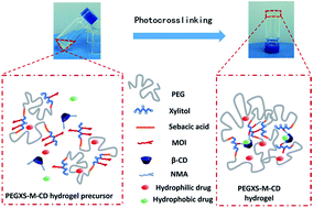
A drug delivery system (DDS) which can achieve a sustained release of both hydrophilic and hydrophobic drugs is highly beneficial for biomedical applications. In this study, we present an injectable photocurable composite hydrogel based on methacrylated poly(ethylene glycol)-co-poly(xylitol sebacate) (PEGXS-M) and acrylamidomethyl-β-cyclodextrin (β-CD-NMA) for both hydrophilic and hydrophobic drug release. The PEGXS-M copolymers were synthesized by the polycondensation of PEG, xylitol and sebacate, and then incorporation of a methacrylate group. The PEGXS-M-CD composite hydrogels were prepared by the photocrosslinking of a PEGXS-M and β-CD-NMA mixed solution under UV irradiation, and the swelling ratio, rheological properties and degradation behavior of the hydrogels were investigated. The incorporation of β-CD-NMA into the PEGXS-M hydrogel decreased the swelling ratio and meanwhile improved the mechanical properties. Adipose-derived mesenchymal stem cells (ADMSCs) were cultured in the presence of hydrogels and the cultivation results indicated good biocompatibility of these hydrogels. Furthermore, theophylline and phenethyl alcohol were chosen as hydrophilic and hydrophobic model drugs to be encapsulated within PEGXS-M-CD hydrogels for drug release investigation. The good sustained drug release efficiency of these hydrogels has been successfully confirmed, and the addition of adamantanamine hydrochloride enhanced the release of drugs from the hydrogels. These results suggest that the PEGXS-M-CD composite hydrogels have a great potential in drug delivery for the sustained release of both hydrophilic and hydrophobic drugs.


1. Introduction

Over the past several decades, more and more attention has been devoted to the development of drug delivery systems (DDS) to improve the conventional administration through various approaches such as enhancing drug loading, reducing drug toxicity and extending the release time.1–6 The materials used for the design of drug carriers play a critical role in drug delivery, and recently a number of biomaterials involving biodegradable micro-7,8 and nanoparticles,9–12 lipids,13 nanofibers,14,15 polymeric micelles16,17 and hydrogels18–22 have been developed for DDS. Among these materials, hydrogels are widely used as drug carriers due to their excellent chemical and physical characteristics including high water content, easily tunable mechanical properties, biocompatibility and biodegradability.23–26 Especially, injectable hydrogels with remarkable advantages such as minimal invasiveness and in situ gelation, have been extensively used in various biomedical fields and also exhibit potential in drug delivery applications.27–30

Recently, a series of injectable hydrogels have been developed based on various gelation approaches and crosslinking methods. For instance, some studies show that the injectable hydrogels can be prepared through the environmental stimuli such as pH31 or temperature.32 However, most of pH and temperature sensitive injectable hydrogels could not exhibit stable physical and chemical properties for a long term in vivo use due to the non-covalent crosslinking, which would limit their application in DDS. Contrastively, photopolymerization, a covalent crosslinking process, is a convenient and efficient approach to prepare injectable hydrogels in situ.33–36 Among a series of photocurable hydrogels, poly(ethylene glycol) (PEG)-based photocrosslinkable hydrogels have been widely investigated due to the hydrophilicity, good biocompatibility and the ability to be easily modified with functional groups,36–41 and therefore most of hydrophilic drugs can be easily encapsulated within PEG-based photocurable hydrogels due to the good hydrophilicity of PEG polymer network. However, the weak biodegradation of PEG is a major limitation for its practical applications, and furthermore in most cases many PEG-based hydrogels can not be used for hydrophobic drugs release applications because of the poor solubility of hydrophobic drugs within the hydrogel precursor solution. Therefore, developing a biodegradable photocurable hydrogel that can encapsulate both hydrophilic and hydrophobic drugs will be beneficial for drug delivery applications.

The incorporation of biodegradable polymer segments into PEG chain is a simple and effective approach to develop degradable PEG-based photocurable hydrogels.42 For example, Wu et al.36 synthesized the hydrogels based on PEG and poly(glycerol sebacate) exhibited an approximately linear degradation rate. Furthermore, the incorporation of hydrophobic polyols segments into the hydrophilic polymer chains for hydrogel designing can easily perform the hydrophobic domains, which would enhance the affinity for hydrophobic drugs and lead to the potential applications for DDS. In addition, polyols such as xylitol, sorbitol, and mannitol have many hydroxyl groups which can be easily modified with other functional groups for various biomedical applications.43–45 In order to improve the drug biocompatibility, enhance the hydrophobic drug solubility, reduce the side effects, meliorate the stability and modulate the release behavior, cyclodextrins (CDs) have been chosen to combine with hydrogels based on synthetic polymers.46–50 CDs with a large amount of hydroxyl groups play an important role in DDS due to their remarkable ability to form inclusion complexes with a variety of drugs.51–54 The hydrophilic outer surface and the hydrophobic inner cavity of CDs contribute to the host–guest interactions between CDs and guest molecules.54,55 However, CDs are likely to escape from the networks easily during the period of drug release when they are just physically mixed within hydrogels, which dramatically decreases and limits the sustained drug release time. Therefore, designing an injectable hydrogel that grafting CDs molecules with covalent bonds is necessary and beneficial for sustained drug release applications.56

Herein, we design and synthesize a series of photocurable injectable hydrogels based on the methacrylated poly(ethylene glycol)-co-poly(xylitol sebacate) (PEGXS-M) and acrylamidomethyl-β-cyclodextrin (β-CD-NMA) for both hydrophilic and hydrophobic drug release applications. The swelling ratio, rheological properties and degradation behavior of the PEGXS-M and PEGXS-M-CD hydrogels were investigated. The cytocompatibility of the hydrogels was evaluated using adipose-derived mesenchymal stem cells (ADMSCs). Furthermore, the drug release behavior of PEGXS-M and PEGXS-M-CD hydrogels was investigated in phosphate buffer saline (PBS) (pH 7.4) at 37 °C using theophylline as hydrophilic model drug and phenethyl alcohol as hydrophobic model drug, respectively. In addition, adamantanamine hydrochloride (ADH) was added into the drug release system to evaluate the response of PEGXS-M-CD hydrogels to the addition of competitive compound.

2. Materials and methods

2.1. Materials

Poly(ethylene glycol) (6000 g mol−1), sebacic acid (analytical grade, 99% pure) and xylitol (analytical grade, 99% pure), 2-(methacryloyloxy)ethyl isocyanate (MOI), stannous octoate (Sn(Oct)2), tetrahydrofuran (THF) (anhydrous, ≥99.9%), β-cyclodextrin, theophylline and polyethylene glycol maleate (PEGMA) with a molecular weight of 526 were purchased from Sigma-Aldrich. N-(Hydroxymethyl)acrylamide (NMA), phenethyl alcohol and adamantanamine hydrochloride (ADH) were supplied by J&K; and 2,2′-azobis[2-methyl-N-(2-hydroxyethyl)propionamide] (VA-086) was purchased from Wuhan Fude Chemical (China). Phosphate buffer saline (PBS) and deionized water were analytical grade.

2.2. Synthesis of methacrylated poly(ethylene glycol)-co-poly(xylitol sebacate) copolymers (PEGXS-M)

The synthesis scheme of poly(ethylene glycol)-co-poly(xylitol sebacate) copolymers (PEGXS) was shown in Fig. 1. First, sebacic acid (SAA) and poly(ethylene glycol) (PEG) with different ratios (Table 1) reacted at 125 °C under N2 for 8 h. Polycondensation was accelerated through a reduction of pressure to 5 kPa. After 24 h, an appropriate amount of xylitol (XLT) (Table 1) was added under nitrogen, then the reaction continued under the pressure of 5 kPa for 24 h at 125 °C. The products were dissolved in THF and precipitated in diethyl ether, and then dried at room temperature over 24 h.
image file: c5ra11902b-f1.tif
Fig. 1 Synthesis scheme of PEGXS-M copolymers (a), β-CD-NMA (b) and PEGXS-M-CD hydrogels (c).
Table 1 Feed ratio of the PEGXS and PEGXS-M polymers
Samples PEG6000 (w/w) Molar ratio PEG[thin space (1/6-em)]:[thin space (1/6-em)]SAA[thin space (1/6-em)]:[thin space (1/6-em)]XLT PEG (g) SAA (g) XLT (g) PEGXS-80% (g) MOI (g)
PEGXS-85% 85% 5[thin space (1/6-em)]:[thin space (1/6-em)]17[thin space (1/6-em)]:[thin space (1/6-em)]12 9.000 1.031 0.548    
PEGXS-80% 80% 5[thin space (1/6-em)]:[thin space (1/6-em)]23[thin space (1/6-em)]:[thin space (1/6-em)]18 9.000 1.369 1.092    
PEGXS-75% 75% 1[thin space (1/6-em)]:[thin space (1/6-em)]7[thin space (1/6-em)]:[thin space (1/6-em)]5 9.000 2.124 1.141    
PEGXS-M-1           1.000 0.120
PEGXS-M-2           1.000 0.150
PEGXS-M-3           1.000 0.180


The hydroxyl groups of PEGXS copolymers were functionalized with vinyl groups by using MOI. PEGXS copolymers were dissolved in anhydrous THF, then different amounts of MOI and catalyst Sn(Oct)2 were added (Table 1). The reactions were carried out under nitrogen at 62 °C for 12 h (Fig. 1). The products were precipitated in diethyl ether, and were dried at room temperature over 24 h.

2.3. Synthesis of N-acrylamidomethyl-β-cyclodextrin (β-CD-NMA)

β-CD and NMA (molar ratio 1[thin space (1/6-em)]:[thin space (1/6-em)]20) were added to 1% HCl solution. The reaction was carried out at 80 °C for 30 min. Then the products were precipitated in acetone and dried for 48 h at room temperature.

2.4. Preparation of hydrogels

PEGXS-M polymers (250 mg) were dissolved in 1 mL deionized water, and β-CD-NMA monomer (15 mg) and photo-initiator VA-086 (10 mg) was added under stirring. Then 120 μL hydrogel precursor was placed in a cylindrical silastic mould (inner diameter 1.1 cm) and exposed to 365 nm UV light (20 mW cm−2) to initiate the crosslinking reaction. After the photopolymerization for 3 minutes, the hydrogel was obtained.

2.5. Characterization

Fourier transform infrared (FT-IR) spectra of PEGXS, PEGXS-M, β-CD and β-CD-NMA were recorded in an FT-IR spectrometer (NICOLET 6700, Thermo) in the range of 4000–650 cm−1.

The proton nuclear magnetic resonance (1H NMR) (AVANCE III HD, Bruker) of PEGXS and PEGXS-M polymers were obtained by using CDCl3 (4% w/v) as solvent, while β-CD and β-CD-NMA monomers were dissolved in deuterated water (1% w/v).

The molecular weight of PEGXS polymers was determined by gel permeation chromatography (GPC) (Waters-2414, Waters) using THF as the solvent.

2.6. Swelling ratio

The dried PEGXS-M and PEGXS-M-CD hydrogels were immersed in deionized water at 37 °C until swelling equilibrium (∼4 h). Then equilibrated hydrogels were taken out and weighed after the removal of superfluous water. The equilibrium swelling ratio (ESR) was determined by the following equation:
 
ESR = (WsWd)/Wd × 100% (1)
where Ws and Wd represent the weight of the hydrogels at the swollen and at the dry states, respectively. The experiments were carried out in triplicate.

2.7. Rheological assay

The rheological experiments of PEGXS-M and PEGXS-M-CD hydrogels, swollen to equilibrium in deionized water, were carried out triplicate at 22 °C, using 0.1% strain and a dynamic frequency sweep test covered the angular frequency 0.1–100 rad s−1 in a strain-controlled DHR-2 Hybrid Rheometer (TA Instruments) with the parallel-plate configuration (plate diameter 20 mm, gap 1 mm).

2.8. Scanning electron microscope (SEM)

Morphological evaluations of hydrogels were investigated by using a scanning electron microscope (SEM) (SU-8010, Hitachi). The hydrogels were lyophilized and then the fractured surface was sputter-coated with gold before observation.

2.9. In vitro cell compatibility

The adipose-derived mesenchymal stem cells (ADMSCs) were isolated and expanded as our previously described.5 As our previously reported, a direct contact test between the hydrogels and ADMSCs was employed to evaluate the potential cytotoxicity of the PEGXS-M-CD hydrogels on cells. In brief, the PEGXS-M copolymers (250 mg), β-CD-NMA monomer (15 mg) and photoinitiator (VA-086) (10 mg) were dissolved in 1 mL Dulbecco's phosphate buffered saline (DPBS, GIBCO), and sterilized by filtration (0.22 μm filter, Millipore). After that, 1 mL of the monomer solution was poured into a sterile dish (diameter 35 mm) and photopolymerized under UV irradiation with a light intensity of 20 mW cm−2 for 3 minutes to form the hydrogel films. PEGMA hydrogel films used as a control group were prepared by a similar procedure with a PEGMA monomer concentration of 25%. The hydrogel films were cut into small disks (diameter 5 mm, thickness 1 mm) by using a Harris Uni-Core™ (size 5.0 mm) and washed with Dulbecco's modified eagle medium (DMEM) for twice. Followed that, ADMSCs were seeded into a 96-well plate (Costar) with the inoculate density of 6000 cells per cm2, and the hydrogel discs were transferred into the plate after the cells adhering onto the tissue culture polystyrene plate. The medium was refreshed every two days.

The proliferation and viability of the ADMSCs clinging to the hydrogels were determined by alamarBlue® and LIVE/DEAD® viability/cytotoxicity assays, respectively. When cultured for 1, 3 and 5 days, 10 μL of alamarBlue® reagent in 100 μL DMEM was added into each well after removing the hydrogel disks. The plate was incubated in a humidified incubator containing 5% CO2 for 4 h at 37 °C. 100 μL of the medium in each well was transferred into a 96-well black plate (Costar) and read by a microplate reader (Molecular Devices) employing 530 nm as the excitation wavelength and 600 nm as the emission wavelength. The proliferation performance was repeated four times for each group. As to the LIVE/DEAD® viability/cytotoxicity assays, a LIVE/DEAD® viability/cytotoxicity kit (Molecular Probes) was used to stain the hydrogel and control groups. The cell adhesion and proliferation were observed under an inverted fluorescence microscope (IX53, Olympus) after being washed three times with DPBS and stained for 45 min at 37 °C.

2.10. Drug loading and drug release

Theophylline (TP) and phenethyl alcohol (PA) were selected as hydrophilic and hydrophobic model drugs. TP and PA were thoroughly dissolved or dispersed in the PEGXS-M-3 polymer (with or without β-CD-NMA) solutions, with a concentration of 16 mg mL−1 and 22.5 μL mL−1, respectively. After that, photoinitiator VA-086 was added to the mixture and photopolymerized in the silastic mould used before. The disks of hydrogels were dried at room temperature for 3 days.

The hydrogels loading with drugs were divided into three groups. The in vitro release experiment of the first group, PEGXS-M hydrogels, were carried out in 50 mL phosphate buffer saline (PBS, pH 7.4). The second group is PEGXS-M-CD hydrogel, which was performed at the same condition. The third group is also PEGXS-M-CD hydrogel, but the release medium is PBS (pH 7.4) containing competitive compound, adamantanamine hydrochloride (ADH) (0.1% w/v). The temperature was maintained at 37 °C, and the stirring rate was kept at 100 rpm. 1 mL of the dissolution medium was taken out periodically and was replaced by 1 mL fresh PBS, which was preheated at 37 °C. The concentration of the drugs was analyzed by the UV-Vis spectrophotometer (PerkinElmer Lambda 35). The λmax of TP and PA were 272 nm and 258 nm, respectively.

2.11. Statistical analysis

The experimental data were presented as mean ± standard deviation (SD) (n = 3). The results were analyzed by two-tailed Student's t-test. P ≤ 0.05 was considered as statistical significance.

3. Results and discussion

3.1. Synthesis and characterization of PEGXS-M copolymers and β-CD-NMA

As shown in Fig. 1, PEGXS-M polymers were synthesized by three steps. In brief, PEG and sebacic acid were polymerized between the hydroxyl group from PEG and carboxyl group from sebacic acid to form a linear PEGS polymer chain. PEGXS prepolymers were then synthesized by the polycondensation with the addition of xylitol. Thirdly, PEGXS prepolymers were methacrylated via the reaction between the isocyanate groups of MOI and hydroxyl groups of PEGXS copolymers. The water-solubility of PEGXS prepolymers increased with an increase in the molar ratio of PEG in polymer chain. However, the increase in PEG segments will cause the decrease of xylitol contents, representing the reduction of available hydroxyl groups and the decline of methacrylation degree. To reach a balance between available hydroxyl groups and good water-solubility, PEGXS-80% prepolymers (containing 80 wt% PEG, Mn = 9574 g mol−1, PDI = 1.1) were chosen for further study. The degree of methacrylation can be tuned by adjusting the molar ratio of PEGXS-M copolymer and MOI molecule, thus a series of PEGXS-M copolymers were synthesized. The chemical structure of PEGXS-M copolymers was confirmed by 1H NMR and FT-IR spectra. Fig. 2 shows the 1H NMR spectra of the PEGXS and PEGXS-M copolymers. In Fig. 2(a), the peaks at 1.24 ppm (peak a), 1.55 ppm (peak b), and 2.26 ppm (peak c) corresponded to the methylene protons of sebacic acid, and the peaks at 3.58 (peak d) and 4.16 (peak e) ppm were attributed to the methylene protons of PEG and xylitol, respectively, indicating that the copolymerization between PEG and sebacic acid and xylitol were successful. The occurrence of the new peak at 1.88 ppm (peak f) in Fig. 2(b) assigned to the protons of methylic terminal groups, while the peaks at 5.54 ppm (peak g) and 6.06 ppm (peak h) were attributed to the protons of vinyl groups, indicating that MOI was successfully grafted on PEGXS polymers. In addition, as shown in Fig. 2(c), the absorption peak at 1733 cm−1 is corresponded to the carbonyl (C[double bond, length as m-dash]O) stretching vibrations of the ester groups in PEGXS and PEGXS-M copolymers. Furthermore, the broad absorbance band at 3450 cm−1 is correlated to hydroxyl groups (–OH). The appearance of the absorption at 1637 cm−1 is attributed to the vinyl groups (C[double bond, length as m-dash]C), which confirms the incorporation of MOI into PEGXS copolymers. On the other hand, after photocrosslinking, the decrease of absorption band at 1637 cm−1 corresponding to the vinyl groups suggests that the PEGXS-M hydrogels were successfully synthesized.
image file: c5ra11902b-f2.tif
Fig. 2 Characterization of PEGXS and PEGXS-M copolymers. (a) 1H NMR spectra of PEGXS and (b) PEGXS-M copolymers. (c) FT-IR spectra of PEG6000, PEGXS, PEGXS-M copolymers and PEGXS-M hydrogels.

β-CD-NMA was synthesized with the NMA/CD molar ratio of 20 at 80 °C in 1% hydrochloric acid following the previous report.57 The reaction was carried out for 30 min to maximize the double bond content and avoid the degradation of β-CD-NMA. The formation of β-CD-NMA was confirmed by 1H NMR. As shown in Fig. 3(b), the new peaks at 5.77 ppm and 6.22 ppm are attributed to the vinylic protons of acrylamidomethyl groups in NMA. The double bond content within β-CD-NMA was calculated by comparing the integrals of the peaks at 4.68 ppm (anomeric proton H-1 of β-CD58) and 5.92 ppm (vinyl groups on NMA segment), as shown in Fig. 3(a), and the integration result suggests that approximately 2.5 double bonds were incorporated with each β-CD-NMA molecule, which shows a similar reaction efficiency compared with the previous report.57


image file: c5ra11902b-f3.tif
Fig. 3 1H NMR spectra of β-CD (a) and β-CD-NMA (b).

3.2. Preparation and characterization of hydrogels

VA-086 is a kind of biocompatible photoinitiator, and has been widely used to prepare photocurable hydrogels for biomedical applications.59,60 Therefore, VA-086 was chosen as the photoinitiator in this study. PEGXS-M-1, PEGXS-M-2 and PEGXS-M-3 copolymers were easily dissolved in deionized water and then the hydrogels were obtained under UV irradiation for three minutes in the presence of VA-086. When preparing the precursor solution of PEGXS-M-3-CD hydrogels, β-CD-NMA was first dissolved into deionized water, and then PEGXS-M-3 copolymers and photoinitiator were both dissolved into the β-CD-NMA solution completely (Fig. 4(a)). PEGXS-M-3-CD hydrogels were formed after photocrosslinking under UV irradiation for three minutes (Fig. 4(b)). These data show that both PEGXS-M and PEGXS-M-3-CD hydrogels can be formed within a short time, which indicates their potential as injectable hydrogel for in vivo applications. Furthermore, the porous micro-structure of PEGXS-M-3-CD hydrogels before and after swelling in deionized water were clearly observed by SEM observation (Fig. 4(c) and (d)), respectively. The hydrogel after swelling was lyophilized, and during lyophilization, the ice nucleation and crystallization may change the physical microenvironment of hydrogels leading to the increase of stresses that may cause some collapses.61,62 After swelling, the pore size within hydrogels (Fig. 4(d)) was still larger than that before swelling (Fig. 4(c)), which suggests that the drug release would increase after hydrogel swelling due to the larger porous microstructure.
image file: c5ra11902b-f4.tif
Fig. 4 PEGXS-M-CD hydrogels formed by photocrosslinking. PEGXS-M and β-CD-NMA precursor solution (a), PEGXS-M-CD hydrogels (b), the SEM image of PEGXS-M-CD hydrogels before swelling (c) and after swelling (d).

The equilibrated swelling ratios (ESR) of PEGXS-M and PEGXS-M-CD hydrogels were determined in deionized water at 37 °C. All of these hydrogels reached a swelling equilibrium within 4 h. As shown in Fig. 5(a), the ESR of hydrogels decreased from 8.2 ± 0.4 to 6.1 ± 0.2 with the increase of methacrylation degree from PEGXS-M-3 to PEGXS-M-1. In addition, the ESRs of PEGXS-M-3-CD hydrogels are lower than that of PEGXS-M-3 hydrogels, due to the relatively low hydrophilicity of β-CD-NMA and higher crosslinking density of the PEGXS-M-3-CD hydrogels. Furthermore, the mechanical properties of PEGXS-M and PEGXS-M-CD hydrogels were determined by rheological assay. Fig. 5(b) shows that storage modulus (G′) of all hydrogels ranges from 1000 Pa to around 6000 Pa with the independence of angular frequency, which is the evidence of a well-structured crosslinked network. The storage moduli of PEGXS-M hydrogels were increased with the increase in methacrylation degree due to the increase of crosslinking density. In addition, PEGXS-M-3-CD hydrogels have a significantly higher storage modulus compared with PEGXS-M-3 hydrogels, due to the higher crosslinking density from the incorporation of crosslinker β-CD-NMA. These results indicate that the swelling ratio and mechanical properties of these PEGXS-M hydrogels can be easily adjusted by the methacrylation degree.


image file: c5ra11902b-f5.tif
Fig. 5 Properties of PEGXS-M and PEGXS-M-CD hydrogels. Equilibrated swelling ratio of PEGXS-M and PEGXS-M-CD hydrogels (a) and rheological properties of PEGXS-M and PEGXS-M-CD hydrogels (b). Data were represented as mean ± SD. n = 3.

3.3. In vitro degradation of hydrogels

For in vivo degradation, the biomaterials were not just degraded by the hydrolyzation but also the enzymolysis. Especially, lipase is the major contribution to degradation of polyesters in vivo, which is dominated by the hydrolysis of ester bonds.63 The data of in vitro degradation of biodegradable polyester hydrogels in the presence of lipase can be used to predict the in vivo erosion. Therefore, the in vitro degradation of PEGXS-M and PEGXS-M-CD hydrogels was carried out in PBS (pH 7.4) containing lipase (0.4 U mL−1) at 37 °C. Fig. 6 shows that PEGXS-M-1 hydrogels lost approximately 40% of their initial weight in seven days, and over 30% of their initial weight lost on the first day due to the rapid hydrolysis of ester linkages. Moreover, the degradation rates increased with the decrease of the methacrylation degree and crosslinking density. It seems that the incorporation of β-CD-NMA has little effect on the degradation rates of hydrogels. This might be because that the main driving force of the polyester hydrogels' enzymatic degradation is the cleavage of the ester linkage. β-CD-NMA is insensitive to lipase and PEGXS-M-CD hydrogel has a very low content of β-CD-NMA. Therefore, the incorporation of β-CD-NMA has negligible contribution to the enzymatic degradation of the hydrogels.
image file: c5ra11902b-f6.tif
Fig. 6 The degradation profile of PEGXS-M and PEGXS-M-CD hydrogels in PBS (pH 7.4) containing lipase (0.4 U mL−1) at 37 °C. Mean for n = 3 ± SD.

3.4. In vitro cytocompatibility

In order to evaluate the cytocompatibility of the PEGXS-M-CD hydrogels, a direct contact test was employed to determine the ADMSCs viability and proliferation by performing alamarBlue® assay and LIVE/DEAD® Viability/Cytotoxicity Kit assay. Cells contacting with the PEGMA hydrogels were used as the control group for the excellent cytocompatibility of PEGMA hydrogels.64 The direct contact test can suggest the influence of hydrogels' surface properties (e.g. charge density, hydrophilicity and adhesion) on cell adhesion, proliferation and morphology. Moreover, the hydrogels were immersed in the medium for a long time, therefore the direct contact test can also reveal the influence of hydrogel extract on cell behavior. In Fig. 7(a), when cultured for 1 day, the cell proliferation of PEGXS-M-CD hydrogels group showed a cell number of 100% that of control group (P > 0.05). After incubating for 3 days, compared with the cell proliferation results on day 1, both the two groups showed a significant cell proliferation (P ≤ 0.05), and PEGXS-M-CD hydrogel group had a cell proliferation result of 103% that of control group (P > 0.05). Continuing to incubate to the fifth day, compared with proliferation results on day 3, all the groups showed a continuous cell proliferation (P ≤ 0.05). Meanwhile, PEGXS-M-CD hydrogel group possessed a cell number of 98.8% compared to that of control group (P > 0.05). The LIVE/DEAD® assay result was consistent with the alamarBlue® assay. As shown in Fig. 7(b), when incubated for 1 day, both the experimental group and control group showed few dead cells (red color stands for dead cells stained with EthD-1) and the live cells (green color stands for live cells stained with calcein AM) in experimental group had a spindle-like morphology similar to the control group. After culturing to the third day, both the two groups showed about 85% confluence. When incubated to the fifth day, the two groups had reached a 100% confluence, which still existed few dead cells in the two groups. This might be because of normal cell metabolism and apoptosis. The results suggest that there is no significant difference in the proliferation of ADMSCs between PEGXS-M-CD hydrogels and PEGMA hydrogels. Furthermore, the two groups showed the similar cell adhesion and morphology. Thus, the PEGXS-M-CD hydrogels are non-cytotoxic and have good cytocompatibility, which suggests that the hydrogels are promising carriers for drug controlled release.
image file: c5ra11902b-f7.tif
Fig. 7 Cytocompatibility of PEGXS-M-CD hydrogels. (a) Proliferation of ADMSCs cultured in the presence of PEGXS-M-CD hydrogels and PEGMA hydrogels. Mean for n = 3 ± SD. *P ≤ 0.05. (b) Cultivation of ADMSCs in the presence of PEGXS-M-CD hydrogels and PEGMA hydrogels.

3.5. In vitro drug release

PEGXS-M-CD hydrogels composed of PEG segments which are quite hydrophilic and CD section which can encapsulate hydrophobic drugs. Therefore, the release of hydrophilic and hydrophobic drugs from PEGXS-M-CD hydrogels was performed in this work. As a hydrophilic model drug, theophylline (TP) is a methylxanthine drug used for the treatment of asthma, infant apnea and chronic obstructive pulmonary disease (COPD). Fig. 8 shows the cumulative release profiles of TP from PEGXS-M and PEGXS-M-CD hydrogels in PBS (pH 7.4, with or without ADH). The TP release from PEGXS-M hydrogels was about 38% for the first hour without an obvious release and it reached 70% after 3 h, and 90% after 8 h release. The release of TP from PEGXS-M-CD hydrogels was slower than PEGXS-M hydrogels. This might be because TP is a kind of methylxanthine derivatives which can form inclusion complexes with β-CD.65,66 The carbonyl group of TP molecules can form hydrogen bonds with the hydroxyl group of β-CD,67 and the interaction between β-CD and TP slowed down the release of TP from PEGXS-M-CD hydrogels. ADH was chosen as a competitive compound, due to the strong host–guest interaction between β-CD units and adamantyl groups. The release rates from PEGXS-M-CD hydrogels were accelerated by the presence of ADH, suggesting that the release of TP from PEGXS-M-CD hydrogels can be triggered by the chemical stimulants of ADH.
image file: c5ra11902b-f8.tif
Fig. 8 In vitro release profiles of TP from PEGXS-M and PEGXS-M-CD hydrogels.

Phenethyl alcohol (PA), a colorless liquid, exists in various essential oils. As a kind of additive, it is widely used in food and cosmetics industry, because of its pleasant floral odor. Fig. 9(a) shows that λmax of PA aqueous solution is 258 nm, while the peak in PA/β-CD aqueous solution exhibited a slight red shift to 259 nm, indicating that the host–guest interaction occurred between β-CD and benzene ring.46 Furthermore, the phenyl ring of PA in the cavity of β-CD is located toward the secondary rim, while the polar group –CH2CH2OH protrudes through the primary rim.54 Thus PA was selected as a hydrophobic model drug to evaluate the chemical-responsive release properties of PEGXS-M-CD hydrogels. As shown in Fig. 9(b), the release rate from PEGXS-M hydrogels for 1 h and 6 h was about 25% and 75%, and PEGXS-M-CD hydrogels showed a low release rate because the addition of CD in the hydrogels. When the competitive molecule ADH was added in the release system, the PA release rate was 36% for 1 h and 90% for 6 h, indicating that the release of PA can be triggered by chemical stimulant. This means that the hydrogels can be used for on-demand release system.


image file: c5ra11902b-f9.tif
Fig. 9 UV spectra of PA and PA/β-CD (a) and in vitro release of PA from the hydrogels (b).

4. Conclusions

A series of photocurable injectable PEGXS-M-CD hydrogels were designed and successfully synthesized for hydrophilic and hydrophobic drugs release applications. The PEGXS-M-CD composite hydrogels were formed by the photocrosslinking of PEGXS-M and β-CD-NMA mixing solution. The incorporation of β-CD-NMA into PEGXS-M hydrogels increased the crosslinking density of polymer network, therefore decreased the swelling ratio and meanwhile improved the mechanical properties of these composite hydrogels. The good biocompatibility of these hydrogels were successfully confirmed by the ADMSCs culturing in the presence of hydrogels. Furthermore, the theophylline and phenethyl alcohol as model for hydrophilic and hydrophobic drugs were encapsulated within PEGXS-M-CD hydrogels in situ, and the release results showed that the incorporation of CD into the hydrogels decreased the release rate of both drugs compared to PEGXS-M hydrogels. The release of both drugs was accelerated by adding adamantanamine hydrochloride in the release medium. These data suggest the PEGXS-M-CD composite hydrogels show a great potential for controlled delivery applications for both hydrophilic and hydrophobic drugs.

Acknowledgements

The authors thank “the Fundamental Research Funds for the Central Universities” (Grant No. xjj2013029), and the Scientific Research Foundation for the Returned Overseas Chinese Scholars for financial support of this work.

References

  1. A. C. Anselmo and S. Mitragotri, J. Controlled Release, 2014, 190, 15–28 CrossRef CAS PubMed.
  2. U. Siemoneit, C. Schmitt, C. Alvarez-Lorenzo, A. Luzardo, F. Otero-Espinar, A. Concheiro and J. Blanco-Méndez, Int. J. Pharm., 2006, 312, 66–74 CrossRef CAS PubMed.
  3. L. Wei, C. Cai, J. Lin and T. Chen, Biomaterials, 2009, 30, 2606–2613 CrossRef CAS PubMed.
  4. K. Park, J. Controlled Release, 2014, 190, 3–8 CrossRef CAS PubMed.
  5. J. Zhao, X. Zhao, B. Guo and P. X. Ma, Biomacromolecules, 2014, 15, 3246–3252 CrossRef CAS PubMed.
  6. B. Guo, J. Yuan and Q. Gao, J. Mater. Sci.: Mater. Med., 2007, 18, 753–757 CrossRef CAS PubMed.
  7. E. Quinlan, A. López-Noriega, E. Thompson, H. M. Kelly, S. A. Cryan and F. J. O'Brien, J. Controlled Release, 2015, 198, 71–79 CrossRef CAS PubMed.
  8. J. Zhao, B. Guo and P. X. Ma, RSC Adv., 2014, 4, 17736–17742 RSC.
  9. A. Umerska, O. I. Corrigan and L. Tajber, Int. J. Pharm., 2014, 477, 102–112 CrossRef CAS PubMed.
  10. B. Guo, A. Finne-Wistrand and A.-C. Albertsson, Chem. Mater., 2011, 23, 1254–1262 CrossRef CAS.
  11. B. Guo, A. Finne-Wistrand and A. C. Albertsson, Chem. Mater., 2011, 23, 4045–4055 CrossRef CAS.
  12. B. L. Guo, J. F. Yuan and Q. Y. Gao, Polym. Int., 2008, 57, 463–468 CrossRef CAS PubMed.
  13. T. Lajunen, L. Viitala, L.-S. Kontturi, T. Laaksonen, H. Liang, E. Vuorimaa-Laukkanen, T. Viitala, X. le Guével, M. Yliperttula and L. Murtomäki, J. Controlled Release, 2015, 203, 85–98 CrossRef CAS PubMed.
  14. J. Sharma, M. Lizu, M. Stewart, K. Zygula, Y. Lu, R. Chauhan, X. Yan, Z. Guo, E. K. Wujcik and S. Wei, Polymers, 2015, 7, 186–219 CrossRef CAS PubMed.
  15. X. J. Ma, J. Ge, Y. Li, B. L. Guo and P. X. Ma, RSC Adv., 2014, 4, 13652–13661 RSC.
  16. W. Lan, C. Chunhua, L. Jiaping and C. Tao, Biomaterials, 2009, 30, 2606–2613 CrossRef PubMed.
  17. B. Guo, J. Yuan and Q. Gao, J. Appl. Polym. Sci., 2007, 104, 1279–1284 CrossRef CAS PubMed.
  18. Z. Wu, X. Zou, L. Yang, S. Lin, J. Fan, B. Yang, X. Sun, Q. Wan, Y. Chen and S. Fu, Colloid Surface. B, 2014, 122, 90–98 CrossRef CAS PubMed.
  19. L. Zhang, L. Wang, B. Guo and P. X. Ma, Carbohydr. Polym., 2014, 103, 110–118 CrossRef CAS PubMed.
  20. B. Guo, J. Yuan and Q. Gao, Colloid Polym. Sci., 2008, 286, 175–181 CAS.
  21. L. Zhang, Y. Li, L. C. Li, B. L. Guo and P. X. Ma, React. Funct. Polym., 2014, 82, 81–88 CrossRef CAS PubMed.
  22. L. C. Li, J. Ge, B. L. Guo and P. X. Ma, Polym. Chem., 2014, 5, 2880–2890 RSC.
  23. N. Annabi, A. Tamayol, J. A. Uquillas, M. Akbari, L. E. Bertassoni, C. Cha, G. Camci-Unal, M. R. Dokmeci, N. A. Peppas and A. Khademhosseini, Adv. Mater., 2014, 26, 85–124 CrossRef CAS PubMed.
  24. B. Guo, L. Glavas and A.-C. Albertsson, Prog. Polym. Sci., 2013, 38, 1263–1286 CrossRef CAS PubMed.
  25. B. Guo and Q. Gao, Carbohydr. Res., 2007, 342, 2416–2422 CrossRef CAS PubMed.
  26. B. Guo and P. X. Ma, Sci. China: Chem., 2014, 57, 490–500 CrossRef CAS.
  27. A. L. Z. Lee, V. W. L. Ng, S. Gao, J. L. Hedrick and Y. Y. Yang, Biomacromolecules, 2015, 16, 465–475 CrossRef CAS PubMed.
  28. K. Ren, C. He, C. Xiao, G. Li and X. Chen, Biomaterials, 2015, 51, 238–249 CrossRef CAS PubMed.
  29. G. Xu, X. Wang, C. Deng, X. Teng, E. J. Suuronen, Z. Shen and Z. Zhong, Acta Biomater., 2015, 15, 55–64 CrossRef CAS PubMed.
  30. Y. Wu, B. Guo and P. X. Ma, ACS Macro Lett., 2014, 3, 1145–1150 CrossRef CAS.
  31. L. Zhao, L. Zhu, F. Liu, C. Liu, D. Shan, Q. Wang, C. Zhang, J. Li, J. Liu, X. Qu and Z. Yang, Int. J. Pharm., 2011, 410, 83–91 CrossRef CAS PubMed.
  32. Y. Lee, H. J. Chung, S. Yeo, C.-H. Ahn, H. Lee, P. B. Messersmith and T. G. Park, Soft Matter, 2010, 6, 977–983 RSC.
  33. T. Potta, C. Chun and S.-C. Song, Macromol. Rapid Commun., 2010, 31, 2133–2139 CrossRef CAS PubMed.
  34. C. M. Valmikinathan, V. J. Mukhatyar, A. Jain, L. Karumbaiah, M. Dasari and R. V. Bellamkonda, Soft Matter, 2012, 8, 1964–1976 RSC.
  35. B. Guo, A. Finne-Wistrand and A.-C. Albertsson, J. Polym. Sci., Part A: Polym. Chem., 2011, 49, 2097–2105 CrossRef CAS PubMed.
  36. Y. Wu, L. Wang, B. Guo and P. X. Ma, J. Mater. Chem. B, 2014, 2, 3674–3685 RSC.
  37. H. Ayhan and F. Ayhan, Turk. J. Biochem., 2014, 39, 403–415 Search PubMed.
  38. H. Cong Truc, N. Minh Khanh and D. S. Lee, Chem. Commun., 2012, 48, 10951–10953 RSC.
  39. A. K. Gaharwar, C. Rivera, C.-J. Wu, B. K. Chan and G. Schmidt, Mater. Sci. Eng., C, 2013, 33, 1800–1807 CrossRef CAS PubMed.
  40. C.-Y. Ko, C.-Y. Yang, S.-R. Yang, K.-L. Ku, C.-K. Tsao, D. C.-C. Chuang, I. M. Chu and M.-H. Cheng, RSC Adv., 2013, 3, 25769–25779 RSC.
  41. J. A. Wieland, T. L. Houchin-Ray and L. D. Shea, J. Controlled Release, 2007, 120, 233–241 CrossRef CAS PubMed.
  42. I. Pomerantseva, N. Krebs, A. Hart, C. M. Neville, A. Y. Huang and C. A. Sundback, J. Biomed. Mater. Res., Part A, 2009, 91, 1038–1047 CrossRef PubMed.
  43. J. P. Bruggeman, C. J. Bettinger, C. L. E. Nijst, D. S. Kohane and R. Langer, Adv. Mater., 2008, 20, 1922–1927 CrossRef CAS PubMed.
  44. J. P. Bruggeman, B.-J. de Bruin, C. J. Bettinger and R. Langer, Biomaterials, 2008, 29, 4726–4735 CrossRef CAS PubMed.
  45. S. Selvam, M. V. Pithapuram, S. P. Victor and J. Muthu, Colloids Surf., B, 2015, 126, 35–43 CrossRef CAS PubMed.
  46. J. Zhang and P. X. Ma, Adv. Drug Delivery Rev., 2013, 65, 1215–1233 CrossRef CAS PubMed.
  47. J. Zhang and P. X. Ma, Nano Today, 2010, 5, 337–350 CrossRef CAS PubMed.
  48. R. Machín, J. R. Isasi and I. Vélaz, Carbohydr. Polym., 2012, 87, 2024–2030 CrossRef PubMed.
  49. C. Rodriguez-Tenreiro, C. Alvarez-Lorenzo, A. Rodriguez-Perez, A. Concheiro and J. Torres-Labandeira, Eur. J. Pharm. Biopharm., 2007, 66, 55–62 CrossRef CAS PubMed.
  50. J.-F. R. dos Santos, R. Couceiro, A. Concheiro, J.-J. Torres-Labandeira and C. Alvarez-Lorenzo, Acta Biomater., 2008, 4, 745–755 CrossRef CAS PubMed.
  51. D. C. Bibby, N. M. Davies and I. G. Tucker, Int. J. Pharm., 2000, 197, 1–11 CrossRef CAS.
  52. J. Li and X. J. Loh, Adv. Drug Delivery Rev., 2008, 60, 1000–1017 CrossRef CAS PubMed.
  53. T. R. Thatiparti and H. A. von Recum, Macromol. Biosci., 2010, 10, 82–90 CrossRef CAS PubMed.
  54. K. A. Connors, Chem. Rev., 1997, 97, 1325–1357 CrossRef CAS PubMed.
  55. M. V. Rekharsky and Y. Inoue, Chem. Rev., 1998, 98, 1875–1917 CrossRef CAS PubMed.
  56. A. Concheiro and C. Alvarez-Lorenzo, Adv. Drug Delivery Rev., 2013, 65, 1188–1203 CrossRef CAS PubMed.
  57. M. H. Lee, K. J. Yoon and S. W. Ko, J. Appl. Polym. Sci., 2001, 80, 438–446 CrossRef CAS.
  58. H. J. Schneider, F. Hacket, V. Rudiger and H. Ikeda, Chem. Rev., 1998, 98, 1755–1785 CrossRef CAS PubMed.
  59. P. Occhetta, N. Sadr, F. Piraino, A. Redaelli, M. Moretti and M. Rasponi, Biofabrication, 2013, 5, 035002 CrossRef CAS PubMed.
  60. T. Billiet, E. Gevaert, T. de Schryver, M. Cornelissen and P. Dubruel, Biomaterials, 2014, 35, 49–62 CrossRef CAS PubMed.
  61. R. F. Weska, W. C. Vieira Jr, G. M. Nogueira and M. M. Beppu, Mater. Res-Ibero–Am. J., 2009, 12, 233–237 CAS.
  62. X. L. Tang and M. J. Pikal, J. Pharm. Res., 2004, 21, 191–200 CrossRef CAS.
  63. W. Wang, J. Liu, C. Li, J. Zhang, J. Liu, A. Dong and D. Kong, J. Mater. Chem. B, 2014, 2, 4185–4192 RSC.
  64. F. X. Hu, K. G. Neoh, L. Cen and E. T. Kang, Biomacromolecules, 2006, 7, 809–816 CrossRef CAS PubMed.
  65. Y. L. Wei, L. H. Ding, C. Dong, W. P. Niu and S. M. Shuang, Spectrochim. Acta, Part A, 2003, 59, 2697–2703 CrossRef.
  66. H. O. Ammar, M. Ghorab, S. A. ElNahhas, S. M. Omar and M. M. Ghorab, Pharmazie, 1996, 51, 42–46 CAS.
  67. W. F. Zhang, X. G. Chen, P. W. Li, Q. Z. He and H. Y. Zhou, J. Appl. Polym. Sci., 2007, 103, 1183–1190 CrossRef CAS PubMed.

This journal is © The Royal Society of Chemistry 2015
Click here to see how this site uses Cookies. View our privacy policy here.