DOI:
10.1039/C5RA11069F
(Paper)
RSC Adv., 2015,
5, 74115-74125
Studies on the electrodeposition and characterization of PTFE polymer inclusion in Ni–W–BN nanocomposite coatings for industrial applications
Received
10th June 2015
, Accepted 18th August 2015
First published on 19th August 2015
Abstract
A Ni–W–BN–PTFE nanocomposite coating with excellent corrosion and friction resistance alongside hardness and a smooth surface was developed. This was achieved by introducing polytetrafluoroethylene (PTFE) polymer to an optimized Ni–W–BN nanocomposite coating deposited on a mild steel substrate by direct current (DC) and pulse current (PC) methods. The deposition was characterized using field-emission scanning electron microscopy (FE-SEM), energy dispersive X-ray analysis (EDAX) and X-ray diffractometry (XRD). The microhardness and friction resistance of the coatings were measured by using a Vicker’s microhardness tester and a TR-101-M4 scratch tester. The contact angle (CA) of a water droplet on the surface of the nanocomposite coating was measured by optical contact goniometry (OCA 35). The corrosion behaviour was measured using Tafel polarization and impedance methods in 3.5% NaCl solution. It was observed that the co-deposition of PTFE solid lubricant particles on the Ni–W–BN nanocomposite coating resulted in a comparatively smooth surface, higher microhardness, a lower friction coefficient, excellent water repellency and enhanced corrosion resistance. The PC method showed enhanced performance over the DC coating due to uniform and smaller grain deposits.
1 Introduction
The electrodeposition method is widely applied for the production of nanocomposite coatings. The amount of incorporated particles determines the properties of the coating and is influenced by the deposition parameters such as the concentration of the particles in the bath, current density, bath agitation and pH.1 Ni–W nanostructured alloys are known to exhibit superior mechanical and chemical properties over Ni coatings.2 Nickel boron nitride [Ni–BN] composites are gaining importance for potential engineering applications due to their high hardness and anti-wear properties.3 The tribological properties of Ni coatings can be successfully improved due to the incorporation of hexagonal (h) boron nitride particles.4,5 Meanwhile the co-deposition of PTFE was studied by Bercot et al.6 Ni–PTFE composite coatings prepared by an electrodeposition method have received much more attention recently for their good water repellency and solid lubrication due to their low surface free energy and friction coefficient.7 Different types of metallic and polymeric coatings used in the automotive industry are widely applied to improve the tribological behaviour of steel based mechanical components. Mechanical components frequently need good corrosion resistance, because often they come into contact with corrosive environments. Generally, pulse plating yields a finer grained deposit, as the current density used in pulse plating can be significantly higher than that of direct current. This results in a higher overpotential and subsequently increases the nucleation rate which leads to the formation of finer grains and smoother surfaces. It has been proven that the application of pulse current in composite plating results in deposits with more particles embedded, a more uniform distribution and a better surface morphology than those obtained under direct current conditions.8 Multicomponent EN–PTFE–SiC composite coatings have demonstrated a promising combination of mechanical and tribological properties as well as a low surface energy.9 The homogeneous distribution of respective second phase particles has led to improved mechanical properties, tribological properties and corrosion resistance as well as improved anti-oxidation properties of the composite coatings.10,11 Polymeric composites offer a wide range of tunable physical properties and are frequently used in high performance applications.12 Fluorine-containing films have been shown more attention because of their chemical inertness, anti-corrosion and anti-sticking properties, and low dielectric constant which makes for promising deposition for various applications.13 The corrosion resistance properties of Ni–P–PTFE coatings are superior to Ni–P coatings.14 All of the above research findings suggest that the incorporation of two or more filler materials, each having a distinct functionality (one providing a lower coefficient of friction and the other corrosion resistance), can result in a composite coating with the potential to enhance the tribological performance. PTFE is a popular polymer solid lubricant because of its chemical inertness, relatively high melting point, low surface tension, and high thermal stability.15 Its coefficient of friction is lower than that of almost any other polymer because of its very low surface energy and excellent non-stick properties. However, studies on polymer nanocomposite coatings have been mostly based on PTFE with hard particles.16–18 No literature is available for PTFE based Ni–W–BN nanocomposite electrodeposition. There are several forms of boron nitride particles available. Hexagonal boron nitride particles were employed in this investigation as they have a structure related to graphite and a more stable and soft form compared to cubic boron nitride particles. Therefore, in this work, the friction and corrosion properties of Ni–W–BN–PTFE nanocomposite coatings deposited by DC and PC methods on mild steel substrates were investigated.
2 Experimental section
Cold rolled mild steel plate cathodes of size 3 × 2.5 × 0.05 cm were polished with fine abrasive paper, degreased with trichloroethylene, and cathodically electro-cleaned in alkaline solution for 2 min and anodically for 30 s. They were rinsed in running water and dipped for 10 s in 5% H2SO4 solutions. An electrolytic nickel plate of size 4 × 2.55 × 0.4 cm was used as the anode for the DC and PC electrodeposition using a myriad bipolar pulsed power supply. Table 1 summarizes the conditions of the electrodeposition procedure. After the deposition, all samples were cleaned with deionized water and dried under ambient conditions. PTFE particles of approximately 1 μm in size were used in this study. Prior to electrodeposition, the PTFE polymer was ultrasonicated for 1 hour. A cetyltrimethyl ammonium bromide (CTAB) surfactant was employed for particle dispersion to prevent the agglomeration of second phase particles in the solution. Additionally, mechanical stirring (600 rpm) was used for thorough mixing of all the components and they were heated to the plating temperature of 65 °C. We kept the inert particles in contact with the bath solution for 24 hours and then followed with the stirring. Certainly, the stirring offered a uniform mass transfer and particle attachment to the cathode. By the use of a magnetic stirrer, the suspended particles were thoroughly stirred in order to attain a uniform distribution of nanoparticles in the bath solution. The hardness of the electrodeposits was measured using a MHG everyone hardness tester (Hong Kong) on the Vicker’s scale. It had a diamond pyramid with a square base and an angle of 136° at the vertex between two opposite faces. The microhardness of the deposit in kgf mm−2 was determined in each case using the formula:| |
 | (1) |
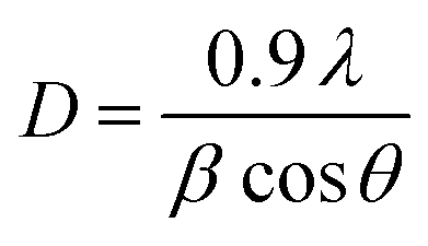
where, L is the load applied in g and d is the diagonal length of the indention (μm). The load applied was 50 g. Scanning electron microscopy (FE-SEM) (FEG-Quanta250, Czech Republic) was used to follow the surface morphology of the composite coatings. The crystalline structure of the plated substrate was identified by X-ray diffraction using a Bruker D8 advance X-ray diffractometer operated with Cu Kα radiation at 40 kV, 20 mA. The scan rate was 0.05 per step with a measured time of 15 per step. The crystallite size of the metal composites was determined using the Scherrer equation:| |
 | (2) |
where, D is the crystalline size, λ is the incident radiation (1.5418 Å), β is the corrected peak width at half-maximum intensity and θ is the angular position. The thickness of the coating was measured from cross-sectional FE-SEM images. Electrochemical polarization studies were carried out using an electrochemical analyzer (EG&G – Auto Lab Analyzer Model: 6310) containing a three electrode cell assembly. The exposed 1 cm2 area of mild steel substrate was used as the working electrode. A rectangular platinum foil and a saturated calomel electrode were used as the auxiliary and reference electrodes respectively. The test solution was 3.5% NaCl kept at 30 °C. Electrochemical impedance measurements were carried out on the electrodeposits after they had attained the steady state potential. The impedance measurements were made in the frequency range of 10 K cs−1 to 10 m cs−1 with a sinusoidal perturbation of 10 mV.19 The surface roughness of the coatings was assessed using a roughness measuring station (make: Mitutoyo, model: Surftest SJ-310). This instrument was used to analyze the average roughness value (Ra) with a measuring speed of 0.5 mm s−1 and a travelling distance of 5 mm. The average value of five readings is reported. Moreover, a TR-101-M4 scratch tester (make: DUCOM) was used to determine the coefficient of friction for the coatings. All the samples were run against 5 N normal loads at 0.2 ms−1speed. The measurement of the water repellency of the Ni–W–BN–PTFE nanocomposite coating was evaluated by determining the static contact angle of water droplets with the help of optical contact goniometry (OCA 35).
Table 1 Plating bath Composition and conditions
| Plating bath |
Composition |
Plating conditions |
| Nickel sulphate |
0.17 M |
Pulse peak c.d 1.2 A cm−2 |
| Sodium tungstate |
0.15 M |
pH = 8, time of 60 min |
| Tri ammonium citrate (TAC) |
0.30 M |
Temperature = 65 °C |
| Ammonium chloride |
0.20 M |
Constant stirring |
| Dimethyl sulphoxide |
0.06 M |
Pulse duty cycle |
| Cetyltrimethyl ammonium bromide (CTAB) |
0.50 g L−1 |
On time – 40 ms |
| Off time – 60 ms |
| BN (h) powder |
6.0 g L−1 |
|
| PTFE |
5–15 g L−1 |
|
3 Results and discussion
3.1. Effect of PTFE particle concentration on the Ni–W–BN nanocomposite deposition
The variation of the PTFE particle concentration in the optimized Ni–W–BN nanocomposite coating is shown in Fig. 1. As the PTFE particle concentration in the bath increases from 5 to 15 g L−1 in the fixed current density of 1.2 A dm−2 at 65 °C, the amount of particles co-deposited also increases. Actually, increasing the concentration of PTFE particles in the solution has produced a higher particle density (particles per liter) and created more opportunities for particle adsorption onto the electrode.20 The cationic surfactant (CTAB) and mechanical stirring were found to be effective in suspending the PTFE particles in the plating solution. For a given cationic surfactant concentration, the coating rate increased with increasing PTFE concentration in solution until it reached a maximum value at approximately 15 g L−1. This can be explained by the two-step adsorption model which has been suggested by Gugliemi.21 The cationic surfactant offers an adhesion force between the PTFE nanoparticles and the cathode resulting in an increase of co-deposited PTFE inert particles. Besides, the cationic surfactant also develops a net positive charge on its surface, which increases its affinity towards the cathode and hence increases the stability of the particle suspension and prevents agglomeration. Like this, it is understood that a cationic surfactant enhances the incorporation of particles in a metal matrix. The zeta potential of the nanoparticles would be increased by the addition of the cationic surfactant CTAB. The positive zeta potential values provide stronger adhesion forces between the inert particles and the cathode. Increasing the concentration of the PTFE nanoparticles (5 g L−1 to 15 g L−1) in the bath enhanced the adsorption rate of PTFE on the cathode surface. A further increase in the concentration (above 15 g L−1) of the PTFE particles in the electrolytic bath did not show any improvement in the adsorption on the cathode surface. Above 15 g L−1 of PTFE particles, the bath solution became saturated and the agglomeration of particles took place. Hence, 15 g L−1 of PTFE particles was taken as the optimum concentration in the electrolytic bath. The PC deposition had more particle incorporation than the DC plating due to the higher nucleation rate in the PC deposition.
 |
| | Fig. 1 Effect of the amount of PTFE in the bath (grams per litre) on the weight percentage of PTFE in the nanocomposite coatings. | |
3.2. Effect of current density on coating thickness
The effect of current density on the Ni–W–BN–PTFE nanocomposite coating thickness is illustrated in Fig. 2. The coating thickness increased with increasing current density up to 1.2 A dm−2 and then it decreased gradually. This may be due to the fact that a current density increased beyond 1.2 A cm−2 results in a more rapid deposition of the nanocomposite coating and a smaller amount of particles are then incorporated in the coating. With increments of current density beyond 1.2 A cm−2, the deposited particles become agglomerated, causing a decrease in coating thickness and an increase in the surface roughness. The coating thickness was slightly higher in PC deposition than DC deposition due to the smaller amount of H2 evolution, the homogeneous distribution of particles and the higher particle incorporation, which have been observed in pulse deposition. In order to create a better understanding of the effect of current density on the electrodeposition of the Ni–W–BN–PTFE nanocomposite coating, the thickness of the coating was determined using cross-sectional SEM images. Fig. 3(a & b) display the typical images from the cross-sectional FE-SEM view of the coatings obtained at a current density of 1.2 A cm2 for the Ni–W–BN–PTFE (DC & PC) nanocomposite coatings. The thickness of the coating was 27 μm and 36 μm for DC and PC deposition respectively.
 |
| | Fig. 2 Effect of current density on coating thickness. | |
 |
| | Fig. 3 Cross sectional FE-SEM images of (a) Ni–W–BN–PTFE (DC) and (b) Ni–W–BN–PTFE (PC) nanocomposite coatings. | |
3.3. XRD measurements
The XRD patterns of the Ni–W–BN and Ni–W–BN–PTFE (DC & PC) nanocomposite coatings are shown in Fig. 4(a–c). The crystalline size of the nanocomposite coatings was calculated from the Debye–Scherrer’s equation. The Ni–W alloy matrix has a face centered crystalline structure. The peaks at 44° and 65° were confirmed from standard XRD reports for Ni–W (JCPDS 65-4828). The grain sizes of the electrodeposited Ni–W–BN and Ni–W–BN–PTFE (DC & PC) nanocomposite coatings were found to be 26 nm, 20 nm and 14 nm respectively. After the incorporation of PTFE particles in the Ni–W–BN nanocomposite coating, the grain size was reduced. This was due to the grain refinement of the PTFE particles and changes in the microstructure of the composite coating. Moreover, the cationic surfactant (CTAB) is one of the most important organic molecules and it has been widely used as a stabilizer and structure-directing agent to control the nucleation, growth and alignment of crystals. The pulse current deposits have a smaller grain size than the direct current electrodeposits. This decrease in the grain size is due to the evenly distributed PTFE particles and it produces crack free deposits. Fig. 4(a–c) show a peak at an angle of 26.8° (JCPDS 85-1068), confirming the incorporation of BN particles. When compared to Fig. 4(a) much less intense BN peaks are seen in Fig. 4(b & c) due to the inclusion of the PTFE particles reducing the peak intensity of the BN particles. According to (JCPDS 47-2217), Fig. 4(b & c) show the peak for PTFE at an angle of 18° in the Ni–W–BN–PTFE nanocomposite coatings.22 The crystalline structure of Ni–W–BN is not affected due to the maintenance of the homogeneity of the bath. The PTFE particles are completely mixed with the bath solution.
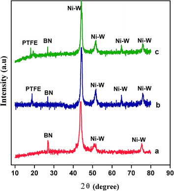 |
| | Fig. 4 XRD patterns for the (a) Ni–W–BN, (b) Ni–W–BN–PTFE (DC) nanocomposite and (c) Ni–W–BN–PTFE (PC) nanocomposite coatings. | |
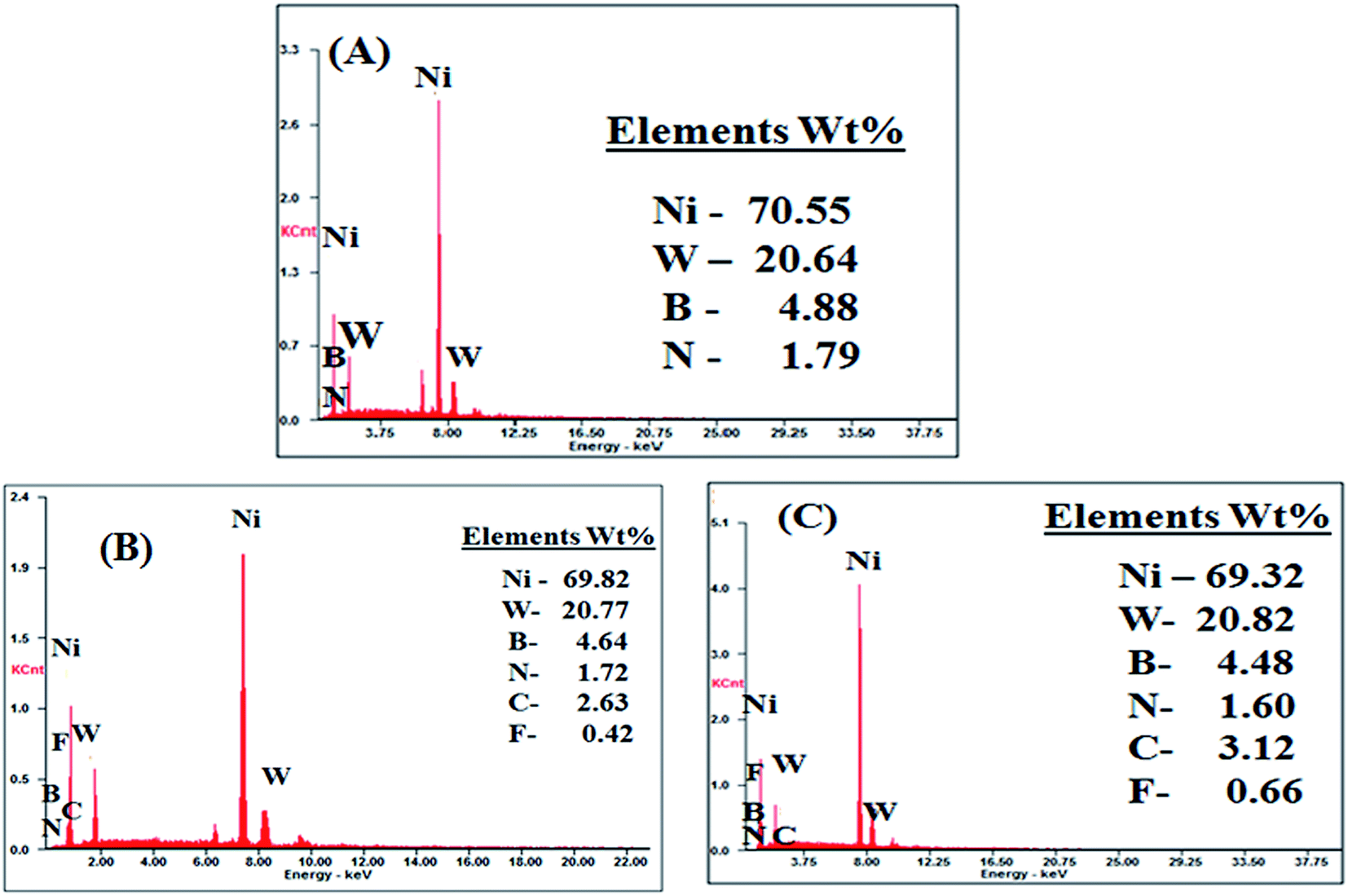
3.4. SEM and EDAX studies
The SEM morphologies of the electrodeposited Ni–W–BN and Ni–W–BN–PTFE (DC & PC) nanocomposite coatings are shown in Fig. 5(a–c). As shown in Fig. 5, the Ni–W–BN nanocomposite coating has long needle shaped deposits. On introducing the PTFE polymer into the deposits, the surface morphology was seen to be covered with distorted spherical particles. The co-deposited PTFE nano-particulates are much more uniformly distributed in the Ni–W–BN–PTFE nanocomposite coating. The deposits obtained from the PC method show bright, smooth and smaller grains compared to the deposits obtained from the DC method. Moreover, the size mentioned from the XRD studies was the same as the size obtained from the FE-SEM results (Fig. 6(a)). The agglomerated Ni–W–BN–PTFE nanocomposite coating is shown in Fig. 6(b). EDAX analysis confirmed the successful electrodeposition of PTFE on the Ni–W–BN nanocomposite coating. The elements Ni, W, B, N, C and F can be precisely detected as shown in Fig. 7. The EDAX analysis gives the % of elements present in the Ni–W–BN–PTFE nanocomposite coatings. When compared to the DC deposition more particles are embedded from the PC deposition. This is because, during DC deposition there is only one diffusion layer, but for PC deposition two diffusion layers are formed due to the pulse. In addition, the longer Toff promotes the transfer of more particles near the cathode and consequently a higher number of particles are incorporated. During the ON time (40 ms pulse), the nucleation and growth of the metal nanoparticles takes place. But the OFF time (60 ms pulse) (zero current or close to the equilibrium potential) allows for compensation of the depletion of the metal ions around the electrode and prevents the overlapping of diffusion zones. Hence, the particle incorporation is higher in the PC method than the DC method. During the Ni–W alloy deposition NiWO4 is formed, which reacts with citrate to form a [(WO4)(Cit)(H)x]x−5 complex. After that, metallic tungsten is formed from the tungstate complex.19 The electrochemical reduction of the WO42− ions occurs as follows:| | |
WO42− + 8H+ + 6e− → W + 4H2O
| (3) |
 |
| | Fig. 5 FE-SEM images of the (a) Ni–W–BN (h), (b) Ni–W–BN–PTFE (DC) nanocomposite and (c) Ni–W–BN–PTFE (PC) nanocomposite coatings. | |
 |
| | Fig. 6 (A) Crystal size of the (a) Ni–W–BN (h), (b) Ni–W–BN–PTFE (DC) nanocomposite and (c) Ni–W–BN–PTFE (PC) nanocomposite coatings. (B) FE-SEM images of the agglomerated Ni–W–BN–PTFE nanocomposite coating. | |
 |
| | Fig. 7 EDAX spectra of the (a) Ni–W–BN (h), (b) Ni–W–BN–PTFE (DC) nanocomposite and (c) Ni–W–BN–PTFE (PC) nanocomposite coatings. | |
The composite electrodeposition mechanism was proposed by Guglielmi’s model. According to this model, the composite electrodeposition procedure proceeds by two steps. The first step is the loose adsorption of particles adhered to the cathode surface by van der Waals forces with a high degree of surface coverage which can be represented by a Langmuir adsorption isotherm. In the second step, the particles are strongly adsorbed onto the surface by coulombic forces under the effect of an applied electric field and are incorporated into the growing metal matrix.
3.5. Tribological study
3.5.1. The effect of hardness. The microhardness profiles of the Ni–W–BN and Ni–W–BN–PTFE coatings are depicted in Fig. 8. The microhardness values were measured for varying concentrations of PTFE in the optimized Ni–W–BN nanocomposite bath. The microhardness value of the Ni–W–BN coating is ∼566 HV. After the inclusion of the PTFE particles, the microhardness value increased gradually. The Ni–W alloy matrix can provide effective protection for the two solid lubricants BN & PTFE which is helpful to improve the coating durability in harsh environments.23 An increase in the microhardness values is due to the dispersion-strengthening and grain refining of the composite particles.24 This strengthening effect is owed to the homogeneous distribution of nanoparticles in the nanocomposite coating.25 Here, the synergistic effect between the PTFE polymer and the Ni–W–BN nanocomposite has enhanced the microhardness value. The microhardness value mainly depends on the co-deposited filler particles. If the co-deposited particles are hard materials like SiC, Al2O3 etc. then the microhardness values increase rapidly. But, if the selection of filler particles is soft materials like h-BN, PTFE and MoS2 then moderate microhardness values are achieved. The PC deposition of the PTFE particles onto the Ni–W–BN nanocomposite coating significantly increased the microhardness values compared to DC deposition due to the fine grain size and compact nature of the coating. During the PC deposition, the higher pulse frequency could make a higher overpotential, providing additional energy for the adsorption of inert particles such as the PTFE polymer. The increase in the microhardness value is also due to the dispersion strengthening effect of second phase particles in the composite coatings which blocks the dislocation motion. Besides, the pulse current coating had a finer grain size, which reduced the porosity and improved the compact structure. The Ni–W alloy has a fine grained fcc crystalline structure. The particles are largely close together and arranged in order. This makes for a uniform and pore free deposition, which enhances the microhardness. In addition, the presence of the multicomponents in the Ni–W–BN–PTFE nanocomposite coating is responsible for the improved microhardness.
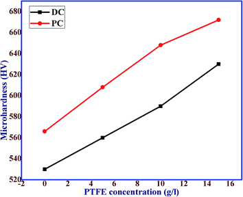 |
| | Fig. 8 Effect of the amount of co-deposited PTFE on the microhardness of the Ni–W–BN–PTFE nanocomposite coatings. | |
3.5.2. Effect of surface roughness. The surface roughness measurements on the electrodeposited coatings were repeated three times at various locations and the average values were recorded. Fig. 9 shows the typical roughness profile of the (a) Ni–W–BN and (b) Ni–W–BN–PTFE nanocomposite coatings. As shown in Fig. 9 the average surface roughness (Ra) value of the Ni–W–BN nanocomposite coating was 0.41 μm. After the inclusion of the PTFE particles (optimized concentration of 15 g L−1) in the Ni–W–BN nanocomposite coating, the average surface roughness (Ra) value was reduced to 0.26 μm. It was observed that the surface roughness values decreased with the addition of PTFE particles in the coatings. Thus, the two solid lubricant fillers play a vital role in reducing the surface roughness. The decrease in roughness is probably due to the dense coating and self lubricating property of the PTFE particles. The PC coatings have lower surface roughness values than the DC coatings due to the uniform and compact surface. The pulse deposited coatings are accomplished with greater cathodic current densities than the DC coatings. It is recognized that a lower overpotential coating generates a deposition with larger surface irregularities on the surface of the cathode; while a higher overpotential deposits generate coatings with smooth surfaces. Hence, the average surface roughness value of the pulse current deposition for the Ni–W–BN–PTFE nanocomposite coating was reduced.
 |
| | Fig. 9 Surface roughness profile of (a) Ni–W–BN (h) and (b) Ni–W–BN–PTFE nanocomposite coatings. | |
3.5.3. Effect of the coefficient of friction. Fig. 10 illustrates the effect of varying amounts of PTFE particle incorporation into the Ni–W–BN nanocomposite coatings on the coefficient of friction. Averages of five readings were noted. The friction coefficient value of the Ni–W–BN nanocomposite coating was found to be 0.28. In order to further decrease the friction coefficient value PTFE was added as the second solid lubricant filler. The inclusion of more solid lubricant has the promising effect of decreasing the friction coefficient.26 Increasing the amount of PTFE in the deposit has led to a decrease in the coefficient of friction. PTFE is a typical solid lubricant and when applied to a metal surface, it imparts non-stick properties and lowers its coefficient of friction. The average coefficient of friction for the Ni–W–BN–PTFE nanocomposite coating had a lowest value in the range of 0.15 indicating excellent friction reduction performance. The decreasing value of the coefficient of friction of the coating is due to the inclusion of two solid lubricant particles such as BN and PTFE. One of the most effective properties of organic coatings is the good adhesion to the substrate. This property of adhesion strength is of great importance to the reduction of the friction coefficient. Besides, rough surfaces usually have lower wear resistance and higher friction coefficients than smooth surfaces. PTFE incorporated into the optimized Ni–W–BN nanocomposite coating reduced the surface roughness value of the coating. Hence, PTFE was an efficient organic filler to reduce the friction. Some interaction between the Ni–W–BN nanocomposite coating and the solid lubricant PTFE polymer reduced the friction coefficient. Based on these results, a lower coefficient of friction in association with the use of the Ni–W–BN–PTFE nanocomposite coating was attained. As we discussed before, using PC deposition increases the surface uniformity and hence, reduces the coefficient of friction significantly compared to DC deposition. This is consistent with the microhardness results as shown in Fig. 8.
 |
| | Fig. 10 Plot of Ni–W–BN and various concentrations of PTFE inclusion to the Ni–W–BN–PTFE nanocomposite coatings versus the friction coefficient. | |
3.6. Electrochemical characterization
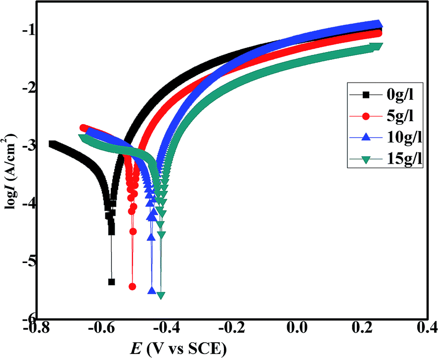
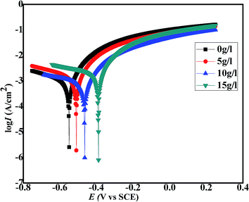
3.6.1. Potentiodynamic polarization study. The detailed corrosion potentials and corrosion current densities of the samples were obtained in 3.5% NaCl solution by electrochemical methods. Fig. 11 signifies the polarization curves obtained for various DC deposits by the potentiodynamic polarization method. The values for corrosion resistance parameters such as corrosion potential (Ecorr) and corrosion current (Icorr) are listed in Table 2. The inclusion of the PTFE particles in the optimized Ni–W–BN nanocomposite coatings has shifted the corrosion potential values positively from −0.561 V to −0.417 V and the corrosion current density values have significantly decreased from 4.3 μA cm−2 to 2.4 μA cm−2.27 This positive shift in the corrosion potential proves the good passivation property of the PTFE polymer. The potential shift towards a more positive value indicates the creation of a more protective passivation layer on the surface of the cathode representing more corrosion resistance. The largest potential shift was seen for 15 g L−1 of PTFE particle inclusion on the optimized Ni–W–BN nanocomposite coating. The positive shift in the corrosion potential demonstrates the better protection of the mild steel substrate against corrosion by the self lubricating nature of the polymer. These statements are evidence of the chemically inert character of the PTFE polymer. When PTFE was added as a fourth component in the Ni–W–BN nanocomposite system a protective oxide layer was formed on the surface of the cathode. The PTFE particles also minimize the defects which act as the active sites for corrosion. On the other hand, the uniformly distributed PTFE particles in the deposit behave as a passive layer between the corrosive media and the deposit surface. Fig. 5 shows that it may be pore free composite surface. Therefore, it has been shown that the presence of PTFE increases the corrosion resistance properties of the nanocomposite coatings. PTFE also behaves as an efficient barrier against corrosive ions and this self lubricating polymer acted as a binder between the substrate and the nanoparticles. The addition of the PTFE particles to the PC deposits results in a lower corrosion current than for the deposits obtained from the DC method (Fig. 12 and Table 3) because the PC method provides smaller crystallites and grain boundaries. Deposits with a smaller grain size have a higher density of nucleation sites for passive films, which leads to a higher fraction of the passive layer and thus a lower passive current density.28 Hence, smaller grain coatings offer enhanced corrosion resistance.29
 |
| | Fig. 11 Potentiodynamic polarization curves for the Ni–W–BN (h) deposit and various amounts of PTFE incorporated into the Ni–W–BN (h) nanocomposite by the DC method. | |
Table 2 Parameters derived from the potentiodynamic polarization curves for the Ni–W–BN and Ni–W–BN–PTFE nanocomposite coatings prepared by the DC method
| PTFE concentration (g L−1) |
Ecorr vs. SCE (mV) |
Icorr (μA cm−2) |
| 0 |
−561 |
4.30 |
| 5 |
−502 |
3.66 |
| 10 |
−459 |
2.81 |
| 15 |
−417 |
2.47 |
 |
| | Fig. 12 Potentiodynamic polarization curves for the Ni–W–BN (h) deposit and various amounts of PTFE incorporated into the Ni–W–BN (h) nanocomposite by the PC method. | |
Table 3 Parameters derived from the potentiodynamic polarization curves for the Ni–W–BN and Ni–W–BN–PTFE nanocomposite coatings prepared by the PC method
| PTFE concentration (g L−1) |
Ecorr vs. SCE (mV) |
Icorr (μA cm−2) |
| 0 |
−548 |
2.54 |
| 5 |
−507 |
1.71 |
| 10 |
−438 |
0.97 |
| 15 |
−386 |
0.75 |
3.6.2. Electrochemical impedance study (EIS). Fig. 13 and Table 4 represent the Nyquist plots for Ni–W–BN and the addition of various amounts of PTFE particles obtained by the DC method. The charge transfer resistance (Rct) values have increased and the double layer capacitance (Cdl) values have decreased with the inclusion of the PTFE particles in the deposit. This indicates that they are influenced by the state of the electrode/solution interface and the real contact area between the electrode and the solution.27 The lower Cdl values result from a reduction in the dielectric constant and/or an increase in the thickness of the electrical double layer. Moreover, the double layer capacitance value is related to the porosity of the coating. The smaller value of double layer capacitance confirmed the less porous nature, which enhances the anticorrosion properties, of the deposits. After the inclusion of the PTFE polymer in the Ni–W–BN nanocomposite coating, the thickness of the coating also increased. The increment of the coating thickness prevented the expansion of the corrosion cavities and increased the anticorrosion properties of the coating. The Nyquist plots obtained for the Ni–W–BN deposits and the addition of various amounts of PTFE particles prepared using the PC method are shown in Fig. 14(a) and Table 5. The proposed equivalent circuit to examine the impedance spectra is shown in Fig. 14(b). As it was noted in the previous sections, a smaller grain size has a higher fraction of the passive layer and a lower corrosion rate due to the high density of nucleation sites for the passive film. Hence, the PC coating was more corrosion resistant than the electrodeposited DC coating.30 All the Ni–W–BN–PTFE nanocomposite coatings have shown higher charge transfer resistance and lower double layer capacitance values than the Ni–W–BN nanocomposite coating. The incorporation of PTFE lowers the chemical activity and will help to reduce the pore size in the nanocomposite coating and prevent the corrosive pits from growing. These data clearly indicate that the chemically inert nature of the PTFE nanoparticles was not affected by a corrosive environment. On the other hand, the hydrophobic character of the PTFE particles hinders the anodic reaction and decreases the corrosion rate. In other words, Ni–W–BN–PTFE nanocomposite coatings have a lower chemical activity and better chemical stability than the Ni–W–BN deposits. The nanocomposite coatings employed in this current study contain four different particles and their combined properties are efficient for the protection of a mild steel surface against corrosion.
 |
| | Fig. 13 Nyquist plots (Z′′vs. Z′) obtained for the Ni–W–BN (h) deposit and various amounts of PTFE incorporated into the Ni–W–BN (h) nanocomposite by the DC method. | |
Table 4 Parameters derived from the electrochemical impedance spectra of the Ni–W–BN and Ni–W–BN–PTFE nanocomposite coatings prepared by the DC method
| PTFE concentration (g L−1) |
Rct (Ω cm2) |
Cdl (μF cm−2) |
| 0 |
616 |
51.70 |
| 5 |
1025 |
31.24 |
| 10 |
1122 |
28.27 |
| 15 |
1314 |
24.33 |
 |
| | Fig. 14 (a) Nyquist plots (Z′′vs. Z′) obtained for the Ni–W–BN (h) deposit and various amounts of PTFE incorporated into the Ni–W–BN (h) nanocomposite by the PC method (b). Equivalent electrical circuit. | |
Table 5 Parameters derived from the electrochemical impedance spectra of the Ni–W–BN and Ni–W–BN–PTFE nanocomposite coatings prepared by the PC method
| PTFE concentration (g L−1) |
Rct (Ω cm2) |
Cdl (μF cm−2) |
| 0 |
1116 |
28.53 |
| 5 |
1250 |
25.23 |
| 10 |
1450 |
21.51 |
| 15 |
1643 |
19.23 |
3.7. Contact angle measurements
To understand the water-repellency of the Ni–W–BN nanocomposite coating and the addition of various concentrations of PTFE particles obtained by PC deposition, the contact angle determination for a water droplet was carried out. Contact angle measurement is a simple quantitative method to analyze the wettability/hydrophobic nature of a solid surface. Surfaces with contact angles of larger than 90° are said to be hydrophobic. The volume of the water droplet was 8 μL on both the Ni–W–BN and Ni–W–BN–PTFE nanocomposite coatings, which are shown in Fig. 15. It was found that the water repellency/hydrophobicity increased noticeably. When the weight percentage of the PTFE concentration was increased the hydrophobic nature also increased. This hydrophobic behaviour is helpful for water removal and makes it difficult for water to adhere to the surface. The surface irregularities were also reduced. This was due to the well dispersed PTFE particles in the bath from ultrasonication and constant stirring. Hence, when the weight percentage of PTFE was increased the water contact angle also increased. The contact angle for the Ni–W–BN nanocomposite coating (A) was about 115°, while that of the PTFE (15 g L−1) polymer incorporated Ni–W–BN–PTFE nanocomposite coating (B) was about 136°, which shows a better water-repellency/hydrophobic nature. This means that the incorporation of the self lubricating PTFE polymer improves the water repellency property of the Ni–W–BN–PTFE nanocomposite coating. This water repellency behaviour of the PTFE polymer was clearly indicated by the hydrophobic nature of the Ni–W–BN–PTFE nanocomposite coating. This hydrophobic behaviour was helpful for water removal and makes it difficult for water adherence which improves the corrosion resistance properties of the nanocomposite coating.
 |
| | Fig. 15 Contact angle measurements of a water droplet (8 μL) on the (a) Ni–W–BN, (b) Ni–W–BN–PTFE (5 g L−1), (c) Ni–W–BN–PTFE (10 g L−1) and (d) Ni–W–BN–PTFE (15 g L−1) nanocomposite coatings. | |
4 Conclusions
The electrodeposition of Ni–W–BN–PTFE nanocomposite coatings by direct and pulse current methods on a mild steel substrate was successfully employed using a watts’ type nickel bath. The cationic surfactant CTAB was used to suspend the PTFE particles well in the bath solution. The inclusion of the PTFE particles in the optimized concentration of the Ni–W–BN nanocomposite coatings significantly improved the microhardness, thickness, corrosion resistance, gave excellent friction reduction and decreased the surface roughness of the nanocomposite coatings. The chemically inert and hydrophobic nature of the PTFE particles decreased the corrosion rate. The solid lubricating nature of the PTFE particles greatly influenced the friction reduction and surface roughness. Contact angle measurements of the Ni–W–BN–PTFE nanocomposite coatings showed a hydrophobic behaviour with CA values of about 136°. The PC deposition of the Ni–W–BN–PTFE nanocomposite coating exhibits better performance than the deposits obtained from the direct current nanocomposite coating due to the uniform, crack free deposits and also the smaller crystalline size.
Acknowledgements
The authors thank the Head of the physics department, Alagappa University, Karaikudi for providing the XRD analysis to carry out this research work and also acknowledge the school of chemistry, Alagappa University, Karaikudi for FE-SEM analysis.
References
- H. Mojtaba, S. H. Mirdamadi and H. R. Rezaie, Electrochim. Acta, 2014, 138, 224–231 CrossRef PubMed.
- M. F. Cardinal, P. A. Castro, J. Baxi, H. Liang and F. H. Williams, Surf. Coat. Technol., 2009, 204, 85–90 CrossRef CAS PubMed.
- E. Pompei, L. Magagnin, N. Lecis and P. L. Cavallotti, Electrochim. Acta, 2009, 54, 2571–2574 CrossRef CAS PubMed.
- Z. Shahri and S. R. Allahkaram, Trans. Nonferrous Met. Soc. China, 2013, 23, 2929–2938 CrossRef CAS.
- Z. Shari, S. R. Allahkaram and A. Zarebidaki, Appl. Surf. Sci., 2013, 276, 174–181 CrossRef PubMed.
- P. Bercot, E. P. Munoz and J. Pagetti, Surf. Coat. Technol., 2002, 157, 282–289 CrossRef CAS.
- W. Feng, A. Susumu and E. Morinobu, Mater. Trans., 2004, 45, 1311–1316 CrossRef.
- S. Kasturibai and G. P. Kalaignan, Bull. Mater. Sci., 2014, 379, 1–87 Search PubMed.
- Y. S. Huang, X. T. Zeng, X. F. Hu and F. M. Liu, Surf. Coat. Technol., 2005, 198, 173–177 CrossRef CAS PubMed.
- K. S. Nabeen, M. Masabumi and S. Tetsuo, Wear, 2003, 254, 555–564 CrossRef.
- E. A. Pavlatou, M. Stroumbouli, P. Gyftou and N. Spyrellis, J. Appl. Electrochem., 2006, 36, 385–394 CrossRef CAS.
- J. R. Vail, D. L. Burris and W. G. Sawyer, Wear, 2009, 267, 619–624 CrossRef CAS PubMed.
- Z. Lifang, W. Fuguo, Q. Li, G. Kaixiong, Z. Bin and Z. Junyan, RSC Adv., 2015, 5, 9635 RSC.
- Q. Zhao, Y. Liu, H. M. Steinhagen and G. Liu, Surf. Coat. Technol., 2002, 155, 279–284 CrossRef CAS.
- W. G. Sawyer, K. D. Freudenberg, P. Bhimaraj and L. S. Schadler, Wear, 2003, 254, 573–580 CrossRef CAS.
- S. Beckford, Y. A. Wang and M. Zou, Tribol. Trans., 2011, 54, 849–858 CrossRef CAS PubMed.
- W. Zhijiang, W. Lina, Q. Yulin, C. Wei and J. Zhaohua, Surf. Coat. Technol., 2010, 204, 3315–3318 CrossRef PubMed.
- N. L. McCook, B. Boesl, D. L. Burris and W. G. Sawyer, Tribol. Lett., 2006, 22, 253–257 CrossRef CAS.
- K. A. Kumar, G. P. Kalaignan and V. S. Muralidharan, Appl. Surf. Sci., 2012, 259, 231–237 CrossRef PubMed.
- Z. Shahri and S. R. Allahkaram, Iran. J. Mater. Sci. Eng., 2012, 9, 1–7 CAS.
- N. Gugliemi, Kinetics of the deposition of inert particles, from electrolytic baths, J. Electrochem. Soc., 1972, 1009–1012 CrossRef PubMed.
- H. Y. Yi, D. G. Chang, L. W. Xiu and P. T. Jiang, Int. J. Electrochem. Sci., 2012, 7, 12440–12455 Search PubMed.
- M. Guozheng, X. Binshi, W. Haidou, W. Xiaohe, L. Guolu and Z. Sen, Surf. Coat. Technol., 2013, 221, 142–149 CrossRef PubMed.
- L. Ruiqian, C. Qingwei and L. Jun, RSC Adv., 2015, 5, 44933 RSC.
- A. Abdel Aal, M. Bahgat and M. Radwan, Surf. Coat. Technol., 2006, 201, 2910–2918 CrossRef CAS PubMed.
- M. Zouari, M. Kharrat, M. Dammaka and M. Barletta, Prog. Org. Coat., 2014, 77, 1408–1417 CrossRef CAS PubMed.
- J. Li, J. Zhen, S. Yi, W. Panpan, P. Qiujie, W. Guoying and G. Hongliang, Int. J. Electrochem. Sci., 2014, 9, 1715–1728 Search PubMed.
- Y. Q. Li, S. L. Jian and J. Qing, Trans. Nonferrous Met. Soc. China, 2010, 20, 82–89 CrossRef.
- K. A. Kumar, P. Mohan, G. P. Kalaignan and V. S. Muralidharan, J. Nanosci. Nanotechnol., 2012, 12, 8364–8371 CrossRef CAS PubMed.
- S. Sangeetha and G. P. Kalaignan, Ceram. Int., 2015, 41, 10415–10424 CrossRef CAS PubMed.
|
| This journal is © The Royal Society of Chemistry 2015 |
Click here to see how this site uses Cookies. View our privacy policy here.