Distinguishing the serum metabolite profiles differences in breast cancer by gas chromatography mass spectrometry and random forest method

Jian-Hua Huanga, Liang Fub, Bin Lia, Hua-Lin Xie*b, Xiaojuan Zhanga, Yanjiao Chena, Yuhui Qina, Yuhong Wanga, Shuihan Zhanga, Huiyong Huanga, Duanfang Liaoa and Wei Wang*a
aTCM and Ethnomedicine Innovation & Development Laboratory, Sino-Luxemburg TCM Research Center, School of Pharmacy, Hunan University of Chinese Medicine, Changsha, 410208, P. R. China. E-mail: wangwei402@hotmail.com; Fax: +86731-8845-8227; Tel: +86731-8845-8240
bCollege of Chemistry and Chemical Engineering, Yangtze Normal University, Chongqing, 408100, China. E-mail: hualinxie@163.com

Received 28th May 2015 , Accepted 29th June 2015

First published on 29th June 2015


Abstract

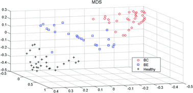
In this study, we proposed a metabolomics strategy to distinguish different metabolic characters of healthy controls, breast benign (BE) patients, and breast malignant (BC) patients by using the GC-MS and random forest method (RF). In the current study, the serum samples from healthy controls, BE patients, and BC patients were characterized by using GC-MS. Then, random forest (RF) models were established to visually discriminate the differences among three groups' metabolites profiles, and further investigate the progress of breast cancer from benign to malignant in patients based on these GC-MS profiles. We successfully discovered the differences between the healthy and breast cancer patients. And the metabolic changes from benign to malignant cancer were obviously visualized. The results suggested that combining GC-MS profiling with random forest method is a useful approach to analyze metabolites and to screen the potential biomarkers for exploring the serum metabolic profiles of breast cancer.


1. Introduction

Breast cancer is the most prevalent malignant disease in women worldwide and a major cause of female death.1 In clinical research, breast cancer is a prognoses disease in which larger tumor size and the presence of lymph node metastasis are associated with worse prognoses.2 The earlier diagnosis is one of the most important strategies to reduce breast cancer morbidity rate and improve the survival rate.2,3 In terms of diagnosis of breast cancer, some current methods can accurately differentiate malignant from normal and benign tissue by identification of malignant tissue characteristic.4,5 Routine breast cancer inspection methods including periodic mammography, physical examination, and blood tests.6 Mammography always misses small tumor that will lead to false positives, resulting in suboptimal sensitivity and specificity and unnecessary biopsies. Furthermore, these conventional BC diagnostics techniques are always expensive and time-consuming; furthermore some patients may feel discomfort during diagnosis process.

Metabolomics is an important platform for quantitative analysis of the metabolites in living systems and their dynamic responses to the changes of both endogenous and exogenous factors by using all kinds of analytical approaches, including gas chromatography-mass spectrometry GC-MS,7–10 high-resolution nuclear magnetic (NMR),11–14 ultra-performance liquid chromatography-mass spectrometry (UPLC-MS).15 Recently, the metabolomics methods were widely used to monitor disease progression, and showed its advantages in various researches, such as diagnosis of human diseases,16 physiological evaluations,17 elucidation of biomarkers,18,19 and drug toxicity.20 The transforming process from normal to malignant cells is always associated with some metabolic disturbances. Therefore, using the metabolomics method for breast cancer research is very suitable. Some previous researches have demonstrated that some volatile organic metabolites could indicate the differences between breast cancer patients and healthy controls,6,21 and some other researchers have reported the serum concentrations of free fatty acids (FFAs) in patients with BC were significantly decreased compared with those in healthy controls.22,23 These researches indicated that using GC-MS metabolites profiles can help breast cancer diagnosis. Besides, GC-MS analysis method has some advantages such as, favorable stability, reproducibility, and sensitivity, and rapid analysis.

Owning to the complexity of these metabolic profiles, multivariate statistical methods are extensively used to deal with these ‘Omics’ data. Principal component analysis (PCA) and partial least squares discriminant analysis (PLS-DA) were the most often used method to visually represent the data information, and some other machine learning methods were applied in the researches more and more frequent.24,25 Random forest (RF) model, one of these machine learning methods, has its own characteristic advantages on dealing with complex metabolomics data. This algorithm has showed its advantages in dealing with these complex metabolomics data, not only distinguish different groups (patients and healthy), but also can help finding the significant changes of metabolites as a potential biomarker, as showed in our previous researches.26–28

Based on these reasons, we established a metabolomics strategy to distinguish different metabolic characters of healthy controls, breast benign patients, and breast malignant patients by using the GC-MS and random forest (RF). The whole experiment contains several steps: firstly, the serum samples from healthy, breast benign patients, and breast malignant patients were profiled by using GC-MS analytical technique; after being pretreated, metabolites information was processed by using RF method; finally, RF model can calculate the sample proximity matrix, by using this sample proximity matrix, not only the differences between the healthy and breast cancer patients were observed, but also the differences between breast benign patients and breast malignant patients were obviously visualized. And some informative metabolites or potential biomarkers have been successfully discovered by means of variable importance ranking in random forest program.

2. Materials and methods

2.1 Samples collection

23 breast benign patients and 30 breast malignant patients were collected from The Tumor Hospital of ChangDe City, Hunan Province. These patients were treated in this hospital, and were diagnosed by the standard methodologies.29 30 healthy controls (who were negative for breast cancer by mammography and ultrasound examination) were selected from volunteers. Total of 83 samples (30 healthy, 23 breast benign patients, and 30 breast malignant patients) were tested in current study. The protocol in this study was approved by the Ethics Committee at The Tumor Hospital of ChangDe City.

2.2 Serum collection and preparation

2 mL of venous blood samples were collected in a blank tube from each individual at 8 o'clock in the morning after overnight fasting. After obtained the serum samples, the samples were kept in −80 °C until analysis. Serum was thawed at 4 °C for 30 minutes. To the 100 μL serum samples, 350 μL methanol (including 1 mg mL−1 of heptadecanoic acid/methanol as internal standard) was added and vortexed for 15 s, and centrifuged for 15 min (15[thin space (1/6-em)]000 rpm, 4 °C). Supernatant was dried by using N2. Then, the supernatant was derivatized by adding methoxyamine/pyridine (20 mg mL−1) mixed for 15 s, and incubated for 1 h (65 °C), followed by addition of 100 μL BSTFA. All the samples were analyzed by using GC-MS at random order after being prepared by described procedure.

2.3 Equipment and reagents

Data was acquired by GC-MS using Agilent 7890A gas chromatography instrument coupled to a 5975C mass spectrometer (Agilent, Santa Clara, California, USA). Methanol (CH3OH) was purchased from Tedia Company (Fairfield, USA). Analytical grade heptadecanoic acid (C17:0), methoxamine, pyridine and bis(trimethylsilyl)-trifluoroacetamide (BSTFA) were purchased from Sigma-Aldrich (St. Louis, MO, USA).

2.4 Gas chromatography-mass spectrometry conditions

GC separation was performed on Agilent DB-5MS equipped with a deactivated fused silica capillary column (0.25 mm × 30 m × 0.25 μm). The oven temperature was maintained at 70 °C for 4 min, programmed to 300 °C (rate of 8 °C min−1), and then held for 3 min. The injection volume of 1 μL was used in the split ratio of 1[thin space (1/6-em)]:[thin space (1/6-em)]10. Helium was used as the carrier gas (flow rate of 1.0 mL min−1). The mass spectrometer was performed using electron impact (EI) ionization source at 70 eV and a 0.90 kV detector voltage in 0.2 s per scan full scan. The mass spectrometer was operated with m/z range from 35 to 650. These analytical conditions were consistent to our previous researches.26

2.5 Principal component analysis (PCA)

Principal component analysis (PCA) was used in current study to exhibit the cluster trend of three groups' samples. The singular value decomposition (SVD) was used to transform raw variables into a set of linearly orthogonal project variables. These project variables contain the almost useful information in the raw signals. The noise signals contain in the raw signals can be eliminated during such decomposing process. We could obtain the Scores and loading by using the SVD. The Scores plot can be used to present the relationships among different samples. The loading values for each variable can be used to select the informative variables. The PCA program used in this study was written by MATLAB in our group.

2.6 Random forest

Random forest model was established by assembling enough classification and regression trees.30,31 The mains implements of RF are based on bagging and random feature selection strategy. The bagging method can ensemble enough tree model in the training process, and two types of datasets are established, the train dataset and “out of bag” dataset. The “out of bag” data also called OOB samples, which can be used to estimate the model precision. This OOB estimation has been proved to be unbiased. In each tree growing process, instead of using all the features to split at each tree node, RF selects only a small subset of features, which makes each tree in the forest is different from each other. Increasing the diversities of trees is an efficient way to increase the classifying and recognition ability of RF method. The detailed RF modeling process can be found in our previous studies.26

Here, two useful tools, the variable importance measure and proximity matrix, in the RF will be introduced, which have showed their advantages in the data interpretation and visualization. The variable importance measure can be used to estimate the importance of each metabolite in the model classification. This information can help us to find the potential biomarkers. In current study, ‘the mean decrease in classification’ measure was adopted. For each tree, the classification accuracy of the OOB samples is determined both with and without random permutation of the variable values one by one. The accuracy of permutation is subtracted from that before permutation, and then averaged over all trees in the forest (calculated as eqn (1)).

 
Importance of j = Accuracyj normal − Accuracyj permuted (1)

The other attractive feature in RF algorithm is the proximity matrix calculation. Proximity values can indicate the similarities among all the samples. In normal situation, samples from the same group always fall into the same or nearby tree node (this is the principle of tree method). In tree method, distance matrix was used to calculate the similarities of samples. In RF method, the proximity between two samples was calculated as the number of times the two samples fall into the same terminal node of a tree, and then divided by the number of trees in the forest.31 After the proximity values are calculated, multi-dimensional scaling (MDS) plot is always used to visualize these analysis results. MDS is a set of related statistical techniques often used to visually explore similarities or dissimilarities in data.32 We can project the first two or three scaling coordinates into low dimensions and obtain the clustering plot of all the samples.

3. Results and discussion

3.1 Data analysis

The typical total ion chromatograms (TICs) of serum metabolic profiles for healthy control (in blue line), BE (in black line), and BC (in red line) were shown in Fig. 1. As could be seen in Fig. 1, the serum metabolites profiles of three groups were similar, but the concentrations of some metabolites were differences. These results suggested that these GC-MS profiles could represent the differences among three groups.
image file: c5ra10130a-f1.tif
Fig. 1 The typical total ion chromatograms (TICs) of healthy control (in blue line), BE (in black line), and BC (in red line).

After these metabolic profiles were collected, qualitative and the quantitative work were carried out, mainly metabolites, including amino acid, organic acid, fatty acid, and carbohydrates were found in the chromatograms (detailed results were listed in Table 1). Then, these metabolites data were input to some pattern recognition algorithms for further analysis.

Table 1 Qualitative and quantitative metabolic profiles of three groups' samples
id trb (min) Endogenous metabolites BC group BE group Healthy
a Identified by standard substances.b Retention time.
1 5.922 Ethylbis(trimethylsilyl)amine 0.2456 ± 0.0705 0.1905 ± 0.0567 0.1958 ± 0.0551
2 6.593 Ethylene glycol 0.0182 ± 0.0020 0.0530 ± 0.0428 0.0746 ± 0.0626
3 6.608 N,N-Diethylacetamide 0.0120 ± 0.0060 0.0220 ± 0.0325 0.0227 ± 0.0302
4 6.84 N,N-Diethyl-acetamide 0.0657 ± 0.0087 0.0476 ± 0.0202 0.0557 ± 0.0107
5 7.716 Lactic acida 0.0872 ± 0.0374 0.0952 ± 0.0592 0.1482 ± 0.2155
6 7.934 Acetic acid 0.0629 ± 0.0140 0.0856 ± 0.0333 0.0412 ± 0.0403
7 10.01 Phosphate 3.1278 ± 1.0173 1.4730 ± 0.7381 1.3767 ± 1.0361
8 10.2 L-Threonine 0.0173 ± 0.0098 0.0108 ± 0.0068 0.0096 ± 0.0065
9 10.297 Acetic acid, phenyl- 0.0047 ± 0.0023 0.0159 ± 0.0133 0.0147 ± 0.0117
10 10.382 Succinic acida 0.0811 ± 0.0429 0.0098 ± 0.0131 0.0119 ± 0.0086
11 10.447 [1,2-Phenylenebis(oxy)]bis[trimethyl- 0.0120 ± 0.0072 0.0078 ± 0.0047 0.0067 ± 0.0039
12 10.503 Glyceric acid 0.0961 ± 0.0266 0.0400 ± 0.0282 0.0183 ± 0.0167
13 10.723 (R*,R*)-2,3-Dihydroxybutanoic acid 0.0267 ± 0.0053 0.0137 ± 0.0014 0.0053 ± 0.0029
14 11.357 2,4-Bis[(trimethylsilyl)oxy]-butanoic acid 0.0147 ± 0.0051 0.0055 ± 0.0030 0.0066 ± 0.0047
15 11.583 (R*,S*)-3,4-Dihydroxybutanoic acid 0.0304 ± 0.0098 0.0132 ± 0.0064 0.0178 ± 0.0107
16 11.797 N-(1-oxobutyl)-glycine 0.0653 ± 0.0244 0.0319 ± 0.0186 0.0274 ± 0.0151
17 12.341 Isovaleroglycine 0.0356 ± 0.0134 0.0160 ± 0.0079 0.0107 ± 0.0073
18 12.483 D-Threitol 0.0214 ± 0.0073 0.0290 ± 0.0130 0.0251 ± 0.0151
19 12.645 N-Crotonylglycine 0.0640 ± 0.0146 0.0207 ± 0.0129 0.0148 ± 0.0099
20 14.53 N-(1-oxohexyl)-glycine 0.0160 ± 0.0072 0.0121 ± 0.0073 0.0132 ± 0.0081
21 14.713 D-Xylose 0.0208 ± 0.0075 0.0082 ± 0.0044 0.0093 ± 0.0063
22 14.823, 15.057 D-Ribose 0.0126 ± 0.0070 0.0152 ± 0.0042 0.0250 ± 0.0110
23 15.509, 15.733 Arabitol 0.0487 ± 0.0364 0.0283 ± 0.0179 0.0278 ± 0.0215
24 16.023 D-Galactose, 6-deoxy-2,3,4,5-tetrakis-O-(trimethylsilyl)- 0.0336 ± 0.0083 0.0177 ± 0.0100 0.0149 ± 0.0104
25 16.087 Mannonic acid 0.0505 ± 0.0177 0.0211 ± 0.0143 0.0168 ± 0.0138
26 16.2 cis-Aconitic acida 0.0435 ± 0.0388 0.0105 ± 0.0079 0.0168 ± 0.0147
27 16.357 Phosphoric acid 0.0414 ± 0.0252 0.0230 ± 0.0141 0.0212 ± 0.0168
28 17.177 Isocitric acida 0.0464 ± 0.0121 0.0340 ± 0.0093 0.0448 ± 0.0838
29 17.563 Hippuric acid 0.0270 ± 0.0126 0.0180 ± 0.0104 0.0156 ± 0.0116
30 17.85, 17.96 D-Fructosea 0.0712 ± 0.0586 0.0471 ± 0.0145 0.0580 ± 0.1031
31 18.087 D-Galactosea 0.0796 ± 0.0214 0.0455 ± 0.0272 0.0389 ± 0.0287
32 18.197, 18.147 D-Glucosea 0.2785 ± 0.0918 0.1741 ± 0.7354 0.1859 ± 0.4136
33 18.507 Altronic acid 0.0202 ± 0.0069 0.0185 ± 0.0100 0.0102 ± 0.0074
34 18.577, 18.65 D-Sorbitola 0.0259 ± 0.0169 0.0254 ± 0.0187 0.0300 ± 0.0275
35 18.983, 19.533 Galactonic acid 0.1213 ± 0.0482 0.0817 ± 0.0328 0.0441 ± 0.0351
36 19.99 Palmitic acid 0.0127 ± 0.0017 0.0148 ± 0.0029 0.0071 ± 0.0025
37 20.403 Myo-inositol 0.0247 ± 0.0128 0.0197 ± 0.0037 0.0334 ± 0.0129
38 25.465 D-Turanose 0.0216 ± 0.0138 0.0197 ± 0.0190 0.0510 ± 0.1099
39 28.125 D-(+)-Lactose monohydratea 0.8475 ± 0.1366 1.0400 ± 0.3349 0.6559 ± 0.2286
40 29.927 Lactose 0.0142 ± 0.0043 0.0143 ± 0.0075 0.0190 ± 0.0163
41 35.223 Cholesterola 0.0107 ± 0.0038 0.0101 ± 0.0021 0.0107 ± 0.0034


Firstly, we used the principal component analysis (PCA) to present the cluster trends of these three groups samples. PCA can project the metabolites profiles into a lower dimensional space to visually evaluate clustering trends. The first three principal components, i.e., PC1, PC2 and PC3, were used to draw the Scores plot (Fig. 2) which can present the samples distribution of three groups. The total contribution of these three PCs accumulated to 94.59% in the total variance of the raw data. As visually observed, the healthy controls are significant different with the BE and BC groups. But the differences of BE and BC group cannot be discriminated, some samples from two groups are overlapped.


image file: c5ra10130a-f2.tif
Fig. 2 The first three principal components from PCA Scores plot of serum profiles for healthy, BE and BC samples.

Therefore, in order to further classify the BE and BC patients, random forest (RF) method was adopted to analyze these metabolites; all the metabolites were used as variables for discrimination. According to the pre-set parameters, RF models were established. During the model training process, the samples proximities are calculated for each pair of cases. As similar samples always fall into the same terminal node or derive from the same parent node. Thus, the samples in the same group always have a larger similarity value than that in other group samples.

To more directly and conveniently observe the patterns in the proximity matrix, multidimensional scaling (MDS) was employed to map the proximity into a lower-dimensional space. From Fig. 3, a good separation between the healthy controls and breast cancer patients could be observed. Furthermore, the differences between BC and BE patients were also emerged. These results sufficiently indicated that the metabolic characters among BC patients, benign patients (BE), and healthy control are distinction. The BE patients were located in the middle of BC patients and healthy controls, and they may develop and progress to malignant tissues. More detailed analysis for each pairs of group has been done in the following sections.


image file: c5ra10130a-f3.tif
Fig. 3 The MDS plot for serum profiles for healthy, BE and BC samples.

3.2 Biomarkers screening between healthy and BE

In metabolomics analysis process, the general aim is to find the best combination of metabolites which can help explain the relevant metabolic pathway. All the 41 compounds in healthy controls and BE patients were used as variables for discrimination analysis. The feature importance for each variable was showed in the Fig. 4. The prediction accuracy, sensitivity, and specificity for current method were 95.65%, 100.0%, and 96.25%, respectively.
image file: c5ra10130a-f4.tif
Fig. 4 The variable importance measures of healthy controls and BE samples obtained by RF models.

Some of metabolites, such as acetic acid, (R*,R*)-2,3-dihydroxybutanoic acid, palmitic acid, and D-(+)-lactose monohydrate, have great contributions to classification accuracy. (R*,R*)-2,3-Dihydroxybutanoic acid is a normal organic acid in human biofluids. Palmitic acid is a saturated fatty acid, may inhibit the metabolic actions of insulin and attenuate insulin signal transduction.33 Moreover, there is a significant direct association between palmitic acid in erythrocyte and risk of breast cancer.34 These metabolites could be considered as potential biomarkers for diagnosing the breast benign patients.

3.3 Biomarkers screening between healthy and BC

We further analyze the differences in metabolites between healthy controls and BC patients. The prediction accuracy, sensitivity, and specificity for healthy controls and BC patients were 100.0%, 96.67%, and 98.33%, respectively. And the feature important for each variable was showed in the Fig. 5.
image file: c5ra10130a-f5.tif
Fig. 5 The variable importance measures of healthy controls and BC samples obtained by RF models.

As could be seen from Fig. 5, several metabolites were consistent with these in BE and healthy controls, such as, (R*,R*)-2,3-dihydroxybutanoic acid and D-(+)-lactose monohydrate. Other metabolites such as D-xylose and galactonic acid were also found larger contribution for the classification. A property of many malignancies, including breast cancer, is constitutive upregulation of glycolysis with persistent glycolysis despite the present of oxygen.35 These metabolites represented with some of changes in metabolic activity of several pathways associated with breast cancer, including amino acid metabolism, glycolysis metabolism. Galactonic acid, is a sugar acid35 and one of the oxidized form of D-galactose. D-Xylose is a five-carbon aldose that can be catabolized or metabolized into useful product by lots of organisms.36,37 These means these metabolites could be considered as potential biomarkers for diagnosing the breast malignant patients.

3.4 Biomarkers screening between BE and BC

This section aims to investigate the metabolic differences between BE and BC patients. This could be seen from Fig. 3, a good separation between BE and BC patients was obtained by using random forest method. In order to evaluate the predictive ability of the proposed method, RF has been employed to classify BE and BC patients. The prediction accuracy, sensitivity, and specificity for current method were 93.33%, 86.96%, and 90.57%, respectively. The feature important for each variable was showed in the Fig. 6.
image file: c5ra10130a-f6.tif
Fig. 6 The variable importance measures of BC and BE samples obtained by RF models.

As could be seen from Fig. 6, three metabolites could be found as the potential biomarkers D-glucose, D-(+)-lactose monohydrate, and D-xylose. Furthermore, the D-xylose is a special metabolite for BC patients, which is different with BE patients and healthy controls. This might be a useful potential biomarker for monitoring the transforming process and metabolic disturbances from benign to malignant cancer. These molecular biomarkers generally can provide prognostic symbols and their diagnostic detection is becoming increasingly important in early diagnosis of breast cancer.

4. Conclusion

The aim of our study was to comprehensively investigate the metabolic profiling changes of healthy control, breast benign patients, and breast malignant patients. The results provided that it was an efficient strategy to use GC-MS coupled with random forest to analyze metabolic fingerprints of the three groups. Changes between the healthy and breast cancer patients' metabolic profiles were revealed. Different metabolites of benign and malignant cancer can be also discriminated by RF analysis. What is more, rapid and reliable determination of malignancy, benign cancers could aid the current clinical approach.

5. Abbreviations

RFRandom forests
GC-MSGas chromatography-mass spectrometry
BCBreast cancer
PCAPrincipal component analysis
PLS-DAPartial least squares discriminant analysis

Acknowledgements

This work was supported by Scientific and Technological Research Program of Chongqing Municipal Education Commission (KJ1401209), and supported by program for the Changsha Science & Technology Bureau (K1205019-31) and Graduate Student Innovation Project of Hunan Province (CX2014B369).

References

  1. J. R. Benson, I. Jatoi, M. Keisch, F. J. Esteva, A. Makris and V. C. Jordan, Lancet, 2009, 373, 1463–1479 CrossRef.
  2. C. Oakman, L. Tenori, L. Biganzoli, L. Santarpia, S. Cappadona, C. Luchinat and A. Di Leo, Int. J. Biochem. Cell Biol., 2011, 43, 1010–1020 CrossRef CAS PubMed.
  3. V. M. Asiago, L. Z. Alvarado, N. Shanaiah, G. N. Gowda, K. Owusu-Sarfo, R. A. Ballas and D. Raftery, Cancer Res., 2010, 70, 8309–8318 CrossRef CAS PubMed.
  4. C. Yang, A. D. Richardson, J. W. Smith and A. Osterman, Pac. Symp. Biocomput., 2007, 12, 181–192 Search PubMed.
  5. W. Lv and T. Yang, Clin. Biochem., 2012, 45, 127–133 CrossRef CAS PubMed.
  6. C. Wang, B. Sun, L. Guo, X. Wang, C. Ke, S. Liu, W. Zhao, S. Luo, Z. Guo and Y. Zhang, Sci. Rep., 2014, 4, 5383 Search PubMed.
  7. J. H. Granger, R. Williams, E. M. Lenz, R. S. Plumb, C. L. Stumpf and I. D. Wilson, Rapid Commun. Mass Spectrom., 2007, 21, 2039–2045 CrossRef CAS PubMed.
  8. Q. Zhang, G. J. Wang, Y. Du, L. L. Zhu and A. Jiye, J. Chromatogr. B: Anal. Technol. Biomed. Life Sci., 2007, 854, 20–25 CrossRef CAS PubMed.
  9. K. K. Pasikanti, P. C. Ho and E. C. Y. Chan, J. Chromatogr. B: Anal. Technol. Biomed. Life Sci., 2008, 871, 202–211 CrossRef CAS PubMed.
  10. H. J. Major, R. Williams, A. J. Wilson and I. D. Wilson, Rapid Commun. Mass Spectrom., 2006, 20, 3295–3302 CrossRef CAS PubMed.
  11. J. C. Lindon, J. K. Nicholson and J. R. Everett, Annu. Rep. NMR Spectrosc., 1999, 38, 1–88 CrossRef CAS.
  12. M. E. Bollard, E. G. Stanley, J. C. Lindon, J. K. Nicholson and E. Holmes, NMR Biomed., 2005, 18, 143–162 CrossRef CAS PubMed.
  13. S. Kochhar, D. M. Jacobs, Z. Ramadan, F. Berruex, A. Fuerhoz and L. B. Fay, Anal. Biochem., 2006, 352, 274–281 CrossRef CAS PubMed.
  14. E. G. Stanley, N. J. C. Bailey, M. E. Bollard, J. N. Haselden, C. J. Waterfield, E. Holmes and J. K. Nicholson, Anal. Biochem., 2005, 343, 195–202 CrossRef CAS PubMed.
  15. R. S. Plumb, K. A. Johnson, P. Rainville, J. P. Shockcor, R. Williams, J. H. Granger and I. D. Wilson, Rapid Commun. Mass Spectrom., 2006, 20, 2800–2806 CrossRef CAS PubMed.
  16. C. Denkert, J. Budczies, T. Kind, W. Weichert, P. Tablack, J. Sehouli, S. Niesporek, D. Koensgen, M. Dietel and O. Fiehn, Cancer Res., 2006, 66, 10795–10804 CrossRef CAS PubMed.
  17. U. Lutz, R. W. Lutz and W. K. Lutz, Anal. Chem., 2006, 78, 4564–4571 CrossRef CAS PubMed.
  18. S. J. Bruce, I. Tavazzi, V. Parisod, S. Rezzi, S. Kochhar and P. A. Guy, Anal. Chem., 2009, 81, 3285–3296 CrossRef CAS PubMed.
  19. M. Oldiges, S. Luetz, S. Pflug, K. Schroer, N. Stein and C. Wiendahl, Appl. Microbiol. Biotechnol., 2007, 76, 495–511 CrossRef CAS PubMed.
  20. S. P. Sawant, A. V. Dnyanmote, M. S. Mitra, J. Chilakapati, A. Warbritton, J. R. Latendresse and H. M. Mehendale, J. Pharmacol. Exp. Ther., 2006, 316, 507–519 CrossRef CAS PubMed.
  21. M. Phillips, J. D. Beatty, R. N. Cataneo, J. Huston, P. D. Kaplan, R. I. Lalisang, P. Lambin, M. B. Lobbes, M. Mundada and N. Pappas, PLoS One, 2014, 9, e90226 Search PubMed.
  22. J. Li, Y. Peng and Y. Duan, Crit. Rev. Oncol. Hematol., 2013, 87, 28–40 CrossRef PubMed.
  23. Y. Zhang, L. Song, N. Liu, C. He and Z. Li, Clin. Chim. Acta, 2014, 437, 31–37 CrossRef CAS PubMed.
  24. S. C. Kalhan, L. Guo, J. Edmison, S. Dasarathy, A. J. McCullough, R. W. Hanson and M. Milburn, Metabolism, 2011, 60, 404–413 CrossRef CAS PubMed.
  25. K. Bryan, L. Brennan and P. Cunningham, BMC Bioinf., 2008, 9, 470 CrossRef PubMed.
  26. Z. Lin, C. M. Vicente Gonçalves, L. Dai, H.-m. Lu, J.-h. Huang, H. Ji, D.-s. Wang, L.-z. Yi and Y.-z. Liang, Anal. Chim. Acta, 2014, 827, 22–27 CrossRef CAS PubMed.
  27. L. Dai, C. M. V. Gonçalves, Z. Lin, J. Huang, H. Lu, L. Yi, Y. Liang, D. Wang and D. An, Talanta, 2015, 135, 108–114 CrossRef CAS PubMed.
  28. J.-H. Huang, R.-H. He, L.-Z. Yi, H.-L. Xie, D.-s. Cao and Y.-Z. Liang, Talanta, 2013, 110, 1–7 CrossRef CAS PubMed.
  29. S. E. Singletary, C. Allred, P. Ashley, L. W. Bassett, D. Berry, K. I. Bland, P. I. Borgen, G. Clark, S. B. Edge and D. F. Hayes, J. Clin. Oncol., 2002, 20, 3628–3636 CrossRef PubMed.
  30. L. Breiman, Mach. Learn., 2001, 45, 5–32 CrossRef.
  31. J.-H. Huang, J. Yan, Q.-H. Wu, M. Duarte Ferro, L.-Z. Yi, H.-M. Lu, Q.-S. Xu and Y.-Z. Liang, Talanta, 2013, 117, 549–555 CrossRef CAS PubMed.
  32. H. Klock and J. M. Buhmann, Pattern Recogn., 2000, 33, 651–669 CrossRef.
  33. M. W. Ruddock, A. Stein, E. Landaker, J. Park, R. C. Cooksey, D. McClain and M.-E. Patti, J. Biochem., 2008, 144, 599–607 CrossRef CAS PubMed.
  34. J. Shannon, I. B. King, R. Moshofsky, J. W. Lampe, D. L. Gao, R. M. Ray and D. B. Thomas, Am. J. Clin. Nutr., 2007, 85, 1090–1097 CAS.
  35. Y. S. Chan and T. B. Ng, PLoS One, 2013, 8, e54212 CAS.
  36. Y.-K. Qiu, D.-Q. Dou, L.-P. Cai, H.-P. Jiang, T.-G. Kang, B.-Y. Yang, H.-X. Kuang and M. Z. Li, Fitoterapia, 2009, 80, 219–222 CrossRef CAS PubMed.
  37. G. Parrilli, R. V. Iaffaioli, M. Martorano, R. Cuomo, S. Tafuto, M. G. Zampino, G. Budillon and A. R. Bianco, Cancer Res., 1989, 49, 3689–3691 CAS.

This journal is © The Royal Society of Chemistry 2015
Click here to see how this site uses Cookies. View our privacy policy here.