One-pot synthesis of gadolinium(III) doped carbon dots for fluorescence/magnetic resonance bimodal imaging

Han Liao ab, Ziyi Wangad, Song Chenae, Hao Wuab, Xiaojun Maa and Mingqian Tan*ac
aDivision of Biotechnology, Dalian Institute of Chemical Physics, Chinese Academy of Sciences, 457 Zhongshan Road, Dalian 116023, China. E-mail: mqtan@dicp.ac.cn; Fax: +86-411-84379562
bUniversity of the Chinese Academy of Sciences, Beijing 100049, China
cSchool of Food Science and Technology, Dalian Polytechnic University, Dalian 116034, China
dThe Second Affiliated Hospital of Dalian Medical University, 467 Zhongshan Road, Dalian 116023, China
eDalian Medical University, 9 Lvshun Southern Road (Western Section), Dalian 116044, China

Received 27th May 2015 , Accepted 28th July 2015

First published on 29th July 2015


Abstract

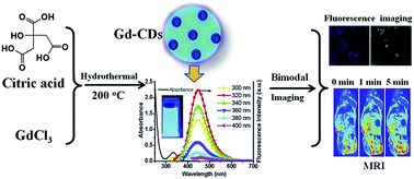
A combination of fluorescence and magnetic resonance imaging (MRI) can provide high-resolution macroscopical anatomical information and high-sensitivity microscopical optical signal simultaneously. In this work, harmless gadolinium(III) doped carbon dots are synthesized in a convenient one-pot hydrothermal approach and used for fluorescence/magnetic resonance multimodal imaging. Unlike typical carbon dots, TEM analysis shows that the carbon dots are particles with irregular shape. Furthermore, Fourier-transform infrared (FTIR) spectra demonstrated structural difference between gadolinium-undoped and doped carbon dots. Nonetheless, the gadolinium-doped carbon dots exhibit strong and stable fluorescence with excitation-dependent emission behavior. Moreover, as an MRI contrast agent, the gadolinium-doped carbon dots represent considerable magnetic resonance properties with a longitudinal relaxation rate of 14.08 mM−1 s−1. In addition, cytotoxicity study reveals that the gadolinium-doped carbon dots show very low toxicity to NCI-H446 cells with an IC50 value of 6.28 mg mL−1. Direct application in cell labeling and in vivo MRI suggests that the gadolinium-doped carbon dots are dual functional fluorescent/MRI probes with excellent biocompatibility.


Introduction

Fluorescent materials have been widely used to study the structure,1,2 status,3,4 characteristics and dynamic processes5,6 of cells. In recent years, with the development of in vivo fluorescence bio-imaging, these fluorescent materials have a great potential to be clinically used in the near future.7,8 Among fluorescent materials, carbon dots (CDs) have attracted more and more interest recently because of their unique stability and biocompatibility. Unlike organic fluorescent dyes, the CDs have excellent chemical stability and photostability.9–11 Without heavy metals, they are far less toxic than semiconductor quantum dots.10,12 In addition, the CDs are high-efficiency vehicles for some compounds or ions which may provide CDs with extra applications due to their abundant surface groups.13,14

Unlike fluorescence imaging, magnetic resonance imaging (MRI) has been used in clinics for a long time. It is a non-invasive diagnostic method with high resolution of soft tissue. More anatomical and hemodynamic information can be obtained by using MRI contrast agents. Due to unique paramagnetic property, gadolinium ions are ideal metal ions for MRI contrast agents. Currently, most of clinical MRI contrast agents are gadolinium chelates because free gadolinium ions are highly toxic.15,16 A combination of fluorescence imaging and MRI can obtain cellular-level information of particular tissue and high-resolution anatomical structure of a whole body simultaneously. Many previous studies introduced some intricately-assembled fluorescent/MRI contrast agents, which were composed of gadolinium chelates and fluorescent dye or semiconductor quantum dots.17,18 However, further application of these agents might be handicapped by their considerable cytotoxicity, or tedious preparing processes.

Doping the gadolinium during the preparation of CDs may provide a new strategy for the development of fluorescent/MRI dual-modality probe. However, it remains relatively less studied compared to the single modality (fluorescence) imaging agents owing to its complicated synthesis process. In 2012, Bourlinos and his co-workers reported a method to synthesize small molecular gadolinium chelates modified CDs to obtain fluorescent/MRI bimodal contrast agents.19 Lately, Xu et al. developed an approach to prepare gadolinium-containing CDs for MR and fluorescent dual-modality imaging.20 This inspired us to explore more efficient approach for facile preparation of fluorescence/magnetic resonance bimodal imaging agent by using simple raw materials. This study reported hereby will show a convenient one-pot method of preparing fluorescent Gd-CDs for dual-modality bio-imaging. Only citric acid was selected as carbon source of the CDs because it can provide more carboxyl groups which may chelate gadolinium ions during and after formation of CDs than other nontoxic acid. Morphological structure, optical character, fluorescent property and magnetic relaxometric efficiency of the Gd-CDs were observed or measured to evaluate the possibility for bio-imaging. Fluorescence imaging of living cells and MRI of anesthetized mice were employed to confirm biocompatibility and fluorescence-magnetic resonance dual responses of the Gd-CDs.

Experimental

Materials

Gadolinium(III) chloride hydrate (GdCl3, 99.9% Gd) was purchased from Strem Chemicals (MA, USA). Citric acid was purchased from Damao Chemicals Ltd (Tianjin, China). Sephadex gel G-25 was obtained from Yuanye Institute of Biotechnology (Shanghai, China). 3-(4,5-Dimethylthiazol-2-yl)-2,5-diphenyltetrazolium bromide (MTT) were from Beyotime Institute of Biotechnology (Shanghai, China). All reagents were used without any further purification. NCI-H446 cell line was a gift kindly presented by Department of Pharmaceutical Chemistry, College of Pharmacy, Dalian Medical University. DMEM medium was purchased from Gibco (NY, USA), and fetal bovine serum (FBS) was purchased from TransGene Biotech (Beijing, China).

Instrumentation

The CDs were deposited on 400 mesh carbon-coated copper grids to observe their shape and size with a JEOL model JEM-2000 EX transmission electron microscope (TEM). Fluorescence spectra were recorded by a PerkinElmer LS55 fluorescence spectrometer, while UV-visible spectra were recorded on a UV-2550 UV-visible spectrometer (Shimadzu, Japan) at room temperature. Infrared absorption spectra of citric acid, the CDs and Gd-CDs were measured using a Bruker Tensor27 Fourier transform infrared (FTIR) spectrometer with KBr pellet assay. Cells were observed by Olympus FV1000MPE confocal microscopy system. Magnetic relaxation rate measurement and MRI were preceded using MINIMR-RAT MRI system (Shanghai Niumag Corporation, China).

Preparation of CDs

CDs and Gd-CDs were prepared via hydrothermal method. Briefly, 1.05 g (5.0 mmol) of citric acid and 66 mg (0.25 mmol) of GdCl3 were dissolved into 25 mL of water with gentle stirring. The solution was transferred into Teflon-lined stainless autoclave (50 mL), and heated at 200 °C for 8 h. After reaction, the solution was concentrated by lyophilization and then refined using Sephadex G-25 to obtain Gd-CDs solution. Eriochrome black T was used as an indicator of free gadolinium ions during purification. The solid product was obtained after lyophilization and stored at 4 °C. Citric acid as raw material, undoped CDs were prepared using the same synthesis and purifying methods.

Fluorescence quantum yield (Φ) measurement

Quinine sulfate as a reference, fluorescence quantum yield (Φ) of Gd-CDs was calculated with the following equation:21
Φx = Φref(Ix/Iref)(Aref/Ax)(ηx/ηref)2
where Φ is fluorescence quantum yield; “I” is the fluorescence intensity; “A” is the optical absorbance at excitation wavelength; “η” is the refractive index of the solvent. The subscript “ref” refers to the reference (quinine sulfate in 0.1 M H2SO4), and “x” refers to the sample (Gd-CDs in water). Absorption values of the reference and the sample were kept below 0.1 at excitation wavelength to reduce reabsorption.

pH effect on fluorescence intensity of the Gd-CDs

Solutions with different pH values (from 2 to 11) were prepared by various proportion of 0.2 M NaOH and Britton–Robinson buffer, which contained 0.04 M H3PO4, 0.04 M CH3COOH and 0.04 M H3BO3. To 1.9 mL of the Britton–Robinson buffer solution was added with 0.1 mL of the Gd-CDs solution (0.5 mg mL−1) and then the fluorescence intensity at 445 nm of the mixture was recorded by using a 325 nm wavelength as excitation.

Ionic strength effect on fluorescence intensity of the Gd-CDs

Sodium chloride was used as model salt for preparing solutions with various ion concentrations in this study. 5.0, 2.5, 2, 1.5, 1.0 and 0.5 M NaCl was diluted with equal volume of the Gd-CDs solution (25 μg mL−1), respectively. The fluorescence intensity at 445 nm of the solution was recorded using a 325 nm wavelength as excitation.

Cytotoxicity assay

Cytotoxicity of the Gd-CDs was investigated with human lung cancer cell line NCI-H446. NCI-H446 cells (8 × 10³ cells per well) were seeded into a 96-well plate with 0.1 mL of DMEM supplemented with 10% fetal bovine serum and incubated at 37 °C in a humidified 5% CO2 atmosphere. After attachment for 24 h, the cells were exposed to various concentrations of the Gd-CDs solution (dissolved in the medium and neutralized by NaHCO3) and further incubated for 48 h. Following, the cells were carefully washed by PBS for 3 times and treated with 50 μL MTT (1 mg mL−1, dissolved in Hank's solution) for another 4 h. A mixture of DMSO and DMF (1[thin space (1/6-em)]:[thin space (1/6-em)]1 v/v, 200 μL per well) was employed to dissolve formazan crystal. The optical absorbance at 570 nm of each well was measured on a Wellscan Mk3 microplates reader (Labsystems Dragon Ltd, Finland). Proliferation values were calculated as follow:
Proliferation value = (AsampleAblank)/(AcontrolAblank)

In this equation, A is absorbance of each well. Subscript “sample” refers to the wells containing cells treated with the Gd-CDs; “control” refers to the wells containing intact cells; “blank” refers to the wells containing medium only.

In vitro fluorescence imaging

NCI-H446 cells (2 × 10⁴ cells per dish) were seeded in thin-bottom dishes with 1 mL of DMEM complete medium containing 10% (v/v) fetal bovine serum and incubated at 37 °C in a 5% CO2 incubator. After attaching for 24 h, the cells were treated with Gd-CDs contained (5 mg mL−1, neutralized by NaHCO3) medium for 48 h. Before observed by laser confocal microscope, the cells were carefully washed by Hanks solution for 3 times and soaked in 1 mL Hanks solution. With excitation laser at 405, 488 and 543 nm, fluorescence images from blue (425–475 nm), green (500–530 nm) and red (560–600 nm) channels were obtained. Bright field images were also captured for ensuring the location of fluorescence signal.

Relaxometric measurement

Before relaxometric measurement, the gadolinium in the Gd-CDs was quantified by inductively coupled plasma-optical emission spectroscopy (ICP-OES 7300DV, PerkinElmer, JAP). The Gd-CDs solution was then diluted according to the concentration of gadolinium: 0.4, 0.2, 0.1, 0.05 and 0.025 mM Gd. The longitudinal and transverse relaxation time of all the diluted Gd-CDs solution samples were measured with Inversion Recovery and Carr–Purcell–Meiboom–Gill sequence respectively. In vitro MRI of these diluted Gd-CDs solutions was performed with Spin Echo sequence. The MRI parameters were set as follows: T = 32.00 °C, TR = 300 ms, TE = 19 ms, slide gap = 0.5 mm and slide width = 2.5 mm.

In vivo MRI

Kunming mice (18–22 g) were purchased form Specific Pathogen Free Animal Center, Dalian Medical University. The mice were anesthetized by isoflurane to avoid artifacts caused by autonomic movement. After an adaptation period about 10 min, warm Gd-CDs solution (gadolinium concentration: 3.16 mM, neutralized by NaHCO3) was intravenously injected at the dosage of 10 mL kg−1. The injection process takes about 30 s. Coronal MRI of the mice was performed at 1, 5, 10, 20, 30 and 60 min after injection with Spin Echo sequence, and signal intensity values of kidney, bladder, liver, lung and muscle of the mice were measured by OsiriX software (version 6.5). The MRI parameters were set as follows: T = 32.00 °C, TR = 500 ms, TE = 19 ms, slide gap = 0.5 mm and slide width = 2.5 mm. The authors state that all animal studies were carried out at Specific Pathogen Free Animal Center at the Dalian Medical University according to the animal protocols approved by the Animal Ethics Committee (AEC).

Results and discussion

Preparation of Gd-CDs

CDs are carbon nanoparticles with fluorescence,22 that can be artificially obtained using various physical or chemical methods such as laser ablation,23 electrochemical oxidization,24 hydrothermal carbonization25 and pyrolysis.19 Herein, only the mixture of citric acid and GdCl3 were used to prepare the Gd3+-containing Gd-CDs that possessed the capability for both fluorescent and MR imaging (Scheme 1). Since only citric acid was selected as carbon source for the preparation of Gd-CDs using one-pot hydrothermal method in this study, we hoped that the synthesis and purification processes would be simple and the resulted Gd-CDs might exhibit good biocompatibility. Before biological use, the Gd-CDs were neutralized by sodium bicarbonate to avoid unpredictable effect caused by high H+ concentration. As we know that the cytotoxicity of Gd-CDs was mainly relevant to the carbon sources and strong complexation ability to the Gd3+. Since only citric acid and GdCl3 were involved in the synthesis, purification of the Gd-CDs was carried out by gel-chromatography to remove the unreacted citric acid, free Gd3+ and other impurities. Because free Gd3+ ions are highly toxic, a test of free Gd ions is necessary before biological use. Herein, we used Eriochrome black T as an indicator of free Gd ions and found that the Gd-CDs could not react with the Eriochrome black T, demonstrating that free Gd3+ ions were isolated from the Gd-CDs. The conditions of the medium where the test was performed in terms of pH, solvents and salt concentrations were listed in Table S1. As shown in Fig. S1, the Eriochrome black T solution can react with free gadolinium ions (GdCl3) to form red substance when pH was 7, 9 and 11 at 0, 24 and 48 h, respectively. While the Gd-CDs and water could not react with it to form red substance at the investigated conditions. The results indicated that the binding between the Gd3+ and CDs was stronger than that between the Gd3+ and Eriochrome black T. No leakage of Gd3+ ions was found from the Gd-CDs at different pH values when they were stored at room temperature up to 48 h.
image file: c5ra09948j-s1.tif
Scheme 1 Design and preparation of the Gd-CDs for fluorescence/magnetic resonance bimodal imaging.

Structure of the Gd-CDs

After purification and lyophilization, light brown solid Gd-CDs were collected. FTIR spectra further indicated the presence of carboxyl groups on the particles (Fig. 1). Partly carbonized citric acid residues should be the source of these carboxyl groups. As illustrated in FTIR spectra, two absorption peaks located at 3498 cm−1 and 3294 cm−1 were disappeared after carbonization. The spectral changes suggested that hydroxyl groups on citric acid might participate in carbonization. Comparing with the undoped CDs, the Gd-CDs showed less absorption from C–H bond (3700 cm−1–3100 cm−1, 1450 cm−1–1200 cm−1 and 650 cm−1–400 cm−1), because the presence of gadolinium ions might enhance the degree of dehydrogenation. In other words, gadolinium ions might facilitate the carbonization of citric acid in hydrothermal condition. In addition, comparing with that of undoped CDs, the red-shifted peaks of C[double bond, length as m-dash]O (1562 cm−1) and C–O bonds (1388 cm−1) of Gd-CDs suggested that carboxyl groups might chelated with gadolinium ions.
image file: c5ra09948j-f1.tif
Fig. 1 FTIR spectra of citric acid, undoped CDs and Gd-CDs.

The size and shape of the undoped CDs and Gd-CDs observed by TEM were completely different. The undoped CDs were spherical particles with smaller size (Fig. 2A) like other CDs described previously,9,10,12 while the Gd-CDs were irregular particles with much larger size (Fig. 2B). The Gd-CDs might be formed by aggregating of smaller CDs. Complexation reaction between gadolinium ions and carboxyl groups on the CDs might be a reason of aggregation. However, the Gd-CDs can be completely dissolved in water and PBS medium, respectively, at a concentration of 50 mg mL−1 at 4 °C for a week. The Gd-CDs solution (50 mg mL−1 in water) was very stable and no precipitation was found when it was stored at 4 °C for a week (Fig. S2). Fig. S3 shows the Gd-CDs dissolved in water, PBS, and Dulbecco's Modified Eagle Medium (DMEM) medium, respectively, at room temperature for 48 h. The Gd-CDs were well dissolved in these solvents and no obvious precipitation of the Gd-CDs with the time was observed. However, aggregation of the Gd-CDs was found when they were dissolved in DMEM medium for dynamic light scattering (DLS) analysis, probably due to the amino acid interaction with the Gd-CDs in DMEM medium (Fig. S4). The XPS spectrum in Fig. 3A suggested the presence of Gd3+ peak at 140.3 ev in the Gd-CDs with a doping ratio of approximately 0.53%.


image file: c5ra09948j-f2.tif
Fig. 2 TEM images of undoped CDs and Gd-CDs.

image file: c5ra09948j-f3.tif
Fig. 3 XPS spectrum (A), ultraviolet-visible absorption and photoluminescence emission spectra (B), pH stability (C) and cytotoxicity of the Gd-CDs.

Fluorescence property

The refined Gd-CDs were analyzed by the ultraviolet-visible and fluorescence spectrophotometry. The UV-vis spectrum of the CDs consisted of only one peak at 336 nm. In addition, a strong background till 430 nm was observed. Like CDs reported previously,9,12 the Gd-CDs exhibited an excitation-dependent emission behavior (Fig. 3B). The emission spectra of the Gd-CDs were recorded at excitation wavelengths of 300, 320, 340, 360, 380 and 400 nm, respectively. At excitation wavelength of 320 nm, the CDs showed a maximum emission peak at 446 nm with a Stokes shift of about 126 nm. When the excitation wavelength shifted from 300 to 400 nm, the emission peak showed a red shift from 445 to 454 nm. The fluorescence intensity of the emission peak was also depended on excitation wavelength. CDs are mixed nanoparticles, and the variance of fluorescence structures in CDs may cause this broad emission property.9,26 The fluorescence quantum yield of the Gd-CDs at 320 nm was 4.06%, which was higher than previously reported Gd-CDs synthesized by pyrolysis at 250 and 300 °C.20

The fluorescence property of the Gd-CDs could be influenced by changing the pH values. As illustrated in Fig. 3C, the fluorescence intensity obviously increased when the pH value changed from 2 to 7 and reached the maximum value at pH 7, keeping unchanged up to pH 11. The results demonstrated the fluorescence could not be influenced by aggregation at higher pHs (up to pH 11). The Gd-CDs with higher fluorescence intensity within physiological pH range might be suitable for bio-imaging. The DLS results of Gd-CDs at different pHs in a Britton–Robinson buffer was shown in Fig. S5. The hydrodynamic size of Gd-CDs was significantly affected by changing pH values. Aggregation of Gd-CDs was observed at pH 2–6, and the size decreased from pH 7 to 9 and increased when pHs changed from 10 to 11.


image file: c5ra09948j-f4.tif
Fig. 4 Bright field and fluorescence microscope images of human lung cancer cells incubated with the Gd-CDs for 48 h.

image file: c5ra09948j-f5.tif
Fig. 5 In vivo T1-weighted MR images of mice (A) and quantification of magnetic resonance signal intensity in their kidney and bladder (B) before and after intravenous injection of Gd-CDs (n = 3).

In addition, to investigate the salt concentration on fluorescence intensity of the Gd-CDs, sodium chloride was used in this experiment because it exists in most of organisms.27 Our data revealed that ionic strength had no effect on fluorescence intensity of the Gd-CDs (Fig. S6). The good stability of the Gd-CDs in sodium chloride solution was of benefit to their further applications in biomedical field.

Cytotoxicity

To minimize the toxicity of CDs themselves, citric acid, a nontoxic organic polyprotic acid, was used as carbon source for synthesis of Gd-CDs. The Gd-CDs did not show any obvious toxicity like free gadolinium ions because the ions were complexed with the carboxyl groups of the Gd-CDs. Cytotoxicity of the Gd-CDs against NCI-H446 cells was evaluated using a MTT assay. As shown in Fig. 3D, the Gd-CDs demonstrated very low toxicity to NCI-H446 cells with a calculated IC50 value of 6.28 mg mL−1, which was greater than that (0.121 mg mL−1) of the Gd-CDs prepared by the pyrolysis of gadolinium chelates.19 Raw materials and fabrication processes of carbonization might play a significant role in cytotoxicity of synthetic CDs, and citric acid was an appropriate carbon source for synthesizing Gd-CDs with hydrothermal assay.

In vitro fluorescence imaging

To investigate potential application of the Gd-CDs as fluorescent probes, in vitro uptaking test was performed using human lung cancer cell line NCI-H446. As illustrated in Fig. 4, obvious fluorescent signal from the Gd-CDs treated cells could be observed on blue, green and red channel as compared with the control cells without adding the Gd-CDs. The excitation dependent fluorescence of the Gd-CDs made it possible for multiple color emission in cells imaging by using Gd-CDs only. The fluorescence signal was mainly distributed in cytoplasm indicated that the Gd-CDs could penetrate cell membrane instead of nuclear membrane. According to previous studies, the cytoplasm specific property of CDs should be related with endocytosis.1,28 The results indicated that the Gd-CDs were able to be used for in vitro tumor cells labeling via a simple incubation method.

Relaxation properties

The longitudinal and transverse relaxivity curves of the Gd-CDs showed a linearly dependence between relaxation index and concentration of gadolinium ions (Fig. S7). The estimated value is 20 Gd3+ attached per carbon dot based on the ICP-OES analysis. The longitudinal relaxation rate (r1) and transverse relaxation rate (r2) of the Gd-CDs was 14.08 mM−1 s−1 and 15.85 mM−1 s−1, respectively. The longitudinal relaxation rate was higher than other Gd-CDs described previously.29,30 The in vitro imaging revealed that T1-weighted MRI signal intensity enhanced with the increase of gadolinium concentration (Fig. S7A), but signal intensity of T2-weighted MRI was saturated when gadolinium concentration was higher than 0.1 mM (Fig. S7B). These results indicated that the Gd-CDs could be used as a promising candidate for T1-weighted MRI contrast agents. Previous studies demonstrated that the longitudinal relaxation rate of a certain gadolinium contained contrast agent could be enhanced when the gadolinium ions were confined within limited space.31,32 Comparing with gadolinium chelates, the Gd-CDs have a superior magnetic resonance relaxation rate because the doped gadolinium ions were restricted by CDs.

In vivo MRI

The Gd-CDs had good biocompatibility which can be directly used for in vivo MRI in mice. Fig. 5 shows the T1-weighted 2D images of mice before and after the injection of Gd-CDs agent. Unlike previously reported Gd-CDs,30 the Gd-CDs were quickly excreted by kidney and accumulated in bladder after 30 minutes intravenous injection into the mice. The remaining Gd-CDs might be eliminated within 3 h. Strong contrast enhancement was observed in the kidney and urinary bladder, indicating that the Gd-CDs agent could be readily excreted via renal filtration (Fig. 5A and S8). In comparison, the Gd-CDs agent resulted in relatively weak enhancement in liver and lung post-injection. No obvious accumulation of the Gd-CDs was found in muscle (Fig. S9). According to the signal from the bladder, the Gd-CDs were mainly eliminated via kidneys within 30 minutes and the remaining Gd-CDs might be eliminated within 2 to 3 h for estimation. The fast clearance rate of the Gd-CDs indicated that they might be a short-acting contrast agent that can reduce the cumulative or long-term toxicity. The Gd-CDs had similar magnetic resonance property and clearance rate with Gd2O3 nanoparticles.33 Fig. 5B shows the quantitative MR enhancement in the kidneys and bladders before and after the injection of the Gd-CDs agent. The result further evidenced the Gd-CDs underwent bladder clearance from the body.

Conclusions

In summary, our current study described the synthesis, property and bimodal bio-imaging of the Gd-CDs by using citric acid as carbon source. The Gd-CDs showed strong and stable fluorescence property with excitation-dependent emission behaviour, together with higher longitudinal relaxation rate. Different from the preceding paper on Gd(III)-doped carbon dots,30 the Gd-CDs prepared in this work exhibited greater relaxation rate by using only citric acid and GdCl3 as precursors. They were successfully used for both cell imaging and in vivo MR imaging without any further modification. The strategy developed here may provide a facile method for the preparation of multimodal nanoprobes for both fluorescence/MR imaging.

Acknowledgements

This work was supported by the National Nature Science Foundation of China (91227126), National Special Fund for Key Scientific Instrument and Equipment Development (2013YQ17046307) and the Nature Science Foundation of Liaoning Province, China (2013020177).

Notes and references

  1. L. Cao, X. Wang, M. J. Meziani, F. Lu, H. Wang, P. G. Luo, Y. Lin, B. A. Harruff, L. M. Veca, D. Murray, S. Y. Xie and Y. P. Sun, J. Am. Chem. Soc., 2007, 129, 11318–11319 CrossRef CAS PubMed.
  2. R. Wombacher, M. Heidbreder, S. van de Linde, M. P. Sheetz, M. Heilemann, V. W. Cornish and M. Sauer, Nat. Methods, 2010, 7, 717–719 CrossRef CAS PubMed.
  3. M. Oheim, M. van 't Hoff, A. Feltz, A. Zamaleeva, J. M. Mallet and M. Collot, Biochim. Biophys. Acta, 2014, 1843, 2284–2306 CrossRef CAS PubMed.
  4. Y. Urano, D. Asanuma, Y. Hama, Y. Koyama, T. Barrett, M. Kamiya, T. Nagano, T. Watanabe, A. Hasegawa, P. L. Choyke and H. Kobayashi, Nat. Med., 2009, 15, 104–109 CrossRef CAS PubMed.
  5. L. Feng, Z. M. Liu, J. Hou, X. Lv, J. Ning, G. B. Ge, J. N. Cui and L. Yang, Biosens. Bioelectron., 2014, 65, 9–15 CrossRef PubMed.
  6. Q. Xue, Y. Lv, S. Xu, Y. Zhang, L. Wang, R. Li, Q. Yue, H. Li, X. Gu, S. Zhang and J. Liu, Biosens. Bioelectron., 2015, 66, 547–553 CrossRef CAS PubMed.
  7. E. de Boer, N. J. Harlaar, A. Taruttis, W. B. Nagengast, E. L. Rosenthal, V. Ntziachristos and G. M. van Dam, Br. J. Surg., 2015, 102, e56–72 CrossRef CAS PubMed.
  8. H. Kobayashi and P. L. Choyke, Acc. Chem. Res., 2011, 44, 83–90 CrossRef CAS PubMed.
  9. B. de and N. Karak, RSC Adv., 2013, 3, 8286 RSC.
  10. H. U. Lee, S. Y. Park, E. S. Park, B. Son, S. C. Lee, J. W. Lee, Y. C. Lee, K. S. Kang, M. I. Kim, H. G. Park, S. Choi, Y. S. Huh, S. Y. Lee, K. B. Lee, Y. K. Oh and J. Lee, Sci. Rep., 2014, 4, 4665 Search PubMed.
  11. S. S. Wee, Y. H. Ng and S. M. Ng, Talanta, 2013, 116, 71–76 CrossRef CAS PubMed.
  12. C. Jiang, H. Wu, X. Song, X. Ma, J. Wang and M. Tan, Talanta, 2014, 127, 68–74 CrossRef CAS PubMed.
  13. W. Dong, Y. Dong, Y. Wang, S. Zhou, X. Ge, L. Sui and J. Wang, Spectrochim. Acta, Part A, 2013, 116, 209–213 CrossRef CAS PubMed.
  14. P. Huang, J. Lin, X. Wang, Z. Wang, C. Zhang, M. He, K. Wang, F. Chen, Z. Li, G. Shen, D. Cui and X. Chen, Adv. Mater., 2012, 24, 5104–5110 CrossRef CAS PubMed.
  15. J. M. Idée, M. Port, C. Robic, C. Medina, M. Sabatou and C. Corot, J. Magn. Reson. Imag., 2009, 30, 1249–1258 CrossRef PubMed.
  16. A. J. Spencer, S. A. Wilson, J. Batchelor, A. Reid, J. Rees and E. Harpur, Toxicol. Pathol., 1997, 25, 245–255 CrossRef CAS PubMed.
  17. R. Bakalova, Z. Zhelev, I. Aoki, K. Masamoto, M. Mileva, T. Obata, M. Higuchi, V. Gadjeva and I. Kanno, Bioconjugate Chem., 2008, 19, 1135–1142 CrossRef CAS PubMed.
  18. J. Liu, K. Li, J. Geng, L. Zhou, P. Chandrasekharan, C.-T. Yang and B. Liu, Polym. Chem., 2013, 4, 1517–1524 RSC.
  19. A. B. Bourlinos, A. Bakandritsos, A. Kouloumpis, D. Gournis, M. Krysmann, E. P. Giannelis, K. Polakova, K. Safarova, K. Hola and R. Zboril, J. Mater. Chem., 2012, 22, 23327 RSC.
  20. X. Ren, L. Liu, Y. Li, Q. Dai, M. Zhang and X. Jing, J. Mater. Chem. B, 2014, 2, 5541–5549 RSC.
  21. Z. Qian, J. Ma, X. Shan, L. Shao, J. Zhou, J. Chen and H. Feng, RSC Adv., 2013, 3, 14571 RSC.
  22. P. G. Luo, F. Yang, S.-T. Yang, S. K. Sonkar, L. Yang, J. J. Broglie, Y. Liu and Y.-P. Sun, RSC Adv., 2014, 4, 10791 RSC.
  23. Y. P. Sun, B. Zhou, Y. Lin, W. Wang, K. A. Fernando, P. Pathak, M. J. Meziani, B. A. Harruff, X. Wang, H. Wang, P. G. Luo, H. Yang, M. E. Kose, B. Chen, L. M. Veca and S. Y. Xie, J. Am. Chem. Soc., 2006, 128, 7756–7757 CrossRef CAS PubMed.
  24. Q. L. Zhao, Z. L. Zhang, B. H. Huang, J. Peng, M. Zhang and D. W. Pang, Chem. Commun., 2008, 5116–5118 RSC.
  25. H. Yan, M. Tan, D. Zhang, F. Cheng, H. Wu, M. Fan, X. Ma and J. Wang, Talanta, 2013, 108, 59–65 CrossRef CAS PubMed.
  26. Q. Li, T. Y. Ohulchanskyy, R. Liu, K. Koynov, D. Wu, A. Best, R. Kumar, A. Bonoiu and P. N. Prasad, J. Phys. Chem. C, 2010, 114, 12062–12068 CAS.
  27. C. L. Fox Jr, J. M. Winfield, L. B. Slobody, C. M. Swindler and J. K. Lattimer, JAMA, J. Am. Med. Assoc., 1952, 148, 827–833 CrossRef CAS PubMed.
  28. C. Liu, P. Zhang, X. Zhai, F. Tian, W. Li, J. Yang, Y. Liu, H. Wang, W. Wang and W. Liu, Biomaterials, 2012, 33, 3604–3613 CrossRef CAS PubMed.
  29. N. Gong, H. Wang, S. Li, Y. Deng, X. Chen, L. Ye and W. Gu, Langmuir, 2014, 30, 10933–10939 CrossRef CAS PubMed.
  30. Y. Xu, X. H. Jia, X. B. Yin, X. W. He and Y. K. Zhang, Anal. Chem., 2014, 86, 12122–12129 CrossRef CAS PubMed.
  31. M. Botta and L. Tei, Eur. J. Inorg. Chem., 2012, 2012, 1945–1960 CrossRef CAS PubMed.
  32. J. J. Law, A. Guven and L. J. Wilson, Contrast Media Mol. Imaging, 2014, 9, 409–412 CrossRef CAS PubMed.
  33. M. W. Ahmad, W. Xu, S. J. Kim, J. S. Baeck, Y. Chang, J. E. Bae, K. S. Chae, J. A. Park, T. J. Kim and G. H. Lee, Sci. Rep., 2015, 5, 8549 CrossRef CAS PubMed.

Footnotes

Electronic supplementary information (ESI) available. See DOI: 10.1039/c5ra09948j
These two authors contributed equally to this paper.

This journal is © The Royal Society of Chemistry 2015
Click here to see how this site uses Cookies. View our privacy policy here.