DOI:
10.1039/C5RA09020B
(Paper)
RSC Adv., 2015,
5, 53623-53628
Synthesis of bulk and supported nickel phosphide using microwave radiation for hydrodeoxygenation of methyl palmitate
Received
14th May 2015
, Accepted 10th June 2015
First published on 10th June 2015
Abstract
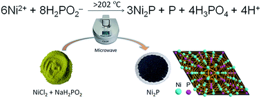
In this paper, we proposed a novel method for preparing bulk and supported Ni2P catalysts under mild conditions. Ni2P and Ni2P/SiO2 were synthesized from nickel hypophosphite precursors at 230 °C for 5 min using a CEM Discover microwave reactor, and the initial reaction temperature is about 202 °C. The catalysts were characterized using XRD, TEM, SEM, XPS, BET, carbon monoxide chemisorption, and the catalytic performance was tested for hydrodeoxygenation (HDO) of methyl palmitate in a fixed-bed reactor. Interestingly, microwave irradiation does not result in sintering of Ni2P particles. The principal products of the HDO reaction for both catalysts are pentadecane and hexadecane. Isomerization products were not detected, and other by-products content is very low (<1%). The HDO results demonstrate that the catalyst prepared using a microwave has better activity than that prepared using calcination.
1. Introduction
In recent years, synthesis of renewable fuel from biomass has attracted considerable attention. The HDO reaction is an important reaction for the preparation of renewable fuel. However, conventional hydrogenation catalysts (such as CoMo/Al2O3 and NiMo/Al2O3) do not possess sufficient stability under HDO reaction conditions, because the sulfur content will be gradually decreased during the reaction.1 Therefore, more and more studies focus on seeking noble metal catalysts and other novel catalytic materials to promote HDO efficiently.
Metal phosphides, a class of interstitial compounds, have attracted considerable attention for their unexpected properties in catalysis, magnetic applications, and applications in telecommunications, optoelectronic devices, lithium batteries, and solar cells.2,3 In particular, Ni2P used as hydrodesulfurization catalyst shows excellent activity and resistance to sulfur poisoning performance.2 In recent studies, Ni2P as a highly effective HDO catalyst shows good activity and stability in converting biomass to renewable oil.4–10 J. Moon and coworkers studied the active sites of Ni2P/SiO2 catalyst for HDO of guaiacol by using XAFS and DFT study. They revealed that the active site of Ni2P is composed of a threefold hollow Ni and P sites which lead to adsorption of H or OH groups.7 A. Iino and coworkers studied the reaction mechanism of Ni2P/SiO2 catalyst for HDO of 2-methyltetrahydrofuran at a medium pressure of 0.5 MPa. They concluded that Ni2P/SiO2 showed 100% conversion and 85% selectivity to n-pentane, with higher oxygen removal activity and less C–C bond cracking activity than commercial noble metal Ru/C and Pd/Al2O3 catalysts based on the same amount of active sites.8
As a class of new catalytic materials, the synthesis and applications of metal phosphides underwent tremendous development in the recent decades. A variety of methods for synthesizing metal phosphides have been reported.11–26 Generally, the synthesis of metal phosphide has become highly convenient, which has broken through the limitations of high temperature and heating rate.19,20 In this paper, we proposed an alternative path to the synthesis of nickel phosphide. Ni2P and Ni2P/SiO2 were synthesized from nickel hypophosphite precursor at 230 °C for 5 min using a CEM DISCOVER microwave reactor. Interestingly, microwave radiation does not result in sintering of Ni2P particles. The HDO results show that the catalyst prepared using microwave having better activity than that prepared using calcination.
Microwave is an electromagnetic wave with a frequency of 2.45 GHz and commonly used in materials synthesis.27–29 It is mainly through the polarization of electromagnetic waves on the polar molecules to achieve a uniform temperature inside the sample. In our previous studies, bulk and supported Ni2P could be synthesized from the decomposition of nickel hypophosphite precursor in a static protecting gas atmosphere at 250 °C.20 Herein, we use microwave radiation instead of traditional tube furnace to synthesize bulk and supported Ni2P. The reaction may occur at a high temperature is expressed in ionic formula as follows:
|
6Ni2+ + 8H2PO2− = 3Ni2P + P + 4H3PO4 + 4H+
| (1) |
2. Experimental methods
SiO2 was purchased from Tianjin Chemist Scientific Ltd., China. It has a specific surface area of 279 m2 g−1, a pore volume of 0.8 cm3 g−1. Other reagents were analytical pure grade and purchased from Alfa Aesar.
2.1. Synthesis of bulk and supported nickel phosphides
2.1.1. Calcination method. The preparation of bulk Ni2P is illustrated as follows. Nickel chloride (NiCl2·H2O) and sodium hypophosphite (NaH2PO2·H2O) were dissolved in deionized water under stirring with a mole ratio of P/Ni = 1.5/1. After stirring for 1 h, the solution was evaporated slowly to dehydrate, and was dried at 80 °C for 8 h to obtain the precursor. At the beginning of the thermal decomposition, the air in the reactor was removed by flowing Ar, after which the precursor was treated at 250 °C for 30 min in a static Ar atmosphere (heating rate: 5–20 °C min−1). The product was cooled to ambient temperature under Ar and was washed several times with deionized water to remove ion impurities, after which the wet material was dried at 120 °C for 3 h.The preparation of Ni2P/SiO2 is illustrated as follows. NiCl2·H2O and NaH2PO2·H2O were dissolved in deionized water under stirring with a mole ratio of P/Ni = 1.5/1. After stirring for 1 h, the solution was impregnated onto the SiO2 using incipient wetness impregnation. Then, the wet solid were evaporated slowly to dehydrate, and were dried at 80 °C for 8 h to obtain the precursor. At the beginning of the thermal decomposition, the air in the reactor was removed by flowing Ar, after which the precursor was treated at 250 °C for 30 min in a static Ar atmosphere (heating rate: 5–20 °C min−1). The product was cooled to ambient temperature under Ar and was washed several times with deionized water to remove ion impurities, after which the wet material was dried at 120 °C for 3 h.
2.1.2. Microwave method. The preparation of bulk Ni2P is illustrated as follows NiCl2·H2O and NaH2PO2·H2O were dissolved in deionized water under stirring with a mole ratio of P/Ni = 1.5/1. After stirring for 1 h, the solution was evaporated slowly to dehydrate, and was dried at 80 °C for 8 h to obtain the precursor. At the beginning of microwave radiation, the air in the reactor was removed by flowing Ar, after which the precursor was radiated at 230 °C for 5 min in a static Ar atmosphere (heating power of CEM Discover reactor is 250–290 W). The product was cooled to ambient temperature under Ar and was washed several times with deionized water to remove ion impurities, after which the wet material was dried at 120 °C for 3 h.The preparation of Ni2P/SiO2 is illustrated as follows. NiCl2·H2O and NaH2PO2·H2O were dissolved in deionized water under stirring with a mole ratio of P/Ni = 1.5/1. After stirring for 1 h, the solution was impregnated onto the SiO2 using incipient wetness impregnation. Then, the wet solid were evaporated slowly to dehydrate, and were dried at 80 °C for 8 h to obtain the precursor. At the beginning of microwave radiation, the air in the reactor was removed by flowing Ar, after which the precursor was radiated at 230 °C for 5 min in a static Ar atmosphere (heating power of CEM Discover reactor is 250–290 W). The product was cooled to ambient temperature under Ar and was washed several times with deionized water to remove ion impurities, after which the wet material was dried at 120 °C for 3 h.
2.2. Characterization
The characterization was performed for passivated sample, which was exposed to the air and without any reduction treatment. Powder X-ray diffraction (XRD) was performed on a Bruker D8 focus diffractometer, with Cu Kα radiation at 40 kV and 40 mA. The transmission electron microscope (TEM) images were purchased using a Philips Tecnai G2 F-20 field emission gun TEM. Scanning electron microscope (SEM) images were obtained using a Tescan Vega3 SBH. Brunauer–Emmett–Teller (BET) specific surface areas were obtained using nitrogen adsorption/desorption measurements at 77 K with a BELSORP-Mini instrument. Samples were degassed at 300 °C for 3 h before measurement. The surface area was calculated using a multi-point BET model. X-ray photoelectron s pectroscopy (XPS) was carried out using a Kratos Axis Ultra DLD spectrometer employing a monochromated Al-Kα X-ray source (hν = 1486.6 eV), hybrid (magnetic/electrostatic) optics and a multi-channel plate and delay line detector (DLD). All XPS spectra were recorded using an aperture slot of 300 × 700 microns, survey spectra were recorded with a pass energy of 80 eV, and high-resolution spectra with a pass energy of 40 eV. In order to subtract the surface charge effect, the C1s peak was fixed at a binding energy of 284.6 eV. The CO chemisorption was performed with Micromeritics Chemisorb 2750 gas-adsorption equipment. The sample was loaded into a quartz reactor and pretreated in 10% H2/Ar at 450 °C for 3 h. After cooling in He, pulses of 10% CO/He in a He carrier (25 mL (NTP) min−1) were injected at 35 °C through a loop tube.
2.3. Catalytic activity test
HDO catalytic activities were evaluated using 30 wt% methyl palmitate in decalin. HDO reaction was carried out in a fixed-bed microreactor. The catalyst was pelleted, crushed, and sieved with 20–40 mesh; 1.0 g of the catalyst was diluted with SiO2 to a volume of 5.0 mL in the reactor. Prior to the reaction, catalysts were pretreated in situ with flowing H2 (100 mL min−1) for 3 h. The testing conditions for the HDO reaction were 1 MPa, weight hourly space velocity (WHSV) = 6 h−1, and H2/oil = 1000. Liquid products were collected every hour after a stabilization period of 6 h. Both feed and products were analyzed with an Agilent 7890A/5975C GC-MS equipped with a flame ionization detector and a HP-5 column. The TOF was calculated using eqn (2): |
 | (2) |
where FA0 is the molar rate of reactant fed into the reactor (μmol s−1), W is the catalyst weight (g), COuptake is the uptake of chemisorbed CO (μmol g−1), and XA is the reactant conversion (%).
3. Results and discussion
3.1. Synthesis of bulk and supported nickel phosphides
Similarly, NaH2PO2·H2O and NiCl2·6H2O were used as a raw material. Fig. 1a shows the XRD patterns of the samples prepared at different temperatures for 10 min from precursors with a P/Ni mole ratio of 1.5. The result indicates that Ni2P can be synthesized at 230 and 250 °C. The strong diffraction peaks of NaCl were detected when the temperature is 200 °C, which indicates the precursor was not decomposed and 200 °C is lower than the lowest reaction temperature (Fig. 1b). From the temperature curves of 230 and 250 °C, we can clearly see that this is an exothermic reaction, and the initial reaction temperature is between 202 and 206 °C. When a decomposition reaction occured, the measured temperature suddenly increased, and then rapidly came down to the setting values.
 |
| Fig. 1 (a) The XRD patterns of the samples prepared at different temperatures for 10 min from the precursors with a mole ratio of P/Ni = 1.5, (b) microwave temperature patterns of the samples in different setting values. | |
In our previously published paper,20 Ni2P was synthesized in the mole ratio of P/Ni range of 1.5 to 1.75 using a calcination method. To study the influence of the microwave irradiation deeply, a series of samples were prepared with different mole ratios of P/Ni. As shown in Fig. 2, we can clearly see that Ni2P can be synthesized at a mole ratio of P/Ni = 1.5. Similarly, the weak diffraction peaks of Ni12P5 were detected when the mole ratio of P/Ni is 1.34 (eqn (1)). Different with our previous results, the diffraction peaks of Ni5P4 were detected when the mole ratio of P/Ni is 1.75, which indicates that P is a slight excess for the synthesis of bulk Ni2P in this mole ratio. This result also shows that the microwave method has better utilization of phosphorus than calcination method.
 |
| Fig. 2 The XRD patterns of bulk Ni2P prepared with different mole ratios of P/Ni (microwave radiation at 230 °C for 10 min). | |
To study the effect of irradiation time on the bulk Ni2P product, the precursors with different irradiation times at 230 °C were analysed by XRD. The diffraction peaks of Ni12P5 were detected in the precursor after being radiated at 230 °C for 2 min, and these diffraction peaks disappeared after 5 min (Fig. 3). Crystal structure of Ni2P was not damaged when irradiation time extended to 10 minutes. The result indicates that there is a rapid crystal phase transition process from Ni12P5 to Ni2P, and Ni2P can be synthesized in 5 min.
 |
| Fig. 3 The XRD patterns of bulk Ni2P prepared with different microwave radiation time (microwave radiation at 230 °C, P/Ni = 1.5). | |
Fig. 4 shows the SEM images of bulk Ni2P prepared using different methods. Surprisingly, microwave radiation at 230 °C did not cause serious sintering of metal particles, which might because microwave radiation is very uniform. Although irradiation will cause sudden increase in temperature, but the apparent temperature does not exceed 300 °C. On the contrary, the average particle size of Ni2P synthesized using calcination method is significantly larger than that of synthesized using the microwave method.
 |
| Fig. 4 The SEM images of bulk Ni2P prepared using different methods, (a) and (b) prepared using calcination method (P/Ni = 1.5, 250 °C, 30 min), (c) and (d) prepared using microwave method (P/Ni = 1.5, 230 °C, 5 min). | |
Fig. 5 shows the XRD patterns and TEM images of 30 wt% Ni2P/SiO2 catalysts prepared using different methods. Two supported catalysts having similar weak diffraction peaks of Ni2P (PDF# 65-1989). The XRD results show that Ni2P/SiO2 catalysts can be synthesized at a mole ratio of P/Ni = 1.5 using different methods. From the TEM images, we can clearly see that Ni2P particles prepared using microwave method had significantly better dispersion. Subsequent CO uptake and HDO test also show Ni2P particles are well dispersed on SiO2.
 |
| Fig. 5 The XRD patterns and TEM images of 30 wt% Ni2P/SiO2 catalysts prepared using different methods, (a) XRD patterns, (b) prepared using calcination method (P/Ni = 1.5, 250 °C, 30 min), and (c) prepared using microwave method (P/Ni = 1.5, 230 °C, 5 min). | |
XPS analysis was carried out to observe the valence of Ni and P elements on 30 wt% Ni2P/SiO2 surfaces (Fig. 6). The Ni2p spectrum of Ni2P mainly involves two peaks. The first one is centered at 852.9 eV and assigned to Niδ+ of Ni2P, and the second one is at 856.3 eV, corresponding to Ni2+ ions interacting possibly with phosphate ions as a consequence of a superficial passivation. For P2p, binding energy at 129.7 eV is attributed to Pδ− of Ni2P and 133.0 eV is attributed to surface nickel phosphate species.19,26 Some difference is that the binding energy of calcination sample shift slightly to a higher value than that of microwave sample. Temperature and time of synthesis of Ni2P/SiO2 using calcination method are higher than that using the microwave method. The shift of binding energy may be explained by a stronger interaction between Ni2P with SiO2 at high temperature.
 |
| Fig. 6 The XPS spectra of 30 wt% Ni2P/SiO2 catalysts prepared using different methods: (a) Ni2p core level spectra of Ni2P/SiO2, (b) P2p core level spectra of Ni2P/SiO2. | |
3.2. Catalytic activity
To study the effect of two preparation methods on catalyst performance, 30 wt% Ni2P/SiO2 catalysts were synthesized and tested at different temperatures. HDO catalytic activities were evaluated using 30 wt% methyl palmitate in decalin (testing conditions: 1 MPa, WHSV = 6 h−1, H2/oil = 1000). According to published papers,9,30–33 we give a possible HDO route of methyl palmitate over Ni2P/SiO2 (Scheme 1).
 |
| Scheme 1 The HDO route of methyl palmitate over Ni2P/SiO2. | |
As shown in Fig. 7, Ni2P/SiO2 prepared using microwave method has a significantly higher HDO activity at low reaction temperatures. The principal products of the HDO reaction for two catalysts are pentadecane and hexadecane. Isomerization products were not detected, and the content of other by-products is very low (<1%). BET specific surface area of two catalysts was 117, and 120 m2 g−1, respectively (Table 1). However, Ni2P/SiO2 prepared using microwave method has higher CO uptake, which also indicates that catalyst prepared by microwave had a better dispersion of Ni2P particles. Two catalysts have similar TOF value at 290 °C, which is 0.18 and 0.19 s−1. For comparison, 30 wt% Ni/SiO2 were prepared using the incipient wetness impregnation. As shown in Table 1, Ni/SiO2 shows very low conversion (27.0%) and TOF value (0.08 s−1).
 |
| Fig. 7 The catalytic activities of 30 wt% Ni2P/SiO2 catalysts at different temperatures (a) prepared using the calcination method (P/Ni = 1.5, 250 °C, 30 min), and (b) prepared using the microwave method (P/Ni = 1.5, 230 °C, 5 min). | |
Table 1 The physical properties and HDO activities of different catalysts (testing conditions: 1 MPa, 290 °C, WHSV = 6 h−1, H2/oil = 1000)
Samples |
as,BET (m2 g−1) |
CO (μmol g−1) |
Conversion (%) |
TOF (s−1) |
SiO2 |
279 |
— |
— |
— |
30 wt% Ni/SiO2 |
118 |
6.0 |
27.0 |
0.08 |
30 wt% Ni2P/SiO2 (calcination) |
117 |
8.0 |
78.3 |
0.18 |
30 wt% Ni2P/SiO2 (microwave) |
120 |
9.8 |
99.4 |
0.19 |
To clearly understand the advantage of the microwave method, comparison chart of the two methods was given. As shown in Scheme 2, two methods use same precursor and protective gas. The difference is that the microwave method requires lower reaction temperature and shorter time. Including heating and cooling steps, the entire microwave process is only about fifteen minutes, which is far below the calcination method.
 |
| Scheme 2 Pathway of two methods. | |
Conclusions
In this paper, Ni2P and Ni2P/SiO2 were synthesized from nickel hypophosphite precursors at 230 °C for 5 min using a CEM Discover microwave reactor, and the initial reaction temperature is between 202 and 206 °C. The catalysts were characterized using XRD, TEM, SEM, XPS, BET, carbon monoxide chemisorption, and catalytic performance was evaluated for HDO of methyl palmitate in a continuous flow fixed-bed reactor. Interestingly, microwave irradiation does not result in sintering of Ni2P particles. The principal products of the HDO reaction for two catalysts are pentadecane and hexadecane. Isomerization products were not detected, and the content of other by-products is very low (<1%). The HDO results demonstrate that the catalyst prepared using microwave having better activity than that prepared using calcination.
Acknowledgements
This work was financially supported by the NSFC (21376123, U1403293), MOE (IRT-13R30 and 113016A), NSFT (15JCQNJC05500), and the Research Fund for 111 Project (B12015).
References
- D. A. Ruddy, J. A. Schaidle, J. R. Ferrell, J. Wang, L. Moens and J. E. Hensley, Green Chem., 2014, 16, 454–490 RSC.
- S. T. Oyama, J. Catal., 2003, 216, 343–352 CrossRef CAS.
- E. J. Popczun, J. R. McKone, C. G. Read, A. J. Biacchi, A. M. Wiltrout, N. S. Lewis and R. E. Schaak, J. Am. Chem. Soc., 2013, 135, 9267–9270 CrossRef CAS PubMed.
- G. Shi, L. Su and K. Jin, Catal. Commun., 2015, 59, 180–183 CrossRef CAS PubMed.
- S. T. Oyama, T. Onkawa, A. Takagaki, R. Kikuchi, S. Hosokai, Y. Suzuki and K. K. Bando, Top. Catal., 2015, 58, 201–210 CrossRef.
- A. Cho, A. Takagaki, R. Kikuchi and S. T. Oyama, Top. Catal., 2015, 58, 219–231 CrossRef CAS.
- J. S. Moon, E. G. Kim and Y. K. Lee, J. Catal., 2014, 311, 144–152 CrossRef CAS PubMed.
- A. Iino, A. Cho, A. Takagaki, R. Kikuchi and S. Ted Oyama, J. Catal., 2014, 311, 17–27 CrossRef CAS PubMed.
- Y. Yang, C. Ochoa-Hernández, V. A. de la Peña O'Shea, J. M. Coronado and D. P. Serrano, ACS Catal., 2012, 2, 592–598 CrossRef CAS.
- K. L. Li, R. J. Wang and J. X. Chen, Energy Fuels, 2011, 25, 854–863 CrossRef CAS.
- K. L. Stamm, J. C. Garno, G.-Y. Liu and S. L. Brock, J. Am. Chem. Soc., 2003, 125, 4038–4039 CrossRef CAS PubMed.
- X. Wang, P. Clark and S. T. Oyama, J. Catal., 2002, 208, 321–331 CrossRef CAS.
- J. A. Cecilia, A. Infantes-Molina, E. Rodríguez-Castellón and A. Jiménez-López, J. Catal., 2009, 263, 4–15 CrossRef CAS PubMed.
- Y. Xie, H. L. Su, X. F. Qian, X. M. Liu and Y. T. Qian, J. Solid State Chem., 2000, 149, 88–91 CrossRef CAS.
- S. Yang, C. Liang and R. Prins, J. Catal., 2006, 237, 118–130 CrossRef CAS PubMed.
- C. Qian, F. Kim, L. Ma, F. Tsui, P. Yang and J. Liu, J. Am. Chem. Soc., 2004, 126, 1195–1198 CrossRef CAS PubMed.
- A. E. Henkes, Y. Vasquez and R. E. Schaak, J. Am. Chem. Soc., 2007, 129, 1896–1897 CrossRef CAS PubMed.
- A. Wang, M. Qin, J. Guan, L. Wang, H. Guo, X. Li, Y. Wang, R. Prins and Y. Hu, Angew. Chem., Int. Ed., 2008, 47, 6052–6054 CrossRef CAS PubMed.
- Q. Guan, X. Cheng, R. Li and W. Li, J. Catal., 2013, 299, 1–9 CrossRef CAS PubMed.
- Q. Guan, W. Li, M. Zhang and K. Tao, J. Catal., 2009, 263, 1–3 CrossRef CAS PubMed.
- J. Gopalakrishnan, S. Pandey and K. K. Rangan, Chem. Mater., 1997, 9, 2113–2116 CrossRef CAS.
- V. T. da Silva, L. A. Sousa, R. M. Amorim, L. Andrini, S. J. A. Figueroa, F. G. Requejo and F. C. Vicentini, J. Catal., 2011, 279, 88–102 CrossRef PubMed.
- K.-S. Cho, H.-R. Seo and Y.-K. Lee, Catal. Commun., 2011, 12, 470–474 CrossRef CAS PubMed.
- G. Shi and J. Shen, J. Mater. Chem., 2009, 19, 2295–2297 RSC.
- Z. Yao, M. Li, X. Wang, X. Qiao, J. Zhu, Y. Zhao, G. Wang, J. Yin and H. Wang, Dalton Trans., 2015, 44, 5503–5509 RSC.
- H. Song, M. Dai, H. Song, X. Wan and X. Xu, Appl. Catal., A, 2013, 462–463, 247–255 CrossRef CAS PubMed.
- W. Zhu, P. Liu, S. Xiao, W. Wang, D. Zhang and H. Li, Appl. Catal., B, 2015, 172, 46–51 CrossRef PubMed.
- V. Sridhar, I. Lee, H.-H. Chun and H. Park, Carbon, 2015, 87, 186–192 CrossRef CAS PubMed.
- O. Muraza, A. Abdul-lateef, T. Tago, A. B. D. Nandiyanto, H. Konno, Y. Nakasaka, Z. H. Yamani and T. Masuda, Microporous Mesoporous Mater., 2015, 206, 136–143 CrossRef CAS PubMed.
- J. Chen, H. Shi, L. Li and K. Li, Appl. Catal., B, 2014, 144, 870–884 CrossRef CAS PubMed.
- B. X. Peng, X. G. Yuan, C. Zhao and J. A. Lercher, J. Am. Chem. Soc., 2012, 134, 9400–9405 CrossRef CAS PubMed.
- S. Brillouet, E. Baltag, S. Brunet and F. Richard, Appl. Catal., B, 2014, 148–149, 201–211 CrossRef CAS PubMed.
- R. W. Gosselink, S. A. W. Hollak, S.-W. Chang, J. Haveren, K. P. Jong, J. H. Bitter and D. S. van Es, ChemSusChem, 2013, 6, 1576–1594 CrossRef CAS PubMed.
|
This journal is © The Royal Society of Chemistry 2015 |
Click here to see how this site uses Cookies. View our privacy policy here.