Shuai Liua,
Jinkui Feng*a,
Xiufang Bian*a,
Jie Liub and
Hui Xua
aKey Laboratory for Liquid-Solid Evolution and Processing of Materials (Ministry of Education), School of Materials Science and Engineering, Shandong University, Jinan 250061, China. E-mail: jinkui@sdu.edu.cn; xfbian@sdu.edu.cn; Tel: +86 531 88392748
bAdvanced Fibers & Modern Textile Cultivation Base of State Key Lab, Qingdao University, Qingdao 266071, China
First published on 9th July 2015
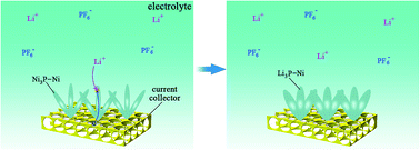
We successfully prepared advanced Ni3P–Ni array electrodes for Li-ion batteries (LIBs) by electroless deposition on 3-D nickel foam. The array structure of Ni3P–Ni can accommodate volume changes during the lithiation/de-lithiation process and promote high-rate capability because the interspaces in such structure can act as ideal volume expansion buffers. It shows excellent electrochemical performance as anode material for LIBs.
The electroless deposition technique of Ni–P alloy coating has been a well known commercial process that has found numerous applications in many fields due to excellent properties of coatings, such as high corrosion resistance, high wear resistance, good lubricity, high hardness, and acceptable ductility.23–25 We have successfully prepared the Ni–P film and Ni–P composite film.23 Herein, we report the synthesis and electrochemical properties of Ni3P–Ni arrays prepared by electroless deposited on 3-D nickel foam. The Ni3P–Ni arrays exhibits high retention capacity and good rate capability for negative electrodes.
The 3-D nickel foam (purchased from lzy battery sales department in China) with a diameter of 14 mm and copper foil (25 mm × 40 mm) (in order to avoid confusion in later analysis) were used as substrates. Specimens were immersed in acetone for 30 min and dried with hair dryer. The plating bath contained 25 g L−1 NiSO4·6H2O, 25 g L−1 NaH2PO2·H2O, 12 g L−1 CH3COONa, and 28 mL L−1 lactic acid (C3H6O3). Coating deposition took place in a thermostated vessel with a capacity of 200 cm3 maintained at 80 °C for 40 min. After the electroless deposition, fresh coatings were annealed at 400 °C for 1 h in vacuum chamber (pressure < 1 × 10⁻³ Pa) and naturally cooled with furnace to room temperature. The structure and surface morphology were characterized by X-ray diffraction (XRD, Rigaku Dmaxrc diffractometer) and scanning emission microscope (SEM, Zeiss SUPRA 55), respectively. Electrochemical performance was measured in 2016 coin-type cells. The as-prepared Ni3P–Ni arrays on 3-D nickel foam was used as the working electrode, Li sheet was used as counter electrode, and Celgard 2400 as the separator, and a mixture of 1 M LiPF6 in ethylene carbonate, dimethyl carbonate, diethyl carbonate (1
:
1
:
1 by volume) as electrolyte. Half-cells were assembled in a glovebox full of argon (purity >99.99%) with the water and oxygen concentrations below 1 ppm at room temperature. Galvanostatic discharge/charge cycles were performed between 0.01 and 3 V on a lithium battery cycler (LAND CT-2001A, Wuhan, China). Cyclic voltammetry (CV) was measured by half coin cell on a CHI 660E (Shanghai China) electrochemical workstation.
Fig. 1 shows the XRD patterns of the electroless deposited precursor film and annealed film with copper foil as substrate. Copper foil was used as the substrate here (Fig. 1a and b) in order to avoid confusion in latter analysis. There is only a wide diffraction peak in the precursor film besides the peaks of Cu substrate indicating that the precursor film is amorphous as shown in Fig. 1a. After annealed at 400 °C for 1 h, the film exhibits excellent crystallization (Fig. 1b). It shows the XRD patterns and EDX (energy-dispersive X-ray) of the electroless deposited film annealed at 400 °C for 1 h. It is clearly seen that all peaks can be well indexed to Ni3P (JCPDS no. 89-2743) (tetragonal structured) and Ni (JCPDS no. 70-0989) (cubic structured). As shown in inset, EDX confirms that the annealed electroless deposited film consists of P and Ni elements. It can be concluded that the annealed electroless deposited film consists of Ni3P and Ni.
![]() | ||
| Fig. 1 (a) XRD patterns of precursor film (electroless deposited Ni–P) with copper foil as substrate (b) XRD patterns and EDX of annealed film (Ni3P–Ni) with copper foil as substrate. | ||
Fig. 2a shows that the surface of uncoated nickel foam substrate (use as current collector) is bare. As shown in Fig. 2d, the corresponding XRD of uncoated Ni foam indicates that the uncoated Ni foam is very pure. The electroless deposited Ni–P coating on Ni foam before annealing is amorphous. After heated at 400 °C for 1 h in vacuum, the coating shows excellent crystallization (composed of Ni3P phase and Ni phase). The SEM images (Fig. 2b and c) of Ni3P–Ni coating show that the morphology and size distribution of the electroless deposited Ni3P–Ni arrays are uniform. The array structure can increase the cyclic performance efficiently because the interspaces in such structure can act as ideal volume expansion buffers.21 The 3-D Ni3P–Ni coated Ni foam is netlike, as shown in Fig. 2e. The nickel foam used as current collector with a three-dimensional network structure not only possesses high electrical conductivity and big surface area, but also it can buffer the large volume change of active materials during lithium insertion and extraction.26 As shown in Fig. 2f, the EDX mapping of elemental P corresponding to Fig. 2e confirms that the distribution of elemental P is homogeneous on 3-D nickel foam.
The lithium storage performance of Ni3P–Ni arrays was characterized by cyclic voltammetry (CV) and static-current charge/discharge. Fig. 3a shows CV curves of the Ni3P–Ni electrode in the 0.01–3 V versus Li/Li+ potential window at a scan rate of 1 mV s−1. The cathodic peak at the potential of about 1 V can be assigned to the successive phase transformation upon lithium-ion insertion via crystalline Ni3P to form Li3P alloy.9,10 During the de-lithiation scan the anodic peak at the potential of about 2.2 V is associated with the re-formation of Ni3P.27 The area of reduction peak is almost equal to that of oxidation peak in the first three CV scans, indicating high reversibility and good cycling stability. The Fig. 3b shows the voltage–capacity curves for the Ni3P–Ni electrode after 1, 10, 20 and 50 cycles at a current density of 0.02 mA cm−2 between voltage limits of 0.01–3 V. The voltage profile with different flat plateaus due to the redox reactions associated with Li+ insertion/extraction could be observed in the first discharge and charge curves. The discharge (Li insertion) and charge (Li extraction) capacities of the Ni3P–Ni were 913.7 mA h g−1 and 395 mA h g−1, respectively, corresponding to a first-cycle efficiency of 43.2% (as shown in Fig. 3c). The irreversible capacity ratio could be assigned to the decomposition of electrolyte, forming a solid/electrolyte interphase (SEI) on the electrode surface,27,28 and to the irreversible insertion of Li ions into the Ni3P–Ni. Moreover, the Ni3P–Ni arrays exhibited excellent capacity retention and maintained a stable capacity of 360 mA h g−1 with a high coulombic efficiency after 100 cycles at a current density of 0.02 mA cm−2. As shown in Fig. 3d, the Ni3P–Ni arrays were allowed to discharge/charge at higher current densities. Highly stable reversible capacities around 398, 352 and 320 mA h g−1 were obtained at current densities of 0.02, 0.04, 0.06 mA cm−2, respectively.
Compared with the morphologies of as-deposited Ni3P–Ni arrays (Fig. 4a), there is a clear volume expansion of the lithiated arrays (Fig. 4b) after 100 cycles at a current density of 0.02 mA cm−2 between voltage limits of 0.01–3 V. The large volume expansion is caused by Li insertion. However, the space between the arrays can accommodate the volume change associated with Li insertion. As shown in the schematic of the lithiation (Fig. 4c pristine and Fig. 4d lithiated), the integrity of the Ni3P–Ni electrodes can be protected by the array structure during lithiation process. In order to confirm that Li+ was actually inserted into the Ni3P–Ni arrays during cycling, XRD scans were performed on a Ni3P–Ni film grown on nickel foam substrate before and after charging with Li+. As shown in Fig. 4e, there is a shift in XRD peak positions. Compared with the Ni3P (231) peak of the as-deposited electrode, the (231) peak of the lithiated electrode has shifted to higher 2θ angles, indicating a lattice change caused by the Li+ insertion.29 The electrochemical conversion mechanism of MxPy with Li+ is commonly considered as:30–32
| MxPy + 3yLi+ + 3ye− → yLi3P + xM | (1) |
Accordingly, the electrode reaction of Ni3P may be expressed as follows:
| Ni3P + (3 − 3x)Li ⇔ (3 − 3x)Ni + (1 − x)Li3P + xNi3Punreacted | (2) |
The Ni dispersed among the Ni3P–Ni can act as the catalyst to facilitate Li3P decomposition (the converse reaction in eqn (2)) to improve the electrochemical properties of the Ni3P–Ni electrode. The material's response to the intensive cycling was examined by XRD and SEM. As shown in Fig. 4f, XRD patterns of Ni3P–Ni structure before and after being cycled for over 100 cycles are essentially unchanged. SEM micrographs of Ni3P–Ni array electrode before (Fig. 4g) and after (Fig. 4h) 100 cycles (galvanostatic discharge/charge at a current density of 0.02 mA cm−2) at full charge state confirm a good morphology stability of the Ni3P–Ni arrays electrode during the lithiation and de-lithiation processes. The morphology stability of the coating can stabilize cycling performance of the electrodes.33 That is to say, the Ni3P–Ni arrays anode for lithium-ion battery have a good cycling performance.
| This journal is © The Royal Society of Chemistry 2015 |