
Design, synthesis and antimicrobial evaluation of dihydropyrimidone based organic–inorganic nano-hybrids†
Tilak Raja,
Beant Kaur Billingb,
Navneet Kaur*b and
Narinder Singh*a
aDepartment of Chemistry, Indian Institute of Technology Ropar (IIT Ropar), Rupnagar, Panjab 140001, India. E-mail: nsingh@iitrpr.ac.in; Tel: +91 1881242176
bCentre for Nanoscience and Nanotechnology (UIEAST), Panjab University, Chandigarh, 160014, India. E-mail: navneetkaur@pu.ac.in; Tel: +91 9815245098
First published on 18th May 2015
Abstract
Substituted dihydropyrimidone derivatives were synthesized through one pot multicomponent Zn(ClO4)2 catalysed Biginelli reactions between differently substituted aromatic aldehydes, urea and ethylacetoacetate. Purified products were characterized and further engineered into metal inorganic conjugates of silver, copper and gold. Size, morphology and stability of nanoaggregates were analysed using FTIR, EDX, TEM, XRD and TGA analysis. Antimicrobial activities of all the synthesized compounds and their metal conjugates were evaluated against various bacterial and fungal strains. Results of investigations revealed that metal nanoaggregate Au@4c has shown maximum inhibition of MIC 8 against bacteria S. aureus. Further, bacterial SEM cell imaging carried out against S. aureus before and after treatment of Au@4c advocates that formation of blebs due to the accumulation of the cytoplasm are possible cause of bacterial cell death.
Introduction
Microbial infections are a leading cause of the large number of deaths every year due to the development of multidrug resistance strains in immune compromised hosts.1–9 Many new drugs have been devised from time to time to combat the microbial infections due to resistant strains. However, pharmaceutical industry faces major challenges in the discovery and development of antimicrobial drugs due to long step organic synthesis and high cost.10 Additionally, use of the harmful chemicals and undesirable side effects are other major drawbacks.11 Therefore, development new antimicrobial agents with significantly low cost and reduced or low toxicity is highly desirable in pharmaceutical industry.12–15Recently, Biginelli single step multicomponent reactions (MCRs) are emerged possible route in synthesis of the dihydropyrimidone derivatives, which possess wide range of the biological activities including antibacterial activities.16–29 At the same time, metal nanoaggregates of silver (Ag), copper (Cu), gold (Au), titanium (Ti), and zinc (Zn) and their conjugates are known for antimicrobial activity30 due to its different in their physical and chemical properties than bulk materials due to its large surface area and quantum sized effects, which makes them highly effective in the treatment.31–42 However, detrimental effect of some metal nanoaggregates to human cells are the major drawback, which can be controlled by using biocompatible metal nanoaggregates and controlling the size of metal nanoaggregates.43
Reported literature on antimicrobial potential of both dihydropyrimidones and metal nanoaggregates, encouraged us to coupled organic dihydropyrimidones with inorganic biocompatible metals44 such as silver (Ag), copper (Cu), gold (Au) to obtain organic–inorganic nanohybrids for evaluation of their antimicrobial activities against various bacterial and fungal strains. To best of our knowledge this is the first report on antimicrobial evaluation on organic metal nanoaggregates of the Biginelli compounds.
Experimental
All the chemicals were purchased from commercial supplier and were used without further purification. 1H NMR and 13C NMR spectra were recorded on JEOL II 400 spectrometer (400 MHz with TMS as internal standard; chemical shifts are expressed in ppm). Fourier transform infrared (FT-IR) of dried compounds were measured on Bruker Tensor 27 spectrophotometer, using a KBr pellet technique. Mass analysis was performed on Shimadzu 79037 GCMS GC-2010 plus. The CHN analysis was performed on Perkin Elmer 2400 CHN Elemental Analyser. Scanning electron microscopic (SEM) studies were conducted with drying the aqueous solutions of materials (10 μM). EDX images were taken with Jeol JSM-6610LV scanning electron microscope, which was operated at 15 keV. TEM images were recorded on Hitachi (H-7500) instrument worked at 80 kV. A 400-mesh carbon-coated copper grid was used for sample preparation. The nanoaggregates were suspended in aqueous medium and single drop of solution is placed onto the grid and draw-off the excess solvent after 1 min. TGA/DSC-I Star system Mettler Toledo was used to record TGA nanoaggregates. The crystal structure of the ZnO powders were analyzed with X-ray diffraction (XRD) with a PANalytical X'PERT PRO diffractometer operated at 45 kV and 40 mA using Ni-filtered Cu Kα radiations with a scan speed of 10° min−1 for 2θ in a range from 10 to 90. For measurement, an air-dried fine powder was taken and made a uniform layer on a zero background sample holder. The average crystalline size was estimated with Dewithe–Scherrer's equation using XRD analysis data.General procedure for synthesis of compounds 4a–e
All the reactions was carried out by reacting differently substituted aromatic aldehydes (15.4 mmol), ethylacetoacetate (15.4 mmol) and urea (23.1 mmol) in 15 ml of methanol by heating at reflux for 8–12 h using Zn(ClO4)2 as a catalyst. Completion of reaction was monitored by TLC. After completion of reaction, solvent was evaporated under reduced pressure and obtained residue was washed three times with methanol to obtain solid product, which was further recrystallized from acetone/water 2![[thin space (1/6-em)]](https://www.rsc.org/images/entities/char_2009.gif) :1 mixture to obtain pure product.
:1 mixture to obtain pure product.
Synthesis of organic nanoaggregates and their metal conjugates
Antimicrobial activities
Antibacterial and antifungal activities of synthesized compounds 4a–e and their organic–inorganic nanohybrids were evaluated using disc-diffusion test for pre-screening of antibacterial and antifungal potential of agents and broth micro-dilution method to determine the minimum inhibitory concentration.Antibacterial activity – materials and methods
The following bacterial strains were used: the gram negative strains used were Escherichia coli (MTCC-119, facultative anaerobic), Pseudomonas aeruginosa (MTCC-741, aerobic) and Shigella flexneri (MTCC-1457, facultative anaerobic). Gram positive strain used is Staphylococcus aureus (MTCC-740, facultative anaerobic) from Microbial Type Culture Collection, IMTECH, Chandigarh, India. Bacteria were cultivated at 37 °C in Nutrient Agar. For MIC determination Nutrient Broth was used. All compounds and standards were dissolved in dimethylsulfoxide (1/10) and applied in different concentrations. Dimethylsulfoxide was used as a negative control and antibiotics (ciprofloxacin) as positive controls.Antibacterial activity
The disk-diffusion assay was applied to determine the growth inhibition of bacteria by compounds to be tested. Further, 4 h grown bacterial cultures in Nutrient Broth (100 ml) were spread onto mueller hinton agar (MHA) and compounds 4a–e were applied to 8 mm disks (Whatman paper no. 1). After 24 h of incubation at 37 °C, the diameter of growth inhibition zones was measured.Antifungal activity – materials and methods
The following fungal strain was used: A. niger (MTCC-281), G. candidum (MTCC-3993), C. albicans (MTCC-227) and C. tropicalis (MTCC-230). Fungus was cultivated at 25 °C on Soyabean Casein Digest Medium (Tryptose Soya Broth) and MIC was determined. The compounds and standard were dissolved in dimethylsulfoxide (DMSO, 1/10) and applied in different concentrations. Dimethylsulfoxide was used as a negative control and antifungal (fluconazole) as positive controls.Antifungal activity
The disk-diffusion assay was applied to determine the growth inhibition of fungi by compounds to be tested. Overnight fungal cultures (100 ml) were spread onto Soyabean Casein Digest Medium (Tryptose Soya Broth). The compounds were applied to 8 mm disks (Whatman paper no. 1). After 48 h of incubation at 25 °C, the diameter of growth inhibition zones was measured.Result and discussions
Chemistry
All the reactions were carried using equimolar concentration of substituted benzaldehyde (1), ethylacetoacetate (2) and urea (3) in refluxing methanol using Zn(ClO4)2 as a catalyst to obtain product (4) in high yield. Progress of reaction was monitored with TLC and after completion of reaction solvent was evaporated under reduced pressure. Solid residue obtained was filtered, washed three times with methanol to get solid product. Finally, pure products were recrystallized from acetone/water 2:1 mixture of solvent (Scheme 1, Table 1). All the products 4a–e were characterized using 1H and 13C NMR, IR, mass (Fig. S1–S4†) and elemental analysis, and in comparison with reported literature data.
 | ||
| Scheme 1 Synthesis of dihydropyrimidone derivatives. | ||
| Entry | Aldehydes | Product | Reaction time (h) | Yield (%) |
|---|---|---|---|---|
| 1 | 1a | 4a | 5 | 80 |
| 2 | 1b | 4b | 4 | 85 |
| 3 | 1c | 4c | 4 | 90 |
| 4 | 1d | 4d | 6 | 86 |
| 5 | 1e | 4e | 8 | 85 |
Synthesized compounds 4a–e were tailored to their organic nanoaggregates and their biocompatible metal conjugates of silver, copper and gold.44 Formation of the metal organic framework leads to the change in physical and chemical properties of the organic compounds (Fig. 1).50 Formation of the nanoaggregates were observed metal nanoaggregates were observed with visible colour change and shift in the IR spectral peaks (Fig. S4–S7†).
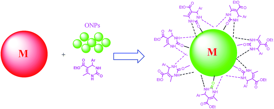 | ||
| Fig. 1 Formation of metal organic nanohybrids. | ||
Size of metal conjugated nanostructures (silver, copper, gold) on ONAs of compound 4a, were dissolved in CHCl3 to separate organic matter from metal particles. Size and morphology of compound 4a on Ag@ONAs was investigated using TEM analysis. Stability of nanoaggregates was determined using TGA analysis (Fig. S8–S10†). TEM images determine the formation of the uniform sized nanoparticles of 100 nm and elemental mapping advocates the presence of silver, nitrogen, carbon and sulphur in the sample (Fig. 2A and B). XRD analysis of the sample indicates the nanoparticle size range 134 nm (Fig. S8†). TGA analysis of the 4a capped nanoaggregates validates the stability (Fig. S9†) with variable temperature range.
 | ||
| Fig. 2 (A) EDS spectra of silver capped compound 4a nanoaggregates. (B) TEM image of compound 4a nanoaggregates. | ||
TEM results revealed the formation of needle shape Cu@ONAs of compound 4a with size 20 nm (Fig. 3A and B). Additionally, EDS profile confirmed the presence of copper, carbon and oxygen in the sample. XRD analysis of the sample indicates the nanoparticle size range 49 nm (Fig. S10†) TGA of the sample confirmed the variable temperature stability range (Fig. S11†).
 | ||
| Fig. 3 (A) EDS spectra of copper capped 4a nanoaggregates, (B) TEM image of copper capped 4a nanoaggregates. | ||
TEM analysis of Au@ONAs of compound 4a indicate the formation nanoaggregates with size range <100 nm (Fig. 4A and B) and XRD nanoparticle size 80 nm (Fig. S12†) with high TGA stability range (Fig. S13†). EDS analysis confirmed the presence of gold, carbon and oxygen in the sample.
 | ||
| Fig. 4 (A) EDS spectra of gold capped compound 4a nanoaggregates. (B) TEM image of compound 4a nanoaggregates. | ||
In vitro antifungal51,52 and antibacterial activities53 of all the synthesized compounds (4a–e) and their metal conjugates i.e., Ag@ONAs, Cu@ONAs and Au@ONAs were carried out against various fungal and bacterial strains.
Initially, in vitro antibacterial studies of all dihydropyrimidones 4a–e including their silver, copper and gold nanoaggregates were carried out against Escherichia coli (MTCC-119, facultative anaerobic), Shigella flexneri (MTCC-1457, facultative anaerobic) and Pseudomonas aeruginosa (MTCC-741, aerobic) and Staphylococcus aureus (MTCC-740, facultative anaerobic) strains by disk-diffusion assay.
The result of the disk diffusion method was utilized to determine the activity of compounds and their metal nanoaggregates against gram negative and gram positive bacteria. Results of the investigations revealed that some of the compounds including metal nanoaggregates are active against different strains of bacteria. Further, MIC values were determined using turbidimetry method (Table 2) to authenticate the results. Ciprofloxacin was used as positive controls. It was observed that some of the compounds have shown good antibacterial activities; when capped over the inorganic nanoaggregates. In the case of aerobic gram positive bacteria Staphylococcus aureus compound 4c showed inhibition of MIC 20 ± 3, however, their metal nanoaggregates of Ag@ONAs and Cu@ONAs showed inhibition of MIC 18 ± 1 and Au@ONAs showed promising MIC 8 ± 0.5. Similarly, organic compound 4a showed inhibition (MIC 15 ± 1) and their Ag@ONAs (MIC 12 ± 0.5) and Au@ONAs (MIC 10 ± 0.5). Compound 4b showed MIC 15 ± 1 and its Ag@ONAs and Au@ONAs (MIC 10 ± 0.5) and Cu@ONAs of MIC 9 ± 1. P. aeruginosa bacteria the maximum growth inhibition observed (MIC 18 ± 1) for 4c, and its Ag@ONAs (MIC 12 ± 1) and Au@ONAs (MIC 10 ± 0.5). In the case of bacteria S. flexneri no significant inhibition was observed however, some of the compounds and their nanoaggregates showed mild inhibition i.e., compound 4abc MIC 71 ± 3, >100, 73 ± 2 and their Au@ONAs MIC 60 ± 2, 72 ± 3 and 52 ± 2, respectively. Compounds 4a had shown maximum inhibition of MIC 72 ± 2 and their Ag@ONAs (MIC 18 ± 2), Cu@ONAs MIC 14 ± 1 and Au@ONAs MIC 10 ± 0.5 against anaerobic gram negative bacteria E. coli.
| Entry | Compounds | MIC (μg ml−1) | |||
|---|---|---|---|---|---|
| S. aureus MTCC-740 | P. aeruginosa MTCC-741 | S. flexneri MTCC-1457 | E. coli MTCC-119 | ||
| a Ag@4, Cu@4 and Au@4 are Ag@ONAs, Cu@ONAs and Au@ONAs nanoaggregates, respectively. | |||||
| 1 | 4a | 15 ± 1 | 30 ± 0.5 | 71 ± 3 | 72 ± 2 |
| 2 | 4b | 15 ± 1 | >100 | >100 | >100 |
| 3 | 4c | 20 ± 3 | 18 ± 1 | 73 ± 2 | 75 ± 2 |
| 4 | 4d | 80 ± 4 | 82 ± 2 | >100 | 90 ± 4 |
| 5 | 4e | 90 ± 2 | 91 ± 2 | >100 | 85 ± 4 |
| 6 | Ag@4aa | 12 ± 0.5 | 40 ± 3 | >100 | 18 ± 2 |
| 7 | Ag@4b | 10 ± 0.5 | >100 | >100 | >100 |
| 8 | Ag@4c | 18 ± 1 | 12 ± 1 | >100 | 22 ± 1 |
| 9 | Ag@4d | 70 ± 2 | 70 ± 2 | >100 | 70 ± 1 |
| 10 | Ag@4e | 72 ± 2 | 75 ± 2 | >100 | 80 ± 2 |
| 11 | Cu@4aa | >100 | >100 | >100 | 14 ± 1 |
| 12 | Cu@4b | 9 ± 1 | >100 | >100 | >100 |
| 13 | Cu@4c | 18 ± 2 | 90 ± 2 | >100 | 40 ± 1 |
| 14 | Cu@4d | 72 ± 3 | >100 | >100 | >100 |
| 15 | Cu@4e | 85 ± 2 | 80 ± 2 | >100 | >100 |
| 16 | Au@4aa | 10 ± 0.5 | 20 ± 1 | 60 ± 2 | 10 ± 0.5 |
| 17 | Au@4b | 10 ± 0.5 | >100 | 72 ± 3 | >100 |
| 18 | Au@4c | 8 ± 0.5 | 10 ± 0.5 | 52 ± 2 | 72 ± 1 |
| 19 | Au@4d | 75 ± 2 | 60 ± 2 | >100 | 80 ± 2 |
| 20 | Au@4e | 82 ± 1 | 75 ± 2 | >100 | 80 ± 1 |
| 21 | Ciprofloxacin | 1.0 | 0.7 | 1.6 | 1.2 |
In order to determine the mechanism of the cell death in the case of S. aureus, cell imaging of bacteria S. aureus was conducted by keeping samples were overnight fixed with glutaraldehyde. Cell suspensions were centrifuged and washed three times with deionised water that had previously been filtered through a 0.22 nm micropore filter. After resuspension in deionised water, one drop (0.025 ml) of each suspension was added to 1 ml of absolute ethyl alcohol, which was applied to a standard metal support plate and allowed to dry in air. All the images are recorded at different interval of time after and were examined in a scanning mode, with Jeol JSM-6610LV scanning electron microscope which operated at 15 keV (Fig. 5).
 | ||
| Fig. 5 SEM Images of the S. aureus at different interval of time: (A) before addition of Au@4c, (B) immediate after treatment of compound with Au@4c, (C) after 12 h incubaction after treatment, (D) 18 h incubaction after treatment, (E) 24 h incubaction after treatment, (F) close cell image of the bacteria after 24 h incubaction. | ||
Results of investigations revealed that bacteria showed regular spherical shape before addition of the Au@4c. Bacterial cell images were taken at different interval of incubation time after addition of compound Au@4c (Fig. 5B–D). Further, after 18 h of the treatment morphological changes were observed in the bacterial cells as shown in Fig. 4D through the formation of blebs54 on the surface of the bacterial cells which might be due to the accumulation of the cytoplasm. After 24 h incubation cells starts rupturing, which lead to the bacterial cell death (Fig. 5E and F).
Antifungal evaluations were also carried out against various fungal strains A. niger (MTCC-281), G. candidum (MTCC-3993), C. albicans (MTCC-227) and C. tropicalis (MTCC-230) using turbidimetry method and determined in terms of MIC (μg ml−1) (Table 3). Few strains have shown marginal activity against fungal strain. In the case of Geotrichum candidum, compound 4a with MIC 40 ± 1 μg ml−1 and its Ag@ONAs (MIC 30 ± 2) and Au@ONAs (MIC 25 ± 2) showed maximum inhibition.
| Entry | Compds | MIC (μg ml−1) | |||
|---|---|---|---|---|---|
| A. niger (MTCC-281) | G. candidum (MTCC-3993) | C. albicans (MTCC-227) | C. tropicalis (MTCC-230) | ||
| a Ag@4, Cu@4 and Au@4 are Ag@ONAs, Cu@ONAs and Au@ONAs of 4a–e, respectively. | |||||
| 1 | 4a | >100 | 40 ± 1 | 99 ± 1 | >100 |
| 2 | 4b | >100 | 60 ± 3 | >100 | >100 |
| 3 | 4c | >100 | >100 | 99 ± 2 | >100 |
| 4 | 4d | >100 | >100 | >100 | >100 |
| 5 | 4e | >100 | >100 | >100 | >100 |
| 6 | Ag@4aa | >100 | 30 ± 2 | 60 ± 2 | >100 |
| 7 | Ag@4b | >100 | 55 ± 4 | >100 | >100 |
| 8 | Ag@4c | >100 | >100 | 95 ± 1 | >100 |
| 9 | Ag@4d | >100 | >100 | >100 | >100 |
| 10 | Ag@4e | >100 | >100 | >100 | >100 |
| 11 | Cu@4aa | >100 | >100 | 99 ± 2 | >100 |
| 12 | Cu@4b | >100 | 45 ± 2 | 94 ± 3 | >100 |
| 13 | Cu@4c | >100 | 97 ± 1 | >100 | >100 |
| 14 | Cu@4d | >100 | >100 | >100 | >100 |
| 15 | Cu@4e | >100 | >100 | >100 | >100 |
| 16 | Au@4aa | >100 | 25 ± 2 | 40 ± 3 | >100 |
| 17 | Au@4b | >100 | 40 ± 4 | >100 | >100 |
| 18 | Au@4c | >100 | 96 ± 1 | >100 | >100 |
| 19 | Au@4d | >100 | >100 | >100 | >100 |
| 20 | Au@4e | >100 | >100 | >100 | >100 |
| 21 | Fluconazole | 42 | 12 | 24 | 20 |
To determine the cytotoxicity of organic–inorganic nanohybrids of Ag@4b, Cu@4b and Au@4c was carried out using MTT assay with HeLa cells cultured in nutrient mixture. As per the standard protocol54 the cells were incubated at 37 °C with 5% CO2 environment for 24 h. Further, on addition of Ag@4b, Cu@4b and Au@4c, only marginal cell death (96–98% cell survival) at concentration (20 μM). However, in order to investigate the potential cytotoxic effects of metal nanoaggregates at higher concentrations, the dose dependent studies were performed. The results indicate that all given organic–inorganic nanohybrids are not much toxic to HeLa cells, if all given organic–inorganic nanohybrids concentrations remains under 200 μM, that is with 84–86% cell survival over 24 h for 200 μM concentration of Ag@4b, Cu@4b and Au@4c.
Conclusion
Biginelli compounds 4a–e were synthesized and processed to organic nanoaggregates (ONAs) for further conversion to their metal conjugates of silver, copper and gold. All the compounds and their metal conjugates were evaluated for antimicrobial activities against different bacterial and fungal strains. Results of the investigations revealed that antimicrobial activity of some of compounds increased upon conversion to their organic–inorganic nanohybrids i.e., organic compound 4c showed MIC 20 ± 3 and their Au@4c showed MIC 08 ± 0.5 against S. aureus bacteria. Further, mechanism of the cell death preliminarily was determined using time dependent SEM analysis. The results of investigations revealed that possible cause of the cell death in the case of S. aureus is due to accumulation of the cytoplasm on the outer membrane of the bacteria leading to bleb formation. These organic–inorganic nanoaggregates can be used as ‘lead’ compounds for further antimicrobial drug development using organic–inorganic nanohybrids.Acknowledgements
Dr Tilak Raj is thankful to UGC, New Delhi, India for Post Doctoral fellowship Grant no. PDFSS-SC-PUN-34.Notes and references
- K. E. Jones, N. Patel, M. Levy, A. Storeygard, D. Balk, J. Gittleman and P. Daszak, Nature, 2008, 451, 990–993 CrossRef CAS PubMed.
- A. J. Lax, Nat. Rev. Microbiol., 2005, 3, 343–349 CrossRef CAS PubMed.
- M. Kamboj and K. A. Sepkowitz, Lancet Oncol., 2009, 10, 589–597 CrossRef.
- M. J. Hill, Eur. J. Cancer Prev., 1995, 4, 127–128 CAS.
- M. N. Alekshun and S. B. Levy, Cell, 2007, 128, 1037–1050 CrossRef CAS PubMed.
- G. D. Wright, Nat. Rev. Microbiol., 2007, 5, 175–186 CrossRef CAS PubMed.
- L. M. Weigel, D. B. Clewell, S. R. Gill, N. C. Clark, L. K. McDougal, S. E. Flannagan, J. F. Kolonay, J. Shetty, G. E. Killgore and F. C. Tenover, Science, 2003, 302, 1569–1571 CrossRef CAS PubMed.
- D. Banerjee and D. Stableforth, Drugs, 2000, 60, 1053–1064 CrossRef CAS PubMed.
- N. Woodford and D. Livermore, J. Infect., 2009, 59, S4–S16 CrossRef.
- A. E. Barnhill, M. T. Brewer and S. A. Carlson, Antimicrob. Agents Chemother., 2012, 56, 4046–4051 CrossRef CAS PubMed.
- (a) M. B. Deshmukh, S. M. Salunkhe, D. R. Patil and P. V. Anbhule, Eur. J. Med. Chem., 2009, 44, 2651 CrossRef CAS PubMed; (b) K. Singh, D. Arora, E. Poremsky, J. Lowery and R. S. Moreland, Eur. J. Med. Chem., 2009, 44, 1997 CrossRef CAS PubMed; (c) C. O. Kappe, Acc. Chem. Res., 2000, 33, 879 CrossRef CAS PubMed.
- W. B. Hugo and A. D. Russell, Types of antimicrobial agents, in principles and practice of disinfection, preservation and sterilization, Blackwell Scientific Publications, Oxford, UK, 1982, pp. 106–108 Search PubMed.
- M. Adolfsson-Erici, M. Pettersson, J. Parkkonen and J. Sturve, Chemosphere, 2002, 46, 1485–1489 CrossRef CAS.
- S. Ghosh, P. Chakraborty, P. Saha, S. Acharya and M. Ray, RSC Adv., 2014, 4, 23251–23261 RSC.
- S. Ghosh, D. Ghosh, P. K. Bag, S. C. Bhattacharyac and A. Saha, Nanoscale, 2011, 3, 1139–1148 RSC.
- (a) M. B. Deshmukh, S. M. Salunkhe, D. R. Patil and P. V. Anbhule, Eur. J. Med. Chem., 2009, 44, 2651 CrossRef CAS PubMed; (b) K. Singh, D. Arora, E. Poremsky, J. Lowery and R. S. Moreland, Eur. J. Med. Chem., 2009, 44, 1997 CrossRef CAS PubMed; (c) C. O. Kappe, Acc. Chem. Res., 2000, 33, 879 CrossRef CAS PubMed.
- (a) R. Gupta, A. K. Gupta, S. Paul and P. L. Kachroo, Indian J. Chem., Sect. B: Org. Chem. Incl. Med. Chem., 1995, 34, 151 Search PubMed; (b) J. Barluengo, M. Tomas, V. Rubio and V. J. Gotor, J. Chem. Soc., Chem. Commun., 1979, 675 RSC.
- (a) R. Ghosh, S. Maiti and A. Chakraborty, J. Mol. Catal., 2004, 27, 47 CrossRef PubMed; (b) R. V. Yarapathi, S. Kurva and S. Tammishetti, Catal. Commun., 2004, 5, 511–513 CrossRef CAS PubMed; (c) K. S. Atwal, G. C. O. Rovnyak, B. C. Reilly and J. Schwartz, J. Org. Chem., 1989, 54, 5898 CrossRef CAS.
- (a) H. Salehi and Q. Guo, Synth. Commun., 2004, 34, 171 CrossRef CAS; (b) S. Tu, F. Fang, S. Zhu, T. Z. Li, X. Hangs and Q. Zhuang, Synlett, 2004, 537 CrossRef CAS PubMed; (c) H. Hazarkhani and B. Karimi, Synthesis, 2004, 8, 1239 Search PubMed.
- T. Matsuda and I. Hirao, J. Chem. Soc. Jpn., 1965, 86, 1195 CAS.
- E. W. Hurst and R. I. Hull, J. Med. Pharm. Chem., 1961, 3, 215 CrossRef CAS.
- (a) S. S. Bokaeva and T. K. Nauch-Issled, Inst. Onkol. Radiol., 1967, 3, 305 (Chem. Abstr., 1968, 68, 103775q) CAS; (b) A. Zidermane, G. Duburs, A. Zilbere, R. Verpele, J. Uldrlkis and K. Kumsars, Latv. PSR Zinat. Akad. Vestis, 1971, 77 (Chem. Abstr, 1971, 75, 47266e) Search PubMed.
- K. Kumsars, A. Velena, G. Duburs, J. Uldrikis and A. Zidermane, Biokhimiya, 1971, 36, 1201 Search PubMed.
- T. Kato, JP Patent, Kokai Tokkyo Koho, Japan, 1984, vol. 59, 190, p. 974, Chem. Abstr. 1985, 102, 132067.
- Y. S. Sadanandam, M. M. Shetty and P. V. Diwan, Eur. J. Med. Chem., 1992, 27, 87 CrossRef CAS.
- K. Cooper, PCT Int. Appl., WO 11, Pfizer Ltd., 1990, pp. 281–20, Chem. Abstr. 1991, 114, 143437f Search PubMed.
- M. Kurono, M. Hayashi, K. Miura, Y. Isogawa, K. Sawai, JP 62, 267, 272, Sanwa Kagaku Kenkyusho Co., Japan, Kokai Tokkyo Koho, 1987Chem. Abstr. 1988, 109, 37832t.
- M. A. Gallop, R. W. Barret, W. J. Dower, S. P. Fodor and E. M. Gordon, J. Med. Chem., 1994, 37, 1233 CrossRef CAS.
- (a) V. V. Dabholkar and T. D. Ravi, J. Serb. Chem. Soc., 2010, 75, 1033 CrossRef CAS; (b) S. Jayakumar and T. K. Shabeer, J. Chem. Pharm. Res., 2011, 3, 1089 CAS; (c) Suresh and J. S. Sandhu, ARKIVOC, 2012, I, 66 Search PubMed.
- C. Malarkodi, S. Rajeshkumar, K. Paulkumar, M. Vanaja, G. Gnanajobitha and G. Annadurai, Bioinorg. Chem. Appl., 2014, 2014, 1–10 CrossRef PubMed.
- H. K. Daima, P. R. Selvakannan, A. E. Kandjani, R. Shukla, S. K. Bhargava and V. Bansal, Nanoscale, 2014, 6, 758 RSC.
- Y. Zhao and X. Jiang, Nanoscale, 2013, 5, 8340 RSC.
- S. Agnihotri, S. Mukherji and S. Mukherji, Nanoscale, 2013, 5, 7328 RSC.
- A. Huczko, Appl. Phys., 2000, 70, 365–376 CAS.
- X. Battle and A. J. Labarta, Appl. Phys., 2002, 35, 15–42 Search PubMed.
- J. P. Ruparelia, A. K. Chatteriee, S. P. Duttagupta and S. Mukherji, Acta Biomater., 2008, 4, 707–716 CrossRef CAS PubMed.
- M. R. Bindhu and M. Umadevi, Mater. Lett., 2014, 120, 122–125 CrossRef CAS PubMed.
- N. G. Kandile, H. T. Zaky, M. I. Mohamed and H. M. Mohamed, Bull. Korean Chem. Soc., 2010, 31, 3530–3538 CrossRef CAS.
- M. L. Schipper, N. Nakayama-Ratchford, C. R. Davis, N. W. S. Kam, P. Chu, Z. Liu, X. Sun, H. Dai and S. S. Gambhir, Nat. Nanotechnol., 2008, 3, 216–221 CrossRef CAS PubMed.
- P. V. AshaRani, G. L. K. Mun, M. P. Hande and S. Valiyaveettil, ACS Nano, 2009, 3, 279–290 CrossRef CAS PubMed.
- R. Duncan, Nat. Rev. Cancer, 2006, 6, 688–701 CrossRef CAS PubMed.
- A. Panacek, L. Kvitek, R. Prucek, M. Kolar, R. Vecerova, N. Pizurova, V. K. Sharma, T. Nevecna and R. Zboril, J. Phys. Chem. B, 2006, 110, 16248–16253 CrossRef CAS PubMed.
- A. M. Schrand, M. F. Rahman, S. M. Hussain, J. J. Schlager, D. A. Smith and A. F. Syed, Wiley Interdiscip. Rev.: Nanomed. Nanobiotechnol., 2010, 2, 544–568 CrossRef CAS PubMed.
- (a) M. A. Omary, Z. Hu, S. Marpu and O. Elbjeirami, US Pat., no. US20100172997 A1, 2009; (b) K. D. Arunachalam, S. Kumar Annamalai and S. Hari, Int. J. Nanomed., 2013, 8, 1307–1315 CrossRef PubMed; (c) C. Singh, R. K. Baboota, P. K. Naik and H. Singh, Adv. Mater. Lett., 2012, 3, 279–285 CrossRef CAS PubMed.
- (a) S. A. Kotharkar, R. R. Nagwade and D. B. Shinde, Ukr. Bioorg. Acta, 2006, 2, 17 Search PubMed; (b) O. Muñoz-Muñiz and E. Juaristi, ARKIVOC, 2003, xi, 16–26 Search PubMed; (c) Y. Huang, F. Yang and C. Zhu, J. Am. Chem. Soc., 2005, 127, 16386 CrossRef CAS PubMed; (d) H. H. Essa, R. S. Daniel and D. H. Ulf, J. Org. Chem., 1998, 63, 3454 CrossRef; (e) A. Stadler and C. O. Kappe, J. Chem. Soc., Perkin Trans. 1, 2000, 1363 RSC; (f) H. A. Stefani and P. M. Gatti, Synth. Commun., 2000, 30, 2165 CrossRef CAS PubMed.
- C. O. Kappe, D. Kumar and R. S. Varma, Synthesis, 1999, 1799–1803 CrossRef CAS PubMed.
- R. O. Al-Kaysi, A. M. Müller, T. S. Ahn, S. Lee and C. J. Bardeen, Langmuir, 2005, 21, 7990 CrossRef CAS PubMed.
- A. Singh, V. Kumar Bhardwaj, G. Kaur, K. Kaur, N. Singh and M. S. Bakshi, RSC Adv., 2014, 4, 21079 RSC.
- S. Avvakumova, P. Verderio, G. Speranza and F. Porta, J. Phys. Chem. C, 2013, 117, 3002 CAS.
- C. D. Mitic, G. B. Vukovic, V. J. Knezevic, S. Stankovic and D. Simic, Arch. Biol. Sci., 2005, 57, 173 CrossRef.
- R. A. Fromtling, J. N. Galgiani, M. A. Pfaller, I. A. Espinel, K. F. Bartizal, M. S. Bartlett, B. A. Body, C. Frey, G. Hall, G. D. Roberts, F. B. Nolte, F. C. Odds, M. G. Rinaldi, A. M. Sugar and K. Villareal, Antimicrob. Agents Chemother., 1993, 37, 39 CrossRef CAS.
- J. Hindler, in Textbook of Diagnostic Microbiology Special Antimicrobial Susceptibility Tests, ed. C. C. Mahon and G. Manuselis, 1995, p. 89 Search PubMed.
- E. L. Chan, R. C. Harris and H. P. Dalton, J. Med. Microbiol., 1987, 23, 149 CrossRef CAS PubMed.
- A. H. Cory, T. C. Owen, J. A. Barltrop and J. G. Cory, Cancer Commun., 1991, 3, 207 CAS.
Footnote |
| † Electronic supplementary information (ESI) available: 1H, 13C and mass spectra of compounds and cytotoxicty results. See DOI: 10.1039/c5ra08765a |
| This journal is © The Royal Society of Chemistry 2015 |