Influence of a blend of guar gum and poly(vinyl alcohol) on long term stability, and antibacterial and antioxidant efficacies of silver nanoparticles

Tamalika Das a, Sabina Yeasmin a, Somanjana Khatua b, Krishnendu Acharya b and Abhijit Bandyopadhyay *a
aDepartment of Polymer Science & Technology, University of Calcutta, 92, A.P.C. Road, Kolkata – 700009, West Bengal, India. E-mail: abpoly@caluniv.ac.in; Tel: +91 033 2350-1397 ext 288 Tel: +91 033 2350-6996 ext 288
bMolecular and Applied Mycology and Plant Pathology Laboratory, Department of Botany, University of Calcutta, 35, Ballygunge Circular Road, Kolkata – 700019, West Bengal, India

Received 5th May 2015 , Accepted 12th June 2015

First published on 15th June 2015


Abstract

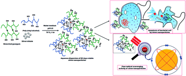
This paper describes a detailed procedure for the synthesis of highly stable (almost for 60 days) and monodispersed silver nanoparticles (average particle size of 16 nm which changed to 42 nm only on the 60th day of standing under refrigeration) in the presence of a blend of a polysaccharide (guar gum) and a hydrophilic synthetic polymer (poly(vinyl alcohol)) where both the polymers provided synergistic effects in reduction and stabilization of incipient nanoparticles. Time dependant stability of aqueous dispersions of silver nanoparticles in various compositions of polymeric matrices was studied from UV-Vis spectroscopy, DLS and zeta potential characterization data. An FTIR spectrum and HRTEM image along with EDX results of the best optimized compositions in respect of particle size and particle size distribution of silver nanoparticles were also obtained. A study displayed how viscosity of the polymeric matrices played a vital role in the long term stabilization of aqueous dispersions of silver nanoparticles. For the first time we are reporting that silver nanoparticles synthesized by a green technique, using a blend of two highly viscous polymers, exhibited excellent stability. The aqueous dispersions of silver nanoparticles also displayed strong antibacterial and antioxidant properties. Hence our synthesized silver nanoparticles may be suitable for commercialization in future.


Introduction

Silver nanoparticles (AgNPs) are prevalently used in various consumer products,1–4 food additives and packaging,5 optoelectronic devices,6,7 biomedical devices,8,9 health care products10 and waste water treatment units11 as one of the essential components. The presence of large proportions of surface particles with high energy due to higher surface area (surface effect) with respect to the bulk of the material and squeezing of electrons into a small area (quantum confinement effect) enable AgNPs to easily adsorb to a variety of molecules or macromolecules even under harsh environmental conditions.12–14 AgNPs exhibit highly distinguishing size and shape tunable properties (optical,15 surface plasmon,16 chemical,17 catalytic activities,17 electrical,18 and thermal19) which make them potential candidates in wide spectrum of products, many of which have already been commercialized.20 The good antimicrobial efficacy of AgNPs even at very low dosage owing to oligo dynamic action21 and reduced problem of development of microbial resistance in any environment, promotes design of many consumer and biomedical products where antimicrobial property in nano-dimension is essential.22,23 AgNPs also exhibit antifungal activities and hence find application in filter beds.24 Interestingly, AgNPs effectively inhibit replication of HIV-1 viruses.25 Other useful properties of AgNPs that provoke more and more research in this field include anti-inflammatory activities,26 antiviral effects,25 metal enhanced fluorescence properties27 and plasmonic heating properties.28 However, exploration of antioxidant properties of AgNPs is quite an overlooked field. Abdel-Aziz et al. in a recent study highlighted antioxidant properties of AgNPs.29 Oxidation is desirable in all living organisms including microbes, for production of energy. However, during oxidative stress in human body, highly reactive radicals (e.g., hydroxyl radicals from splitting of water molecules on exposure to harmful gamma/UV rays or superoxide radicals from biochemical reactions between oxygen and bio-macromolecules like adrenaline, dopamine or some components of mitochondria etc.) are generated which hinders normal functioning of DNA, RNA, proteins and lipids.30 As a consequence of the generation of free radicals in human cells, often cells are injured and cause necrosis or apoptosis leading to various diseases like cancer, cardiovascular problems, diabetes, ageing etc.31 Inclusion of AgNPs in medicinal compositions and various anti-ageing formulations may prove to be beneficial in scavenging any undesirable free radicals. Often packaged foods are spoiled by growth of microorganisms and by free radicals generated from their metabolism processes. However, commercial antioxidants are highly toxic. Thus to deal with the issue of spoilage of food, Kanatt et al. used non toxic extracts of aqueous mint and pomegranate peels as highly efficient antioxidants and antimicrobials in a biodegradable film of chitosan blended PVA for active food packaging.32 Again, isolation of plant extracts always proves to be highly tedious and expensive processes for which most of the greener antimicrobials and antioxidants are deliberately avoided in commercial grounds. On the contrary, commercially viable aqueous dispersions of AgNPs if incorporated in biodegradable and water soluble film formulations may produce cost effective active packaging systems. Often AgNPs tend to leach out from polymeric compositions.33 Although AgNPs are toxic yet when they leach out of the polymer compositions they tend to agglomerate very fast and hence reduce toxicity. Also, AgNPs that leach out of any highly viscous polymer stabilized compositions, are generally in very small concentration and thus are not lethal to human. Hence here we tried to prepare a composition by in situ generation of AgNPs in a biocompatible polymer blend which may be used safely in commercial products. Among the various techniques followed in the in situ synthesis of AgNPs (chemical reduction method using hazardous chemicals,34,35 electrochemical method,36 photochemical method,37,38 radiochemical method,39 sonochemical method,40,41 laser ablation technique42etc.), green synthesis technology is the most favored one owing to the reduced environmental hazards associated with the synthesis procedure and economical viability of the green technology for large scale productions. Natural polymers especially polysaccharides owing to the presence of reactive functional groups (e.g., hydroxyls, aldehydes, amines etc.) are widely used to reduce silver precursors and to stabilize AgNPs. Monodispersed AgNPs were successfully prepared using starch,43 chitosan,44 dextran,45 gum arabic46etc. in a one pot process. An abundantly available edible, non ionic, branched and pH responsive polysaccharide, guar gum (GG) is water soluble owing to the presence of extensive galactose units (i.e.; larger number of hydroxyl groups is present as compared to other polysaccharides). Hence GG may be used as a reducing cum protective agent for synthesis of AgNPs in an aqueous solution. In fact GG was successfully utilized by Pandey et al. to develop AgNPs for ammonia sensing.47 However, till date many safer green synthesis methods of AgNPs have not been commercialized owing to poor stability of stored aqueous dispersions of AgNPs. A research group of Pinto claimed borohydride reduced and citrate stabilized AgNPs were morphologically stable for an extended period of one year.48 Similarly, Popa's group produced AgNPs which were stable for a month in presence of short chains of polyethylene glycol acting as solvent, reducer and stabilizer.49 However, none of the gum or plant extract stabilized aqueous dispersions of AgNPs were stable for an extended period even under refrigeration. In our work, we tried to develop a greener method which could produce long term stable aqueous dispersions of AgNPs. We thought reducing and stabilizing power of GG during in situ preparation of AgNPs could be further enhanced by using another hydrophilic polymer rich in hydroxyl groups and highly compatible with GG. Poly(vinyl alcohol) (PVA) is highly compatible with GG owing to the formation of intra molecular hydrogen bonds. PVA being of synthetic origin would not only increase reducing and stabilizing power of GG by providing more hydroxyl groups to the system but would also impart biocompatibility, biodegradability, good mechanical properties combined with film forming ability to the AgNPs – (GG + PVA) conjugate. Here we prepared highly stable aqueous dispersions of AgNPs (stable nearly for 2 months) by a one pot synthesis approach using an optimized blend of GG and PVA. We then precisely enumerated antibacterial and antioxidant properties of the polymer compositions impregnated with AgNPs.

Experimental details

Materials

GG with an average molecular weight of 1.06 × 10⁶ g mol−1 was obtained as a gift from Hindustan Gum and Chemicals Ltd, Haryana, India. PVA (number average molecular weight 1.15 × 10⁵ g mol−1, degree of hydrolysis ∼98% and purity 99%), AgNO3 (99.9% pure), NaOH pellets, KOH pellets, KH2PO4, concentrated HCl, H2O2, acetone and KBr (spectroscopic grade) were purchased from Merck, India. Bacillus subtilis [MTCC 736] bacterial strains and agar powder were obtained from HiMedia Laboratories Pvt. Ltd, Mumbai, India. Butylated hydroxyl toluene (BHT), ethylenediaminetetraaceticacid (EDTA), ferrous chloride, ferric chloride, ascorbic acid, nitroblue tetrazolium (NBT), trichloroacetic acid (TCA), thiobarbituric acid (TBA), 2-deoxyribose, sodium phosphate, methionine, riboflavin and ferrozine disodium salt were purchased from Sigma chemicals Co. (St. Louis, MO, USA). All reagents and solvents were of analytical grade and were used without further purification. For preparation of any aqueous solution, MilliQ water was used. For synthesis of AgNPs, all glass wares that were used were thoroughly cleaned with chromic acid and double distilled water.
In situ green synthesis of AgNPs using GG, PVA and a combination pack of GG and PVA. In a typical process, an aqueous solution of GG of maximum permissible concentration (1.0 w/v) was prepared under continuous stirring at a temperature of 50 °C. We ensured no lumps of gum were present in the resultant solution of GG and a completely soluble form was obtained only after 48 hours of continuous stirring. We were unable to prepare completely soluble higher concentrated aqueous solution of GG above 1.0 w/v and hence we discarded any higher concentrated solution of GG for synthesis of AgNPs. The aqueous solution of GG was then cooled to room temperature. Three different sets of GG solution in MilliQ (1.0 w/v) were prepared by adjusting pH to ∼4.0, ∼6.0 and ∼8.0 respectively. Finally AgNO3 solution (1 ml, 0.1 (N)) was added to the pH adjusted solution of GG and stirred vigorously for 1 h. The reactions were carried out at 25 °C, 50 °C, 70 °C and 100 °C respectively. At the end of the reactions, the final dispersions containing AgNPs were diluted five times with MilliQ water and centrifuged at 10[thin space (1/6-em)]000 rpm for 30 minutes. The supernatant solutions were carefully isolated and allowed to undergo sonication for further 15 minutes. Finally the aqueous dispersions were stored under refrigeration. Similarly, an aqueous solution of PVA (5.0 w/v) and an aqueous solution of a mixture of GG of strength 1.0 w/v (30 wt%, 50 wt%, 70 wt% w.r.t PVA) and PVA of strength 5.0 w/v (70 wt%, 50 wt% and 30 wt% w.r.t 30 wt%, 50 wt% and 70 wt% GG respectively) were used to synthesize AgNPs dispersions at different pH (∼4.0, ∼6.0 and ∼8.0) and different temperatures (25 °C, 50 °C, 70 °C and 100 °C) by the procedure as described above. All the experiments were also repeated thrice to check reproducibility of the synthesis processes.
Characterization of aqueous dispersion of AgNPs. UV-Vis spectra of aqueous dispersions of polymer stabilized AgNPs (concentration 150 μg ml−1, adjusted after dilution) were recorded against air at 25 °C in a UV-Vis spectrophotometer (CECIL CE7200, 7000 series, UK) from 200 nm to 600 nm using a quartz cuvette of 1 cm optical path length. MilliQ water was used as the blank. Formation of miscible blend of GG with PVA and alteration in structure of the blend on development of AgNPs was studied from FTIR spectra in transmission mode on Shimadzu IR affinity-18000 FTIR spectrometer. Thoroughly dried KBr pellets from the respective samples were prepared for FTIR spectroscopic studies. Hydrodynamic diameter and zeta potential of various compositions of polymer stabilized aqueous dispersions of AgNPs (concentration 150 μg ml−1) in MilliQ was studied on Zeta Sizer Nano Series, Malvern, USA. Particle size and particle size distribution of polymer stabilized AgNPs in the dry state was studied by HRTEM (model JEM, 2010 EM, fitted with a CCD camera) at an accelerating voltage of 50 kV. For HRTEM analysis, an aqueous dispersion of AgNPs70/30/6.1 (concentration 50 μg ml−1) was drop casted on a carbon coated copper grid of 300 mesh size and vacuum dried for 2 days prior to capturing of images. Finally EDX mapping of the same sample was also done using the EDX attachment to HRTEM in order to analyze the exact elemental composition of the dried colloids.
Assay of antibacterial efficacy of GG + PVA stabilized AgNPs. Agar diffusion method using Bacillus subtilis, a gram positive bacteria strain [MTCC 736] was used to analyze the antibacterial properties of AgNPs stabilized by GG and PVA. In a typical process, MTCC 736 bacterial strains were grown on liquid nutrient agar medium for 24 hours just prior to the experiment being seeded by pour plate technique. Four different cups like cavities were made using a cork borer (10 mm) at more or less equal distances. The cavities were chronologically filled with an aqueous solution (1 ml) of (I) AgNO3 (0.1 N) + GG (0.2 mg ml−1), (II) AgNO3 (0.1 N) + PVA (0.2 mg ml−1), (III) AgNO3 (0.1 N) + GG (0.14 mg ml−1) + PVA (0.06 mg ml−1) where 70 wt% GG: 30 wt% PVA ratio was maintained and (IV) an aqueous dispersion of AgNPs70/30/6.1 (0.2 mg ml−1). Finally the plate was incubated at 37 °C for 24 hours.
Assay of superoxide anion radicals scavenging activity of GG + PVA stabilized AgNPs. A method developed by Martinez et al.50 for determination of the superoxide dismutase with modification in the riboflavin-light-nitroblue tetrazolium (NBT) system51 was followed. Each 3 ml reaction mixture contained 50 mM sodium phosphate buffer (pH 7.8), 13 mM methionine, 2 μM riboflavin, 100 μM EDTA, 75 μM NBT and 500 μl test sample solution at a concentration of 2 mg ml−1 (containing aqueous solution of Gum100/0/6.2, Gum0/100/6.1, AgNPs70/30/6.1 or AgNPs50/50/6.1). Reaction was initiated by illuminating sample by a fluorescent lamp. Finally the production of blue formazan was followed by monitoring the increase in absorbance at 560 nm after 10 min of illumination. Identical tubes with the reaction mixture were kept in the dark and served as blank. Percentage scavenging of superoxide anion radicals was studied by comparing absorbance at 590 nm with the solution of control (50 mM sodium phosphate buffer) and the test samples (with antioxidants). A blank test was also performed using test samples without containing any antioxidants.
Assay of hydroxyl radicals scavenging activity of GG + PVA stabilized AgNPs. Here we adopted the work of Halliwell et al. for determination of hydroxyl radicals scavenging efficiency of test samples.52 In a typical process, a freshly prepared reaction mixture (1 ml) contained 2-deoxyribose (2.8 mM), KH2PO4–KOH buffer (20 mM; pH 7.4), FeCl3 (100 mM), EDTA (104 μM), H2O2 (1 mM), ascorbate (100 μM) and 500 μl test sample at a concentration of 2 mg ml−1 (containing aqueous solution of Gum100/0/6.2, Gum0/100/6.1, AgNPs70/30/6.1 or AgNPs50/50/6.1). Reaction was started by addition of H2O2 and then incubated at 37 °C for 1 h. Finally approximately 2 ml TBA–TCA solution (acidified solution of TBA and TCA: 100 ml containing 380 mg TBA, 20 mg TCA) were added in order to inhibit the reaction initiated by H2O2 and then incubated at 100 °C for 15 min. On sufficient cooling of the test sample, absorbance was measured at 535 nm against the sample where TBA–TCA was added prior to reaction mixture. BHT was used as the positive control. Percent inhibition of 2-deoxyribose degradation by the test sample at a concentration of 1 mg ml−1 over the control was measured.
Chelating ability test of ferrous ions GG + PVA stabilized AgNPs. Chelating ability of various samples was determined according to the method of Dinis et al. (1994).53 Aqueous solution of test sample (containing aqueous solution of Gum100/0/6.2, Gum0/100/6.1, AgNPs70/30/6.1 or AgNPs50/50/6.1) was mixed with water (3.7 ml) so that final concentration of the samples became 1 mg ml−1. Finally FeCl2 (0.1 ml, 2 mM) was added to the resultant mixture. The reaction was initiated by the addition of ferrozine (0.2 ml, 5 mM). After 10 min at room temperature, the absorbance of the mixture was determined at 562 nm against a blank (without any test samples but containing another chelating agent, EDTA).

Results and discussions

Preparation and characterization of AgNPs in aqueous solutions of GG, PVA and a blend of GG with PVA

An environment friendly bottom up method was used to prepare highly stable and monodispersed AgNPs. All the compositions of polymer matrices that were varied to synthesize AgNPs are tabulated in Table 1.
Table 1 Sample designation, reaction conditions and various properties of AgNPs of different compositionsa
Sample designation GG (wt%) PVA (wt%) Exact pH maintained during synthesis of AgNPs Conc. Of AgNO3 (N) Time of reaction (h) Time of recording data counted from the end of reaction Average hydrodynamic diameter of AgNPs in dispersions (nm) Zeta potential values of dispersion of AgNPs, ε (mV)
a All the reactions were carried out in water medium at 70 °C. In our work, AgNPs is an abbreviated form of silver nanoparticles in aqueous dispersions.
AgNPs100/0/6.2 100 0 6.2 0.1 1 2 h 950 −0.48
AgNPs0/100/6.1 0 100 6.1 0.1 1 2 h 866 −2.41
AgNPs30/70/6.0 30 70 6.0 0.1 1 2 h 72 −7.88
AgNPs50/50/6.1 50 50 6.1 0.1 1 2 h 37 −9.00
AgNPs70/30/6.1 70 30 6.1 0.1 1 2 h 20 −11.00
AgNPs100/0/8.0 100 0 8.0 0.1 1 2 h 700 −4.93
AgNPs0/100/8.3 0 100 8.3 0.1 1 2 h 1425 −3.43
AgNPs70/30/8.1 70 30 8.1 0.1 1 2 h 230 −6.45
AgNPs70/30/6.1 70 30 6.1 0.1 1 24 h 22 −10.70
AgNPs70/30/6.1 70 30 6.1 0.1 1 7 days 35 −9.96
AgNPs70/30/6.1 70 30 6.1 0.1 1 30 days 48 −9.78
AgNPs70/30/6.1 70 30 6.1 0.1 1 60 days 52 −1.96
AgNPs50/50/6.1 50 50 6.1 0.1 1 30 days 140 −4.40
AgNPs30/70/6.0 30 70 6.0 0.1 1 30 days 153 −2.18


GG being a reducing sugar with high hydroxyl content favors reduction of mono cationic silver precursor molecules and stabilizes the growing AgNPs in aqueous medium. Four different sets of temperature (25 °C, 50 °C, 70 °C and 100 °C) and three sets of pH (∼4.0, ∼6.0 and ∼8.0) of the reaction medium were maintained in order to optimize the composition leading to most stable and finest of AgNPs. Similarly PVA was also used to prepare AgNPs in aqueous medium at different temperatures and pH. In the presence of GG and PVA separately, at 70 °C, color of the aqueous solution containing AgNO3 (at pH ∼6 and ∼8) turned yellow or reddish brown at the end of 1 h which indicated the formation of AgNPs in solution. It was found that when the reaction was carried out at very high temperature (100 °C) either in the presence of GG or PVA, resultant solutions turned black indicating formation of bulk silver with no control over the reduction of silver precursor to silver particles. Interestingly, there was no visible change in the reaction medium when the reduction of AgNO3 was carried out either in the presence of GG or PVA at 25 °C or 50 °C, even at the end of 6 h. This might be due to insufficient activation energy provided to the reaction mixture, which is needed for reduction of Ag+ to Ag0. Hence temperature of the resultant aqueous solution mixtures of all compositions was maintained only at 70 °C during reaction. Again on varying pH of the reaction medium, we observed that at pH ∼4.0, reaction mixture either containing GG or PVA and AgNO3 at a temperature of 70 °C remained colorless even after 6 h of addition of all the reaction components. When the pH of the reaction medium was at ∼6.0 and ∼8.0, reaction mixtures turned colored (from yellow to reddish brown depending upon various compositions of reducing and stabilizing agents used in the synthesis of AgNPs (Fig. 1(III)), indicating formation of smaller sized silver particles. Thus we carried out all the experiments by maintaining the pH of the reaction mixture either at pH ∼6.0 or at ∼8.0 and at a temperature of 70 °C for 1 h. AgNPs100/0/6.2 and AgNPs100/0/8.0 exhibited very low intense peaks at 411 nm (Fig. 1(I)) and 410 nm (Fig. 1(II)) respectively. Again AgNPs0/100/6.1 and AgNPs0/100/8.3 showed a broad low intense spectrum around 420 nm (Fig. 1(I)) and a sharp peak at 418 nm (Fig. 1(II)) respectively.


image file: c5ra08257a-f1.tif
Fig. 1 UV-Vis spectra and digital photograph of aqueous dispersions of AgNPs stabilized by GG, PVA and various blends of GG with PVA (I) & (II) UV-Vis spectra for aqueous dispersion of AgNPs100/0/6.2, AgNPs0/100/6.1, AgNPs30/70/6.0, AgNPs50/50/6.1, AgNPs70/30/6.1, AgNPs100/0/8.0, AgNPs0/100/8.3 and AgNPs70/30/8.1 respectively synthesized at 70 °C. (III) Photograph of aqueous dispersions of (A) AgNPs100/0/6.2, (B) AgNPs0/100/6.1, (C) AgNPs100/0/8.0, (D) AgNPs0/100/8.3, (E) AgNPs30/70/6.0, (F) AgNPs50/50/6.1, (G) AgNPs70/30/6.1 all data were recorded on the same day of the formation of AgNPs. Each test samples were diluted five times with MilliQ water (concentration 150 μg ml−1), sonicated for 1 h, filtered and then analyzed.

Here we believe that both GG and PVA being rich in hydroxyl content, successfully reduced Ag+ to Ag0 through coordination chemistry and stabilized growing AgNPs. Both in neutral and in alkaline aqueous medium, aldehyde groups of the reducing sugar units get oxidized to carboxylic acid and carboxylate groups respectively, by nucleophilic addition of free hydroxyl groups at the carbonyl centers. During the nucleophilic addition reaction, one electron is delivered from one reducing sugar which subsequently reduces Ag+ to Ag0. On the other hand, secondary hydroxyl groups of mannoses, galactoses and PVA (R2CHOH) in neutral as well as in highly basic medium get negatively charged whose number density varies with pH of the medium. Porel et al. showed that negatively charged hydroxyl groups form complexes with positively charged silver ions which progressively dissociate to nucleating particles of silver atoms (reduced form) and carbonyl groups (oxidized form) as depicted through a “polyol route” in the eqn (1).54

 
R2CHOH + AgNO3 → R2CO + H2O + NO2 + Ag0(1)

However, in an acidic medium the reducing groups of GG and PVA remained protonated for which their reducing efficiency dropped and were unable to generate any AgNPs in medium. Now on standing AgNPs, stabilized either by GG or PVA alone in a neutral or alkaline medium, for 7 days, the dispersions turned black indicating rapid agglomeration of AgNPs. In fact, stability of AgNPs for a considerable period of time, prepared by various techniques is one of the gravest problems. Hence in an attempt to prepare highly stabilized AgNPs with better control over particle size and particle size distribution, aqueous solution of a mixture of GG and PVA (pH ∼6.0 and ∼8.0) was used as the reducing cum capping agent for synthesis of AgNPs. Strong absorption spectra (Fig. 1(I)) were recorded at 425 nm, 421 nm and 420 nm for freshly prepared, filtered and well agitated AgNPs70/30/6.1, AgNPs50/50/6.1 and AgNPs30/70/6.0 respectively. Here we propose that GG effectively reduced Ag+ to Ag0 and stabilized the nucleating particles of silver. The presence of PVA enhanced the reducing power of GG and thus helped in generating uniformly distributed AgNPs. From the UV-Vis results, we believe that probably PVA is a poor reducing agent as compared to GG in a medium of pH around 6.0 and hence produced lesser number of AgNPs in medium at the end of 1 h, when 30 wt% GG + 70 wt% PVA blend was used as the polymeric matrix. Blend of 70 wt% GG with 30 wt% PVA and 50 wt% GG with 50 wt% PVA produced significant amount of AgNPs at the end of 1 h in an aqueous medium of pH around 6.0. However blend of 70 wt% GG with 30 wt% PVA produced rapidly agglomerating AgNPs in solution at pH ∼8.1 which might be due to enhanced reducing power of GG as well as of PVA in an alkaline medium. In an alkaline medium, compositions including 30 wt% GG + 70 wt% PVA and 50 wt% GG + 50 wt% PVA reduced Ag+ to Ag0 so fast that the growing silver particles could not be stabilized and thus agglomerated to bulk form of silver at the time of reaction. The hydrodynamic diameter of AgNPs in various stabilizing matrices were also found to be in correlation to the results obtained from UV-Vis spectra of respective samples as depicted in Fig. 2 and tabulated in Table 1.


image file: c5ra08257a-f2.tif
Fig. 2 Particle size distribution of (A) AgNPs70/30/6.1, (B) AgNPs50/50/6.1, (C) AgNPs30/70/6.0, (D) AgNPs100/0/6.2, (E) AgNPs0/100/6.1, (F) AgNPs70/30/8.1, (G) AgNPs100/0/8.0, (H) AgNPs0/100/8.3 all data were recorded by DLS technique on the same day of the formation of AgNPs. Each test samples were diluted five times with MilliQ water (concentration 150 μg ml−1), sonicated for 1 h, filtered and then analyzed.

The hydrodynamic diameter of AgNPs70/30/6.1 was around 20 nm with a polydispersity index of 0.098 which indicated highly monodispersed AgNPs in aqueous medium. Even the hydrodynamic diameter of AgNPs50/50/6.1 and AgNPs30/70/6.0 were recorded as 37 nm and 72 nm with a narrow polydispersity index of 0.13 and 0.198 respectively. However, AgNPs100/0/6.2, AgNPs100/0/8.0, AgNPs0/100/6.1, AgNPs0/100/8.3, AgNPs70/30/8.1 were highly polydispersed and quite larger in size. Thus particle size and size distribution of AgNPs70/30/6.1 was best controlled than those in other compositions. Indeed HRTEM image of dried AgNPs70/30/6.1 displayed highly monodispersed particles with an average particle diameter of 16 nm (Fig. 3). However, average particle diameter acquired by HRTEM varied slightly from those obtained by DLS technique. This might be due to solvent–polymer interaction in aqueous medium owing to presence of large fraction of hydroxyl groups in capping polymers which resulted in greater hydrodynamic diameter of AgNPs. EDX spectrum (Fig. 4A and B) confirmed presence of silver and other elements like carbon (from carbon grid and polymer), oxygen (from polymer coatings) and gold (from instrument) in a typical dried sample of GG and PVA capped AgNPs.


image file: c5ra08257a-f3.tif
Fig. 3 HRTEM image of dried AgNPs70/30/6.1 all data were recorded within 72 hours of the formation of AgNPs.

image file: c5ra08257a-f4.tif
Fig. 4 (A) EDX spectrum and (B) percent distribution of various elements present in dried AgNPs70/30/6.1. All data were recorded within 72 hours of the formation of AgNPs.

Stability of AgNPs, stabilized by various compositions of GG, PVA or both GG and PVA were studied from their respective zeta potential values. As recorded in Table 1 and Figure S1 (in the ESI), zeta potential values of AgNPs70/30/6.1, AgNPs50/50/6.1 and AgNPs30/70/6.0 were found to be −11.0 mV, −9.0 mV and −7.88 mV respectively. However, AgNPs100/0/6.2, AgNPs100/0/8.0, AgNPs0/100/6.1, AgNPs0/100/8.3, AgNPs70/30/8.1 exhibited low zeta potential values of −0.48 mV, −4.98 mV, −2.41 mV, −3.43 mV and −6.45 mV respectively. Undoubtedly, blend of GG and PVA provided better stabilization to the growing AgNPs in a neutral aqueous medium. Zeta potential value of AgNPs with least hydrodynamic particle size i.e.; AgNPs70/30/6.1 being around −11.0 mV implied that AgNPs were electrically neutral and stable. Here we believe that in a neutral aqueous medium (pH ∼6.0), both GG and PVA in a blend exhibited low but enough negative potential owing to the absence of free negatively charged hydroxyl and carboxyl groups, which induced very little positive charge on the surface of the growing AgNPs. Hence the induced charged AgNPs repelled each other and prevented any agglomeration (electrostatic stabilization). For this reason when GG or PVA was used alone as reducer cum stabilizer at pH ∼6.0, they did not provide sufficient free negatively charged hydroxyl and carboxyl groups to stabilize any growing AgNPs by induction effect. However in an alkaline medium, although AgNPs could be better stabilized yet due to very fast reduction of Ag+, particle size and particle size distribution of AgNPs could not be controlled precisely. We would like to add that in our case electrostatic stabilization was not solely responsible for long term stability and narrow particle size distribution of AgNPs. Blend miscibility and viscosity of various mixtures of GG and PVA might also have played a pivotal role in the generation of smaller sized AgNPs and their subsequent stabilization for a prolonged time. In the FTIR spectrum (Fig. 5A), the sharp peaks observed at 3854 cm−1, 3747 cm−1 and 3616 cm−1 corresponds to –OH bond stretching of macromolecular associations (secondary alcoholic groups of GG, primary alcoholic groups of GG and PVA respectively). The band observed at 2926 cm−1 and 2892 cm−1 are assigned to asymmetric and symmetric stretching vibrations of methylene groups of PVA. Other characteristic peaks which appeared at 1643 cm−1, 1516 cm−1, 1445 cm−1, 924 cm−1 and 762 cm−1 are due to C[double bond, length as m-dash]O stretching mode (typical of saccharide absorption due to –COOH group), glycoside bond stretching, C–H bend of methylene, 1–4 linkage of galactose and 1–6 linkage of mannose respectively. Thus FTIR spectrum confirmed that GG formed a highly miscible blend with PVA as there were characteristic peaks of both GG and PVA. Interestingly, FTIR spectrum of dried AgNPs70/30/6.1 (Fig. 5B) exhibited few observable changes. The shift in spectral band from 1643 cm−1 to 1736 cm−1 definitely suggests that COO groups present in GG bound with Ag+ during process of reduction. Thus carboxylate groups of GG and free hydroxyl groups of GG and PVA were responsible for reduction of Ag+ to Ag0 and subsequent stabilization of AgNPs.


image file: c5ra08257a-f5.tif
Fig. 5 FTIR spectrum of (A) blend of 70 wt% GG and 30 wt% PVA (B) dried AgNPs70/30/6.1.

We further studied role of viscosity of the solutions of GG, PVA and GG + PVA on the particle size distribution and stability of AgNPs in aqueous medium. In Table 2, we compared reduced viscosities of various solutions of freshly prepared GG, PVA and GG + PVA at a temperature of 25 °C and found that undoubtedly blend of GG and PVA exhibited higher viscosity than individual solutions of GG or PVA, both at pH around 6.0 and 8.0. Hence any blend of GG and PVA arrested fast movements of growing AgNPs in aqueous medium and prevented their agglomeration for a longer time. However other important factors as discussed earlier in this section were also responsible for stabilization of AgNPs. For this reason, although Gum30/70/6.0 exhibited maximum reduced viscosity yet they did not produce most stable AgNPs.

Table 2 Comparison of reduced viscosities of aqueous solutions of neat GG, neat PVA and various blends of GG and PVA at different pH and at a temperature of 25 °C
Sample designationa GG (wt%) PVA (wt%) pH of aqueous solution Reduced viscosity, ηsp/C (dl g−1)
a All the samples were devoid of silver nanoparticles.
Gum100/0/6.2 100 0 6.2 45.25
Gum0/100/6.1 0 100 6.1 61.08
Gum30/70/6.0 30 70 6.0 70.77
Gum50/50/6.1 50 50 6.1 69.43
Gum70/30/6.1 70 30 6.1 69.22
Gum100/0/8.0 100 0 8.0 42.44
Gum0/100/8.3 0 100 8.3 59.00
Gum70/30/8.1 70 30 8.1 66.55


We also studied reproducibility of the above described synthesis process for generation of AgNPs. Indeed AgNPs70/30/6.2 and AgNPs70/30/6.0 exhibited hydrodynamic diameter in the range 20–30 nm. Thus we may claim that an optimized blend of GG and PVA produces almost monodispersed AgNPs with similar particle size distribution every time we repeated synthesis of AgNPs.

Time variant study on stability of AgNPs stored under refrigeration

Use of two highly viscous polymeric components not only served the purpose of a good reducing agent for Ag+ as comparable to NaBH4 but also stabilized growing AgNPs for 60 days when stored under refrigeration. This is for the first time we are showing such a highly stable aqueous dispersions of AgNPs prepared by an environmental benign technique. We provided a chronological change in morphology of AgNPs70/30/6.1 with time through images, DLS results (Fig. 6A–E) and UV-Vis spectra (Fig. 6F). Interestingly, AgNPs70/30/6.1 showed very gradual change in average particle size from 20 nm to 22 nm, 35 nm, 48 nm and 52 nm at the end of 2 h, 24 h, 7 days, 30 days and 60 days respectively from the moment reduction of Ag+ was stopped. The HRTEM image (Figure S2 in the ESI) which was acquired on the 60th day of the formation of AgNPs revealed quasi spherical and slightly aggregated AgNPs with an average particle diameter of 42 nm. Zeta potential values of the respective samples also dropped accordingly from −11.0 mV to −10.7 mV, −9.96 mV, −9.78 mV and −1.96 mV respectively. Drop in zeta potential values of AgNPs70/30/6.1 indicate that indeed there was certain fraction of agglomeration of AgNPs in the aqueous medium. Nevertheless, AgNPs70/30/6.1 are more stable than any other dispersions of AgNPs, prepared by other methods worldwide so far. Even the UV-Vis spectra of AgNPs70/30/6.1 shifted from 425 nm to 427 nm, 431 nm, 430 nm and 453 nm at the end of 2 h, 24 h, 7 days, 30 days and 60 days respectively from the end of reaction. According to conventional Mie theory, red shifting in UV-Vis spectra of AgNPs70/30/6.1 with time envisage that there were slight distortion in spherical shapes of AgNPs along with aggregation of AgNPs.55 In our system, AgNPs might have grown larger in size by coalescent growth mechanism. AgNPs in the sub 20 nm range are expected to be highly unstable owing to high energy surface area of AgNPs. Hence, in an attempt to reduce energy of the system, smaller sized AgNPs tend to coalesce at the surface to form larger particles with time.56 However, as the viscosity of the medium was very high, the process of coalescence of smaller sized AgNPs was slow. For this reason, slightly modified morphology of AgNPs were obtained even at the end of 60 days. On the other hand, AgNPs50/50/6.1 and AgNPs30/70/6.0 were stable only up to 30 days (results of DLS and zeta potential are tabulated in Table 1). Hence in our case, aqueous solution of 70 wt% GG + 30 wt% PVA produced smallest and monodispersed AgNPs, which were stable nearly for 60 days.
image file: c5ra08257a-f6.tif
Fig. 6 (A–E) Change in particle size distribution of AgNPs70/30/6.1 as recorded by DLS technique at the end of 2 h, 24 h, 7 days, 30 days and 60 days respectively. Inset displays visual change in aqueous medium containing AgNPs70/30/6.1 with respective to time. Images were acquired immediately after taking out of refrigerator. (F) Change in UV-Vis spectrum of AgNPs70/30/6.1 with time. Prior to recording of DLS data and UV spectra, all samples were ultra sonicated for 1 h and filtered.

Antibacterial study of AgNPs stabilized by a blend of GG and PVA

Antibacterial activity of the in situ generated AgNPs by synergistic assistance of GG and PVA was explored by the conventional cup plate method. AgNPs70/30/6.1 indeed exhibited strong antibacterial activity against B. subtilis strains as compared to the behavior of controls (GG + AgNO3, PVA + AgNO3, GG + PVA + AgNO3) used in the experiment against the same bacterial strains. We observed a radial diameter of inhibitory zone of 36 mm, 30 mm, 21 mm and 18 mm respectively around the zone filled with AgNPs70/30/6.1 (0.2 mg ml−1) – nano, PVA + AgNO3 (1 ml) – C2, GG + PVA + AgNO3 (1 ml) – C1 and GG + AgNO3 (1 ml) – C3 as depicted in the Fig. 7. All the results of radial diameter of inhibitory zones around the respective cavities were mean of three different experiments. An inhibitory zone in the agar plate was observed in all the control cavities because of antibacterial power of AgNO3 alone. However the antibacterial efficiency of AgNO3 was suppressed in the presence of GG which is highly susceptible to bacterial attack (C3). Even in the blend of GG and PVA, AgNO3 could not clear the surrounding zone (C1). PVA is less compatible with microbes and hence created a good clearing zone around the cavity in the presence of AgNO3 (C2). Antibacterial effect of AgNPs70/30/6.1 was very high due to high toxicity of metal nanoparticles to any cellular organisms.22 We also believe that there would be hardly any change in antibacterial activity of AgNPs70/30/6.1 with time as we have already reported that morphology and other properties of AgNPs70/30/6.1 almost remained similar even after 60 days.
image file: c5ra08257a-f7.tif
Fig. 7 Antibacterial study on different test samples by agar diffusion method in a petri dish. C-1 contained GG + PVA + AgNO3, C-2 contained PVA + AgNO3, C-3 contained GG + AgNO3, nano contained AgNPs70/30/6.1 at a concentration of 1 mg ml−1. Image clearly showed an inhibitory zone of various diameters around each cavity. Antibacterial study was initiated using 3 days old AgNPs70/30/6.1.

Antioxidant study of AgNPs stabilized by a blend of GG and PVA

Antioxidant activities of best optimized AgNPs (with respect to stability) were assayed by three different methods (superoxide scavenging assay, hydroxyl radical scavenging assay and chelating ability of ferrous ions). Superoxide anion radicals (SO or O2˙) were deliberately generated by photochemical reduction of riboflavin. Subsequently O2˙ reduced NBT and formed blue formazan (NBT2+). Presence of O2˙ in the medium and gradual scavenging of O2˙ by test samples were monitored by recording UV-Vis absorbance at 590 nm which decreased due to reduction in formation of NBT2+ in the absence of free O2˙ in the medium. Percentage inhibition in O2˙ generation in the medium by four different test samples was deduced by using the expression (2):
 
% inhibition in generation of O2˙ = [1 − A/A0] × 100(2)
where A is the absorbance by test samples containing antioxidants and A0 is the absorbance by control.

In the present study, we found that AgNPs70/30/6.1 and AgNPs50/50/6.1 exhibited enhanced scavenging of O2˙ generated in riboflavin-NBT light system as compared to that of Gum100/0/6.2 and Gum0/100/6.1 (Fig. 8I). Both GG and PVA have very low antioxidant property. Presence of AgNPs significantly affected inhibition in the generation of any undesirable O2˙. O2˙ are highly detrimental as they produce large number of secondary highly active radicals like hydroxyl radicals, hydrogen peroxide, singlet oxygen etc. Hence we further tried to assay antioxidant activity of AgNPs against various reactive oxygen species (ROS). Hydroxyl radicals (HR of OH˙) were purposely generated from Fe2+- ascorbate- EDTA-H2O2 system (by Fenton's reaction) which attacked 2-deoxyribose and set off a series of reactions that eventually resulted in the formation of malondialdehyde (MDA), measured as a pink MDA-TBA chromogen at 535 nm. When ferric-EDTA was incubated with H2O2 and ascorbic acid at a pH of 7.4, free OH˙ were formed in the solution and were detected spectrophotometrically by their ability to degrade 2-deoxyribose into fragments that formed a pink chromogen upon heating with TBA at low pH. When the test samples were added to the reaction mixture, they removed OH˙ from the medium and showed decreased absorbance owing to inhibition in the formation of pink chromogen in absence of free OH˙. Finally OH˙ scavenging activity of the test samples were calculated from the general expression (3):

 
% OH˙ scavenging effect = [1 − A1/A0] × 100(3)
where A1 is the absorbance by the test samples containing antioxidants and A0 is the absorbance by the control without any antioxidants.


image file: c5ra08257a-f8.tif
Fig. 8 (I) Superoxide radical scavenging activity (II) hydroxyl radical scavenging activity and (III) ferrous ion chelating ability of (A) AgNPs100/0/6.2, (B) AgNPs0/100/6.1, (C) AgNPs70/30/6.1 and (D) AgNPs50/50/6.1. Results are the mean ± SD of three separate experiments, each in triplicate. All antioxidant studies were initiated using 3 days old AgNPs70/30/6.1 and AgNPs50/50/6.1.

Here we observed that AgNPs70/30/6.1 and AgNPs50/50/6.1 could effectively remove even the highly reactive OH˙ from the system as compared to Gum100/0/6.2 (Fig. 8II). Our studies clearly revealed that Gum0/100/6.1 could not scavenge OH˙. OH˙ are highly active species and thus lead to premature ageing of cells and deterioration of packaged food. Hence highly active antioxidants are often added in many formulations to scavenge OH˙ in order to prevent any further generation of other harmful radicals. Very often OH˙ are generated by reaction of H2O2 with Fe2+ (abundantly present in food and biological systems). Hence undesirable OH˙ generation can be circumvented through formation of complexes with Fe2+ by using suitable chelating agents. Ferrozine quantitatively forms complexes with Fe2+. In the presence of external chelating agent, the complex formation of Fe2+ with ferrozine gets disrupted owing to a competitive reaction between ferrozine and chelating agent for association with Fe2+ and thus there is a reduction in UV-Vis absorbance (in the red zone), which subsequently allows estimation of the chelating ability of the coexisting chelator. The percentage of inhibition of ferrozine–Fe2+ complex formation was calculated from the expression (4):

 
% Fe2+ chelating ability = [1 − A2/A0] × 100(4)
where, A2 is the absorbance of the solution of stabilized AgNPs and A0 is the absorbance of the control.

Here we observed that AgNPs50/50/6.1 and AgNPs70/30/6.1 demonstrated a marked capacity for iron binding ability of 69.61% and 46.24% at a concentration of 1 mg ml−1 respectively (Fig. 8III). We believe that AgNPs in presence of both GG and PVA were able to chelate Fe2+ might be due to presence of carboxyl groups of GG and hydroxyl groups of GG and PVA. Thus presence of AgNPs indeed helped in formation of stronger complexes with ferrous ions adsorption mechanism as compared to neat GG or PVA. From the antioxidant studies we thus demonstrated that GG and PVA capped AgNPs exhibited good antioxidant property. In the present case, two factors might have governed the antioxidant property of the blend of GG and PVA capped AgNPs, one is the inherent antioxidant feature of GG and other is the presence of AgNPs with high surface activity. GG being a derivative of alcohol, is weakly acidic in nature. Hence release of a proton in aqueous medium forms a free radical on the backbone of GG which subsequently combines with a generated free radical and neutralize its effect.57 Moreover the electron withdrawing carboxyl and aldehyde groups present in the GG activate the hydrogen atom of sugar residues, release more free hydrogen atoms to the surrounding medium which in turn combine with the free radicals to form more stable radicals. Also, GG contains many repeated heterocyclic pyrane moieties. The hetero oxygen atoms present in the structure coordinate with Fe2+ ions and form stable five membered chelating rings.58 Thus the capping agent for AgNPs definitely plays a crucial role in developing antioxidant properties in the composition. However, PVA scavenged only less reactive O2˙ (<10%) and formed poor complexes with Fe2+ owing to presence of few hydroxyl groups. Silver is an electropositive metal. Thus if highly reactive radicals are present in the vicinity of silver, silver readily share its free electrons with the unpaired electrons and thereby scavenges the harmful radicals. Again due to very small size of AgNPs, their surface area is very high and can potentially entrap large number of free radicals on their surface. This enhances the antioxidant efficiency of matrix material (GG) in which AgNPs is stabilized. Also, AgNPs being electropositive in nature with very high surface energy can collect more electronegative metal ions like Fe2+. This explains why the chelating ability of blend of GG and PVA rich in AgNPs exhibited such a high chelating ability for Fe2+. It is expected that as morphological stability of AgNPs70/30/6.1 remained unchanged for a prolonged time, then antioxidant property of AgNPs70/30/6.1 would also remain unchanged with time. Hence here we did not perform any time dependant antioxidant studies of AgNPs.

Conclusion

In the present work, we have successfully prepared highly monodispersed AgNPs which were stable for a considerable period of time (almost 60 days). Use of blends of GG and PVA in various proportions as reducer cum stabilizer in an aqueous medium at pH around 6.0, not only encouraged reduction of Ag+ to Ag0 but also stabilized the growing AgNPs for a longer time as compared to AgNPs in either GG or PVA alone. Blend compatibility, high medium viscosity and presence of various functional groups of both GG and PVA highly encouraged generation of AgNPs, arrested the incipient particles to a size until AgNPs acquired sufficient system stability and kept AgNPs in dispersions for a long time. In our study, AgNPs70/30/6.1 in aqueous medium, were smallest in size (16 nm as obtained from HRTEM result), highly monodispersed and stable for 60 days. Thus the current green methodology may be used as an efficient route to develop long term stable and monodispersed AgNPs. AgNPs70/30/6.1 also exhibited stronger antibacterial and antioxidant activities than neat GG (as GG exhibit intrinsic antioxidant properties). Thus here we conclude that in situ prepared AgNPs, reduced and stabilized by two highly viscous polymers may be used in various commercial products.

Acknowledgements

T. D wishes to thank the Department of Polymer Science & Technology (University of Calcutta) for providing financial assistance. The authors also wish to thank CRNN, Kolkata for assistance in HRTEM measurement.

References

  1. S. Kokura, O. Handa, T. Takagi, T. Ishikawa, Y. Naito and T. Yoshikawa, Silver nanoparticles as a safe preservative for use in cosmetics, Nanomedicine, 2010, 6(4), 570–574 CrossRef CAS PubMed.
  2. Y. Xia, J. A. Rogers, K. E. Paul and G. M. Whitesides, Unconventional Methods for Fabricating and Patterning Nanostructures, Chem. Rev., 1999, 99(7), 1823–1848 CrossRef CAS PubMed.
  3. P. Ilana, A. Guy, P. Nina, G. Geoffrey, M. Serguei and G. Aharon, Sonochemical coating of silver nanoparticles on textile fabrics (nylon, polyester and cotton) and their antibacterial activity, Nanotechnology, 2008, 19(24), 245705 CrossRef PubMed.
  4. A. Kumar, P. K. Vemula, P. M. Ajayan and G. John, Silver-nanoparticle-embedded antimicrobial paints based on vegetable oil, Nat. Mater., 2008, 7(3), 236–241 CrossRef CAS PubMed.
  5. E. Fortunati, M. Peltzer, I. Armentano, A. Jimenez and J. M. Kenny, Combined effects of cellulose nanocrystals and silver nanoparticles on the barrier and migration properties of PLA nano-biocomposites, J. Food Eng., 2013, 118(1), 117–124 CrossRef CAS.
  6. A. N. Shipway, E. Katz and I. Willner, Nanoparticle Arrays on Surfaces for Electronic, Optical, and Sensor Applications, ChemPhysChem, 2000, 1(1), 18–52 CrossRef CAS PubMed.
  7. T. L. Temple, G. D. K. Mahanama, H. S. Reehal and D. M. Bagnall, Influence of localized surface plasmon excitation in silver nanoparticles on the performance of silicon solar cells, Sol. Energy Mater. Sol. Cells, 2009, 93(11), 1978–1985 CrossRef CAS.
  8. J. Lin, R. Chen, S. Feng, J. Pan, Y. Li, G. Chen, M. Cheng, Z. Huang, Y. Yu and H. Zeng, A novel blood plasma analysis technique combining membrane electrophoresis with silver nanoparticle-based SERS spectroscopy for potential applications in noninvasive cancer detection, Nanomedicine, 2010, 7(5), 655–663 CrossRef PubMed.
  9. K. Chaloupka, Y. Malam and A. M. Seifalian, Nanosilver as a new generation of nanoproduct in biomedical applications, Trends Biotechnol., 2010, 28(11), 580–588 CrossRef CAS PubMed.
  10. V. Edwards-Jones, The benefits of silver in hygiene, personal care and healthcare, Lett. Appl. Microbiol., 2009, 49(2), 147–152 CrossRef CAS PubMed.
  11. R. Kaegi, A. Voegelin, B. Sinnet, S. Zuleeg, H. Hagendorfer, M. Burkhardt and H. Siegrist, Behavior of Metallic Silver Nanoparticles in a Pilot Wastewater Treatment Plant, Environ. Sci. Technol., 2011, 45(9), 3902–3908 CrossRef CAS PubMed.
  12. A. Bandyopadhyay, T. Das, and S. Yeasmin, Nanoparticles in Lung Cancer Therapy – Recent Trends, SpringerBriefs in Molecular Science 2015 Search PubMed.
  13. S. Patra, A. K. Pandey, D. Sen, S. V. Ramagiri, J. R. Bellare, S. Mazumder and A. Goswami, Redox Decomposition of Silver Citrate Complex in Nanoscale Confinement: An Unusual Mechanism of Formation and Growth of Silver Nanoparticles, Langmuir, 2014, 30(9), 2460–2469 CrossRef CAS PubMed.
  14. E. Saion, E. Gharibshahi and K. Naghavi, Size-Controlled and Optical Properties of Monodispersed Silver Nanoparticles Synthesized by the Radiolytic Reduction Method, Int. J. Mol. Sci., 2013, 14(4), 7880–7896 CrossRef PubMed.
  15. T. Huang and X. H. Nancy Xu, Synthesis and Characterization of Tunable Rainbow Colored Colloidal Silver Nanoparticles Using Single-Nanoparticle Plasmonic Microscopy and Spectroscopy, J. Mater. Chem., 2010, 20(44), 9867–9876 RSC.
  16. T. R. Jensen, M. D. Malinsky, C. L. Haynes and R. P. Van Duyne, Nanosphere Lithography: Tunable Localized Surface Plasmon Resonance Spectra of Silver Nanoparticles, J. Phys. Chem. B, 2000, 104(45), 10549–10556 CrossRef CAS.
  17. R. Xu, D. Wang, J. Zhang and Y. Li, Shape-Dependent Catalytic Activity of Silver Nanoparticles for the Oxidation of Styrene, Chem.–Asian J., 2006, 1(6), 888–893 CrossRef CAS PubMed.
  18. D. Chen, X. Qiao, X. Qiu and J. Chen, Synthesis and electrical properties of uniform silver nanoparticles for electronic applications, J. Mater. Sci., 2009, 44(4), 1076–1081 CrossRef CAS.
  19. K.-S. Moon, H. Dong, R. Maric, S. Pothukuchi, A. Hunt, Y. Li and C. P. Wong, Thermal behavior of silver nanoparticles for low-temperature interconnect applications, J. Electron. Mater., 2005, 34(2), 168–175 CrossRef CAS.
  20. http://www.nanotechproject.org/news/archive/9231/ .
  21. C. W. Nägeli, Ueber oligodynamische Erscheinungen in lebenden Zellen. Druck von Zürcher & Furrer: Zürich, 1893 Search PubMed.
  22. C. Marambio-Jones and E. V. Hoek, A review of the antibacterial effects of silver nanomaterials and potential implications for human health and the environment, J. Nanopart. Res., 2010, 12(5), 1531–1551 CrossRef CAS.
  23. M. Rai, A. Yadav and A. Gade, Silver nanoparticles as a new generation of antimicrobials, Biotechnol. Adv., 2009, 27(1), 76–83 CrossRef CAS PubMed.
  24. S. W. Kim, J. H. Jung, K. Lamsal, Y. S. Kim, J. S. Min and Y. S. Lee, Antifungal Effects of Silver Nanoparticles (AgNPs) Against Various Plant Pathogenic Fungi, Mycobiology, 2012, 40(1), 53–58 CrossRef CAS PubMed.
  25. J. L. Elechiguerra, J. L. Burt, J. R. Morones, A. Camacho-Bragado, X. Gao, H. H. Lara and M. J. Yacaman, Interaction of silver nanoparticles with HIV-1, J. Nanobiotechnol., 2005, 3, 6 CrossRef PubMed.
  26. P. L. Nadworny, J. Wang, E. E. Tredget and R. E. Burrell, Anti-inflammatory activity of nanocrystalline silver-derived solutions in porcine contact dermatitis, J. Inflammation, 2009, 7, 13 CrossRef PubMed.
  27. K. Aslan, Z. Leonenko, J. Lakowicz and C. Geddes, Annealed Silver-Island Films for Applications in Metal-Enhanced Fluorescence: Interpretation in Terms of Radiating Plasmons, J. Fluoresc., 2005, 15(5), 643–654 CrossRef CAS PubMed.
  28. A. G. Skirtach, A. A. Antipov, D. G. Shchukin and G. B. Sukhorukov, Remote Activation of Capsules Containing Ag Nanoparticles and IR Dye by Laser Light, Langmuir, 2004, 20(17), 6988–6992 CrossRef CAS PubMed.
  29. M. S. Abdel-Aziz, M. S. Shaheen, A. A. El-Nekeety and M. A. Abdel-Wahhab, Antioxidant and antibacterial activity of silver nanoparticles biosynthesized using Chenopodium murale leaf extract, J. Saudi Chem. Soc., 2014, 18, 356–363 CrossRef.
  30. A. P. Wickens, Ageing and the free radical theory, Respir. Physiol., 2001, 128(3), 379–391 CrossRef CAS PubMed.
  31. X. Li, Z. Wang, L. Wang, E. Walid and H. Zhang, In Vitro Antioxidant and Anti-Proliferation Activities of Polysaccharides from Various Extracts of Different Mushrooms, Int. J. Mol. Sci., 2012, 13(5), 5801–5817 CrossRef CAS PubMed.
  32. S. R. Kanatt, M. S. Rao, S. P. Chawla and A. Sharma, Active chitosan-polyvinyl alcohol films with natural extracts, Food Hydrocolloids, 2012, 29(2), 290–297 CrossRef CAS.
  33. K. A. Juby, C. Dwivedi, M. Kumar, S. Kota, H. S. Misra and P. N. Bajaj, Silver nanoparticle-loaded PVA/gum acacia hydrogel: Synthesis, characterization and antibacterial study, Carbohydr. Polym., 2012, 89(3), 906–913 CrossRef CAS PubMed.
  34. W. Song, X. Zhang, H. Yin, P. Sa and X. Liu, Preparation and Storage of Silver Nanoparticles in Aqueous Polymers, Chin. J. Chem., 2009, 27(4), 717–721 CrossRef CAS.
  35. M. P. Pileni, Nanosized Particles Made in Colloidal Assemblies, Langmuir, 1997, 13(13), 3266–3276 CrossRef CAS.
  36. B. Yin, H. Ma, S. Wang and S. Chen, Electrochemical Synthesis of Silver Nanoparticles under Protection of Poly(N-vinylpyrrolidone), J. Phys. Chem. B, 2003, 107(34), 8898–8904 CrossRef CAS.
  37. H. H. Huang, X. P. Ni, G. L. Loy, C. H. Chew, K. L. Tan, F. C. Loh, J. F. Deng and G. Q. Xu, Photochemical Formation of Silver Nanoparticles in Poly(N-vinylpyrrolidone), Langmuir, 1996, 12(4), 909–912 CrossRef CAS.
  38. K. Mallick, M. J. Witcomb and M. S. Scurrell, Polymer stabilized silver nanoparticles: A photochemical synthesis route, J. Mater. Sci., 2004, 39(14), 4459–4463 CrossRef CAS.
  39. N. Sheikh, A. Akhavan and M. Z. Kassaee, Synthesis of antibacterial silver nanoparticles by gamma irradiation, Phys. E, 2009, 42(2), 132–135 CrossRef CAS.
  40. R. A. Salkar, P. Jeevanandam, S. T. Aruna, Y. Koltypin and A. Gedanken, The sonochemical preparation of amorphous silver nanoparticles, J. Mater. Chem., 1999, 9(6), 1333–1335 RSC.
  41. M. Darroudi, A. Khorsand Zak, M. R. Muhamad, N. M. Huang and M. Hakimi, Green synthesis of colloidal silver nanoparticles by sonochemical method, Mater. Lett., 2012, 66(1), 117–120 CrossRef CAS.
  42. T. Tsuji, T. Kakita and M. Tsuji, Preparation of nano-size particles of silver with femtosecond laser ablation in water, Appl. Surf. Sci., 2003, 206(1–4), 314–320 CrossRef CAS.
  43. N. Vigneshwaran, R. P. Nachane, R. H. Balasubramanya and P. V. Varadarajan, A novel one-pot “green” synthesis of stable silver nanoparticles using soluble starch, Carbohydr. Res., 2006, 341(12), 2012–2018 CrossRef CAS PubMed.
  44. R. Yoksan and S. Chirachanchai, Silver nanoparticles dispersing in chitosan solution: Preparation by gamma-ray irradiation and their antimicrobial activities, Mater. Chem. Phys., 2009, 115(1), 296–302 CrossRef CAS.
  45. K. P. Bankura, D. Maity, M. M. R. Mollick, D. Mondal, B. Bhowmick, M. K. Bain, A. Chakraborty, J. Sarkar, K. Acharya and D. Chattopadhyay, Synthesis, characterization and antimicrobial activity of dextran stabilized silver nanoparticles in aqueous medium, Carbohydr. Polym., 2012, 89(4), 1159–1165 CrossRef CAS PubMed.
  46. M. A. Ansari, H. M. Khan, A. A. Khan, S. S. Cameotra, Q. Saquib and J. Musarrat, Gum arabic capped-silver nanoparticles inhibit biofilm formation by multi-drug resistant strains of Pseudomonas aeruginosa, J. Basic Microbiol., 2014, 54(7), 688–699 CrossRef CAS PubMed.
  47. S. Pandey, G. K. Goswami and K. K. Nanda, Green synthesis of biopolymer-silver nanoparticle nanocomposite: An optical sensor for ammonia detection, Int. J. Biol. Macromol., 2012, 51(4), 583–589 CrossRef CAS PubMed.
  48. V. V. Pinto, M. J. Ferreira, R. Silva, H. A. Santos, F. Silva and C. M. Pereira, Long time effect on the stability of silver nanoparticles in aqueous medium: Effect of the synthesis and storage conditions, Colloids Surf., A, 2010, 364(1–3), 19–25 CrossRef CAS.
  49. M. Popa, T. Pradell, D. Crespo and J. M. Calderon-Moreno, Stable silver colloidal dispersions using short chain polyethylene glycol, Colloid Surf., A, 2007, 303(3), 184–190 CrossRef CAS.
  50. C. A. Martinez, M. E. Loureiro, M. A. Oliva and M. Maestri, Differential responses of superoxide dismutase in freezing resistant Solanum curtilobum and freezing sensitive Solanum tuberosum subjected to oxidative and water stress, Plant Sci., 2001, 160(3), 505–515 CrossRef CAS PubMed.
  51. S. Chatterjee, G. K. Saha and K. Acharya, Antioxidant activities of extracts obtained by different fractionation from Tricholoma giganteum basidiocarps, Pharmacologyonline, 2011, 3, 88–97 Search PubMed.
  52. B. Halliwell, J. M. C. Gutteridge and O. I. Aruoma, The deoxyribose method: A simple “test-tube” assay for determination of rate constants for reactions of hydroxyl radicals, Anal. Biochem., 1987, 165(1), 215–219 CrossRef CAS PubMed.
  53. T. C. P. Dinis, V. M. C. Madeira and L. M. Almeida, Action of Phenolic Derivatives (Acetaminophen, Salicylate, and 5-Aminosalicylate) as Inhibitors of Membrane Lipid Peroxidation and as Peroxyl Radical Scavengers, Arch. Biochem. Biophys., 1994, 315(1), 161–169 CrossRef CAS PubMed.
  54. S. Porel, N. Venkatram, D. Narayana Rao and T. P. Radhakrishnan, In Situ Synthesis of Metal Nanoparticles in Polymer Matrix and Their Optical Limiting Applications, J. Nanosci. Nanotechnol., 2007, 7(6), 1887–1892 CrossRef CAS PubMed.
  55. G. Mie, Beiträge zur Optik trüber Medien, speziell kolloidaler Metallösungen, Ann. Phys., 1908, 25, 377–445 CrossRef CAS.
  56. N. T. K. Thanh, N. Maclean and S. Mahiddine, Mechanisms of Nucleation and Growth of Nanoparticles in Solution, Chem. Rev., 2014, 114(15), 7610–7630 CrossRef CAS PubMed.
  57. R. B. Xu, X. Yang, J. Wang, H. T. Zhao, W. H. Lu, J. Cui, C. L. Cheng, P. Zou, W. W. Huang, P. Wang, W. J. Li and X. L. Hu, Chemical composition and antioxidant activities of three polysaccharide fractions from pine cones, Int. J. Mol. Sci., 2012, 13(11), 14262–14277 CrossRef CAS PubMed.
  58. D. Arthur, A. Jonathan, P. Ameh and C. Anya, A review on the assessment of polymeric materials used as corrosion inhibitor of metals and alloys, Int. J. Ind. Chem., 2013, 4(1), 1–9 CrossRef.

Footnote

Electronic supplementary information (ESI) available: Zeta potential graphs of AgNPs of different compositions at different time and HRTEM image of 60 days old best optimized AgNPs are provided. See DOI: 10.1039/c5ra08257a

This journal is © The Royal Society of Chemistry 2015
Click here to see how this site uses Cookies. View our privacy policy here.