Phenylalanine ammonia-lyase, a key component used for phenylpropanoids production by metabolic engineering

Jian-Qiang Kong *
Institute of Materia Medica, Chinese Academy of Medical Sciences & Peking Union Medical College, State Key Laboratory of Bioactive Substance and Function of Natural Medicines & Ministry of Health Key Laboratory of Biosynthesis of Natural Products, Beijing, 100050, China. E-mail: jianqiangk@imm.ac.cn; Tel: +86-10-63165169

Received 4th May 2015 , Accepted 17th July 2015

First published on 17th July 2015


Abstract

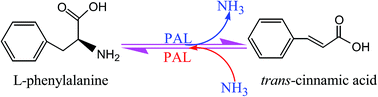
Phenylalanine ammonia-lyase (PAL, EC 4.3.1.24) catalyzes the deamination of phenylalanine to cinnamate and ammonia, the first step of the phenylpropanoid pathway. PALs are ubiquitous in plants and also commonly found in fungi, but have not yet been detected in animals. Typically, PAL is encoded by a small multigene family and the presence of PAL isoforms is a common observation. PAL belongs to the 3,5-dihydro-5-methylidene-4H-imidazol-4-one-containing ammonia-lyase family and has been shown to exist as a tetramer. Both the forward and reverse reactions catalyzed by PALs were of great interest and have potential industrial and medical applications. This review, therefore, covers the recent developments related to the PAL gene distribution, phenylalanine ammonia-lyase gene family, structure and function study of PALs, as well as several potential applications of PALs. As a key gateway enzyme linking the phenylpropanoid secondary pathway to primary metabolism, PALs were extensively applied in heterologous hosts to produce phenylpropanoids. The review thereby highlights the synthetic potentials of PALs as a key component used in metabolic engineering and synthetic biology. Moreover, the other potential PAL applications, like enzyme replacement therapy of phenylketonuria, as a therapeutic enzyme in cancer treatment and microbial production of L-phenylalanine are also discussed in detail. Together these results provide a synopsis of a more global view of potential applications of PALs than previously available.


image file: c5ra08196c-p1.tif

Jian-Qiang Kong

Kong JQ received his Doctor's Degree in 2006 at Peking Union Medical College. Dr Kong's current research involves the metabolic engineering of high-value natural products. Genes related to natural product biosynthesis were isolated from different organisms, like plants, animals and microbes, and then were functionally characterized by in vitro enzymatic reaction. These refined genes were then transformed into varied chassis such as Escherichia coli (E. coli) and Saccharomyces cerevisiae (S. cerevisiae) to rebuild biosynthetic pathways of natural products. The engineered hosts were used to produce natural products on a large scale.


1. Introduction

Phenylalanine ammonia-lyase (PAL, EC 4.3.1.24) catalyzes the conversion of L-phenylalanine (L-Phe) to trans-cinnamic acid (t-CA) by a non-oxidative deamination (Fig. 1).1 This reaction is the first step in the phenylpropanoid pathway and ultimately leads to various phenylpropanoids and their derivatives, such as flavonoids, isoflavonoids, resveratrol, coumarins, stilbenes, anthocyanins, lignin, and other phenolic compounds. In addition to serving as cell wall components (lignin)2 or signaling molecules (acetosyringone)3 in plants, many of these compounds have also displayed promising biological activities, including flower and fruit pigments (anthocyanins),4 UV protectants of plants (apigenin),5 antitumor activities (luteolin, apigenin),6–9 antimicrobial actions (1-methoxyficifolinol, licorisoflavan A, and 6,8-diprenylgenistein),10 as well as anti-inflammatory and neuroprotective effects (rutin, pinocembrin).11,12 Hence, PAL is considered to be the key entry point into the phenylpropanoid pathway, channeling the carbon flow from primary metabolism to secondary metabolism. The PAL enzyme is thereby of particular interest to researchers and has been extensively studied.
image file: c5ra08196c-f1.tif
Fig. 1 Forward reaction catalyzed by PALs.

At present, many PAL genes have been isolated and characterized from plants,13–15 fungi16–18 and prokaryotes.19–21 The structure and mechanism of PAL have been well-characterized.22–24 The activity of PAL is induced dramatically in response to biotic and abiotic stresses.25–30 Moreover, PAL has recently been studied extensively for the aim of potential applications in clinical,19,31,32 industrial,33–36 and biotechnological areas.37–40 As a key gateway enzyme linking the phenylpropanoid secondary pathway to primary metabolism, PAL was therefore an attractive target for metabolic engineering and synthetic biology processes.33,37–39,41–43 Versatile artificial gene clusters carrying PAL were transferred into a variety of heterologous microbial cell factories for the fermentative production phenylpropanoid-derived natural products.44,45 Furthermore, varied PALs may serve in therapeutic applications for treating phenylketonuria (PKU)31,32,46 and cancer.47,48 In addition to medical applications, PAL has synthetic potentials as a biocatalyst for L-phenylalanine production.49–51 Diverse strategies were applied to enhance activity, stability and yield of PALs with the aim of improvement its applications in industrial and medical potentials. There are some reviews focusing on biotechnological production of PALs.52–55 These reviews, however, did not cover the detailed strategies used for functional improvement of PALs in industrial and medical applications. This review, therefore, highlighted the breakthroughs of PALs as a key component used in metabolic engineering and synthetic biology. Furthermore, the advance in medical applications as a therapeutic enzyme for treating PKU and cancer and industrial applications as a biocatalyst for L-phenylalanine production was also discussed in detailed in the present review. Together these introductions provide a synopsis of a more global view of potential applications of PALs than previously available.

2. PAL genes distribution

Due to its central role in phenylpropanoid metabolism, PAL was studied extensively. PAL was first purified from Hordeum vulgare in 1961,1 but was shown to widely exist in plants (Table 1),13–15 fungi16–18 and prokaryotes19–21 afterwards. In plants, PAL occurs widely, including monocots,56–59 dicots,13,14,60,61 gymnosperms,62–65 ferns,66 lycopods,66 liverworts,66 and algae.67 The PALs from plants account for more than 90% of known PALs. PALs are also commonly found in fungi, mainly distributing in Basidiomycetes,16,68–71 Ascomycete,71 and yeast.17,18,29,72–77 The ubiquitous PALs largely reflect the phenylpropanoids are common natural products in plants and fungi, serving diverse physiological functions. In bacteria, PALs are rare. To date, few bacterial counterparts have been identified in Rubrobacter xylanophilus,19 Streptomyces maritimus,78–80 Streptomyces verticillatus,81,82 Photorhabdus luminescens,20,83 and some species of cyanobacteria like Synechocystis sp.,84 Leptolyngbya sp.,84 Oscillatoria sp.,84 Anabaena variabilis,21,31,85 and Nostoc punctiforme.21 The rarity of PALs apparently mirrors the dearth of phenylpropanoids in prokaryotes. In these bacteria, the PAL product trans-cinnamic acid serves as an intermediate in the biosynthesis of antibiotic or antifungal compounds, like enterocin,80,86–88 5-dihydroxy-4-isopropyl-stilbene,89,90 soraphen A91,92 and cinnamamide.82 So far, there are no reports for animal PALs.
Table 1 phenylalanine ammonia-lyase gene family in plant species
Plant species Number of PAL genes Reference
Lemon balm (Melissa officinalis L.) 2 93
Ornithogalum caudatum 2 94
Raspberry (Rubus idaeus) 2 95
Bean (Phaseolus vulgaris L.) 3 175–177
Scutellaria baicalensis 3 178
Trifolium subterraneum 3 179
Salvia miltiorrhiza 3 13 and 180
Arabidopsis thaliana 4 97, 101 and 181–184
Ephedra sinica 4 185
Carrot (Daucus carota) 4 186
Medicago truncatula 4 97
Tobacco (Nicotiana tabacum) 4 115 and 187–189
Populus kitakamiensis 4 190–192
Parsley (Petroselinum crispum) 4 193–197
Artichoke (Cynara cardunculus var. scolymus L.) 4 102
Bambusa oldhamii 4 112–114
Populus trichocharpa 5 97, 198 and 199
Pinus banksiana 5 200
Loblolly pine (Pinus taeda) 5 62, 64 and 201
Cucumber (Cucumis sativus L.) 7 202
Soybean (Glycine max) 8 97 and 203
Sorghum bicolor 9 97
Brachypodium distachyon 9 97
Rice (Oryza sativa L.) 12 15 and 97
Watermelon (Citrullus lanatus) 12 14
Vivis vinifera 16 97
Tomato (Lycopersicon esculentum) 18 204
Maize (Zea mays L.) 20 96 and 97
Potato (Solanum tuberosum L. cv. Datura) 40–50 98


3. Phenylalanine ammonia-lyase gene families

PAL proteins are encoded by a multi-gene family in almost all of the plant species studied (Table 1). The number of PAL genes varied greatly in different plant species, ranging from 2 to a dozen or more copies (Table 1). For example, there are two genes in lemon balm (Melissa officinalis L.),93 Ornithogalum caudatum94 and raspberry (Rubus idaeus).95 20 PAL genes, however, were found in maize (Zea mays L.).96,97 Even in potato (Solanum tuberosum L. cv. Datura), the number of PALs may reach to approximately 40–50.98 Of course, the definite gene number of bona fide PAL in several plant species was uncertain due to prediction results without functional characterization.13,14,99,100

To my knowledge, there are no reports about fungi and bacterial PAL with multi-isoenzymes. The presence of PAL isoforms is a common observation, but the significance of this diversity is still questioned.101

The PAL gene organization in the same gene family exists some variations.14 For example, nine of twelve PAL genes in Citrullus lanatus contained no introns in their ORFs. The ORFs of the other three PAL genes, however, were interrupted by a single intron.14

The isoenzymes of the same PAL gene family are usually expressed differentially in various tissues. Three PAL members from Salvia miltiorrhiza showed differential expression in different tissues. SmPAL1 showed the highest expression in roots, followed by leaves, stems and flowers. SmPAL2 was predominately expressed in stems and flowers. Its expression levels in roots and leaves were very low. SmPAL3 had the highest expression in leaves, less in roots, stems and flowers.13

The individual PAL gene in the same family may respond differentially to biotic or abiotic stresses. The results reported by Hou et al. indicated that all three PALs in Salvia miltiorrhiza were drought- and MeJA-responsive, but the time and degree of reaction differed from one another.13 Although all induced by mechanical wounding, three PAL genes of artichoke showed a gene-specific behaviour with respect to response time, level and duration of expression.102

4. Structure and function of PAL

PAL has the catalytic prosthetic 3,5-dihydro-5-methylidine-4H-imidazol-4-one group (formerly 4-methylideneimidazole-5-one; MIO, Fig. 2),103,104 which is produced by the posttranslational and autocatalytic cyclization of the tripeptide of alanine, serine and glycine (Ala-Ser-Gly triad; ASG motif, Fig. 2) and acts as the electrophile in the reaction mechanism.23,24,104,105 Deamination of L-Phe is dependent upon the MIO prosthetic group in the enzyme. MIO group is conserved in both ammonia-lyases, including PALs, TALs (tyrosine ammonia-lyases) and HALs (histidine ammonia-lyases), and aminomutases, that is L-phenylalanine and L-tyrosine 2,3-aminomutases.52–55 The amino acid residues involved in formation of the MIO moiety were conserved and important to enzyme activity of PALs. Mutation of serine-202, a component of ASG motif, in PAL from parsley (Petroselinum crispum) to alanine resulted in the lack of catalytic activity.106 The same conclusion can be drawn by several reports.19,60,107–109 All of these observations showed the active site domain containing ASG motif played a crucial role in enzyme activity of PALs.
image file: c5ra08196c-f2.tif
Fig. 2 Chemical structure of the MIO cofactor.

PALs had been shown to occur as a homotetramer in vivo110,111 or in vitro.112–114 When various PALs were co-expressed in the single heterologous host, however, the mixed purified proteins formed heterotetrameric enzyme exclusively and preferentially.115

Many studies have shown that the PAL of monocotyledonous plants96,114 and some dicotyledons116 catalyzed the deamination of both L-phenylalanine (PAL activity) and L-tyrosine (TAL activity), which indicated PAL and TAL activities reside in the same polypeptide. This activity is valuable for an alternative route avoiding the reaction catalyzed by cinnamate 4-hydroxylase (C4H).37,117–119 The mechanism of how these bifunctional PALs are able to use both substrates is confirmed experimentally to be existence of substrate selectivity switch.99 Phe is the key residue in the substrate selectivity switch. When mutated to His, this single amino acid functions as a highly efficient substrate selectivity switch, converting the enzyme from exclusively PAL activity to exclusively TAL activity.99 When Phe144 of PAL1 from Arabidopsis was replaced by His, the F144H mutant displayed a marked reduction (30-fold) in affinity for the substrate phenylalanine.99 The observation was supported by different experiments.60,112–114,120,121

5. A key component used in metabolic engineering and synthetic biology process

Up to date, PALs were introduced into different hosts, like Escherichia coli,39,40,42,43,122–124 Saccharomyces cerevisiae,37,38,42,125–127 Pseudomonas putida33,128,129 and Streptomyces80,130–132 to construct varied artificial biosynthetic gene clusters for the fermentative production of flavonoid and flavonoid-related compounds (Fig. 3 and 4). The reconstructed phenylpropanoid pathway in engineered microbes leads to the biosynthesis of a wide range of phenylpropanoid-derived compounds, including phenylpropanoid acids (trans-cinnamic acid (1)33,41 and p-coumaric acid (2)123,124), flavanones ((2RS)-pinocembrin (3),123,124 (2RS)-naringenin (4),123,124 (2S)-pinocembrin (5),40,43,150 (2S)-naringenin (6)43,86,122,126 and eriodictyol (19)133), flavones (chrysin (7)122 and apigenin (8)122), flavonols (galangin (9),122 kaempferol (10)122,127 and quercetin (28)127), curcuminoids (dicinnamoylmethane (11),134,135 6-fluoro-dicinnamoylmethane (11a),135 6,6′-difluorodicinnamoylmethane (11b),135 cinnamoyl-p-coumaroylmethane (12)134 and bisdemethoxycurcumin (13)134), triketide pyrone (14 and 15),134 stilbenoids (trans-resveratrol (18),136,137 pinosylvin (20),137,138 pinosylvin monomethyl ether (21),163 pinostilbene (22),163 pinosylvin dimethyl ether (23)163 and pterostilbene (24)163), chalcone (2′,4′,6′-trihydroxydihydrochalcone (25)37 and phloretin (26)37) and isoflavone (genistein (27)42,155) (Fig. 3, 4 and Table 2), which collectively have diverse biological functions as anticancer activity,79,80,139 neuroprotection effect,140 antiviral potency,132 nephroprotective activity,141 antimicrobial effect,142,143 anti-inflammatory and antioxidant activities.97,134 Moreover, some engineered organisms can produce commodity petrochemical with diverse commercial applications, like styrene (16)37,39,125 and p-hydroxystyrene (17) (Fig. 4 and Table 2).33,144
image file: c5ra08196c-f3.tif
Fig. 3 Schematic overview of all the engineered flavonoids biochemical pathways.

image file: c5ra08196c-f4.tif
Fig. 4 Phenylpropanoids produced by engineered microorganisms containing PALs.
Table 2 Synthetic potentials of PALs in metabolic engineering
No Hosts End products Pathway enzymes Source of PAL References
1 E. coli trans-Cinnamic acid (1) PAL/TAL + 4CL + CHS R. rubra 123 and 124
p-Coumaric acid (2)
(2RS)-Pinocembrin (3)
(2RS)-Naringenin (4)
2 E. coli trans-Cinnamic acid (1) PAL + C4H + 4CL + CHS A. thaliana 117
3 E. coli (2RS)-Pinocembrin (3) PAL + 4CL + CHS A. thaliana 146
4 E. coli (2S)-Pinocembrin (5) PAL + 4CL + CHS + CHI R. glutinis 40
5 E. coli (2S)-Pinocembrin (5) PAL/TAL + 4CL + CHS + CHI R. rubra 43
(2S)-Naringenin (6)
6 E. coli (2S)-Naringenin (6) PAL/TAL + 4CL + CHS + CHI R. glutinis 145
7 E. coli (2S)-Naringenin (6) PAL/TAL + 4CL + CHS + CHI R. glutinis 86
8 E. coli (2S)-Pinocembrin (5) PAL/TAL + 4CL + CHS + CHI R. mucilaginosa 122
(2S)-Naringenin (6)
Chrysin (7) PAL/TAL + 4CL + CHS + CHI + FNS1
Apigenin (8)
Galangin (9) PAL/TAL + 4CL + CHS + CHI + F3H + FLS
Kaempferol (10)
9 E. coli Dicinnamoylmethane (11) PAL/TAL + 4CL + CUS R. rhodotorula 134
Cinnamoyl-p-coumaroylmethane (12)
Bisdemethoxycurcumin (13)
Triketide pyrone (14 and 15)
10 E. coli Dicinnamoylmethane (11) PAL + 4CL + CUS Trifolium pratense 135
6-Fluoro-dicinnamoylmethane (11a)
6,6′-Difluoro-dicinnamoylmethane (11b)
11 E. coli p-Hydroxystyrene (16) PAL/TAL + PDC R. glutinis 144
12 E. coli Styrene (17) PAL + CADC A. thaliana 39
13 E. coli trans-Resveratrol (18) PAL/TAL + 4CL + STS R. glutinis 136
14 E. coli Eriodictyol (19) PAL/TAL + 4CL + CHS + CHI + F3′H + CPR R. glutinis 133
15 E. coli trans-Resveratrol (18) PAL/TAL + 4CL + STS R. rubra 137
Pinosylvin (20)
Pinosylvin
Monomethyl ether (21)
Pinostilbene (22)
Pinosylvin dimethyl ether (23)
Pterostilbene (24)
16 E. coli Pinosylvin (20) PAL + 4CL + STS T. pratense 138
17 E. coli Pinosylvin (20) PAL + 4CL + STS P. crispum 205
A. thaliana
18 E. coli trans-Cinnamic acid (1) PAL/TAL A. thaliana 206
p-Coumarate (2) R. glutinis
19 S. cerevisiae trans-Cinnamic acid (1) PAL + C4H + CPR R. glutinis 41
E. coli p-Coumarate (2)
20 E. coli Genistein (27) PAL/TAL + 4CL + CHS + CHI + IFS R. rubra 42
S. cerevisiae (2S)-Naringenin (4)
21 S. cerevisiae trans-Cinnamic acid (1) PAL + C4H + CPR Poplar hybrid (Populustrichocarpa × P. deltoids) 125
p-Coumarate (2)
4-Vinylphenol (16)
Styrene (17)
22 S. cerevisiae Pinocembrin (3) PAL/TAL + 4CL + CHS R. toruloides 37
Naringenin (4)
Styrene (17)
2′,4′,6′-Trihydroxydihydrochalcone (25)
Phloretin (26)
23 S. cerevisiae p-Coumarate (2) PAL + C4H + CPR Poplar hybrid (P. trichocarpa × P. deltoids) 127
trans-Resveratrol (18) PAL + C4H + CPR + 4CL + RS
(2S)-Naringenin (6) PAL + C4H + CPR + 4CL + CHS + CHI
Genistein (27) PAL + C4H + CPR + 4CL + CHS + CHI + IFS
Kaempferol (10) PAL + C4H + CPR + 4CL + CHS + CHI + FLS + F3H
Quercetin (28) PAL + C4H + 4CL + FLS + CHS + CHI + F3H + F3′H
24 S. cerevisiae trans-Resveratrol (18) PAL + C4H + 4CL + STS R. toruloides 38
p-Coumarate (2)
S. cerevisiae Styrene (17) PAL + FDC A. thaliana 149
25 S. cerevisiae (2S)-Naringenin (6) PAL + C4H + CPR + 4CL + CHS + CHI A. thaliana 126
26 P. putida trans-Cinnamic acid (1) PAL/TAL R. toruloides 129
p-Coumarate (2)
27 P. putida trans-Cinnamic acid (1) PAL/TAL R. toruloides 128
p-Coumarate (2)
28 P. putida trans-Cinnamic acid (1) PAL/TAL + PDC R. toruloides 33
p-Hydroxystyrene (16)
29 S. coelicolor trans-Cinnamic acid (1) PAL S. maritimus 80
30 S. lividans trans-Cinnamic acid (1) PAL S. maritimus 132
31 S. lividans trans-Cinnamic acid (1) PAL S. maritimus 131
32 S. lividans p-Coumarate (2) PAL/TAL Rhodobacter sphaeroides 130


Although various complex chemicals of therapeutic and industrial importance were produced by these engineering microorganisms containing PALs, the yield is in very small amount.37,123,124,126 It is necessary to improve the yield of flavonoids produced by engineered cell factories. Common methods used to improve production from engineered biosynthetic pathways include selection of appropriate hosts,33,38,130,136 enhancing production of pathway enzymes,43,123–125 yield enhancement of the intracellular pool of precursors,43,122 balancing multi-gene expression to optimize flux,40,126,136 alleviating the metabolic burden,33,40,136,145 and optimization of the culture conditions.41,43,136

5.1 Selection of appropriate hosts

According to the characteristic of the pathway enzymes, varied hosts such as E. coli,39–43,86,117,122–124,133,134,136,137,144–147 S. cerevisiae,37,38,125,126 P. putida33,128,129 and Streptomyces80,130–132 were selected as production platforms for the synthesis of flavonoids.

E. coli is a preferred host for flavonoids production due to its fine characteristics like fast growth rate, inexpensive fermentation media and well understood genetics. Even more important, almost all E. coli strains and vectors can be commercially available, facilitating most extensive application in synthetic biology. Up to date, engineered E. coli can be used to produce various flavonoid molecules, such as flavanones ((2S)-pinocembrin (5), (2S)-naringenin (6), eriodictyol (19)), flavones (chrysin (7), apigenin (8)), flavonols (galangin (9), kaempferol (10)), isoflavones (genistein (27)), stilbenes (trans-resveratrol (18), pinosylvin (20)) and so on. Varied E. coli strains were selected to express heterologous proteins based on quality of engineered pathways. For example, when multi-gene pathway under regulations of the same promoter was introduced into E. coli, to prevent a deletion of the repeats as a result of possible homologous recombination, a recA mutant E. coli BLR (DE3) was used to be a host.43,122,134 Moreover, to make more precursors enter the target metabolic flux, precursor-overproducing E. coli strains were usually used as chassises for increased yields.86,145

It is generally accepted that E. coli has a disadvantage in cytochrome P450 expression.117 As the biochemical knowledge regarding cytochrome P450s increases, so does the efficiency of functional P450 protein expression in E. coli through various modifications including N-terminal modifications,133 and using optimized genes.133 Used alone or in combination, these methods can improve protein expression in E. coli, which will undoubtedly extend the application area of E. coli as a host in synthetic biology.148

Although extensively applied in metabolic engineering, E. coli is not always the ideal host due to inability to perform post-translational modifications and formation of inclusion bodies. Other organisms, including S. cerevisiae, are thus selected to produce flavonoids. S. cerevisiae has many inherent properties as a metabolic engineering platform for flavonoid production. Firstly, as a single cell eukaryotic organism, S. cerevisiae has many similar mechanisms to higher eukaryotes, facilitating efficient expression and posttranslational modification of plant-derived flavonoid-biosynthetic proteins.37,38,125,126 Second, S. cerevisiae has long served as a host organism for heterologous expression of proteins. Its genetics and physiology are well documented. Thus, genetic tools of S. cerevisiae for metabolic pathway manipulation are more abundant.149 Third, S. cerevisiae, classified as a GRAS (generally recognized as safe) organism, is easily cultivated in chemically defined medium and exhibits fast growth rates, thus facilitating subsequent application for scaling-up production of flavonoids. Finally, S. cerevisiae can be tolerant to a wide range of physiological stresses such as low pH and high osmotic stress. Collectively, these advantageous traits support the industrial use of yeast for chemicals including flavonoids production.

Normally, P. putida strains may be applied in metabolic engineering and synthetic biology in the presence of toxic chemicals due to its solvent-tolerant nature (Table 2). One of the underlying mechanisms of solvent-tolerance is the presence of solvent efflux pump, which can export hydrophobic molecules, including solvents and toxic products from the interior of whole cells. This mechanism not only protects the cell from toxic effects, but also offers better production by exporting a desired molecule from the cell into the medium.140 A variety of solvent tolerant P. putida strains including S12,33,128,129 DOT-T1E, and IH-2000 have been isolated and used as hosts for the production of toxic end-products. These toxic chemicals mainly refer to aromatic compounds, like p-hydroxystyrene (16, 4-vinyl phenol),33 trans-cinnamic acid (1)129 and p-coumaric acid (2, also known as p-coumarate).128

Streptomyces are well-known antibiotic producer with well-characterized genetic backgrounds. Because Streptomyces have high tolerance towards aromatic compounds150 and powerful biomass-assimilating ability,130,131 they were mainly used for building-block compounds production from biomass nowadays. These Streptomyces strains comprised Streptomyces lividans130–132 and Streptomyces coelicolor.80 These resulting aromatic building blocks contained trans-cinnamic acid (1),80,131,132 p-coumarate (2).130

5.2 Enhancing production of pathway enzymes

As precise control of enzyme expression levels is often critical for the efficient activity of metabolic pathways, the introduction of transcriptional (promoters), translational (ribosome binding site (RBS)) and enzyme (codon-optimized gene) variability to modulate pathway expression levels is essential for generating balanced metabolic pathways and maximizing the productivity of an engineered host.

Increased protein production was achieved by addition of T7 promoter and RBS in front of each gene of reconstructed pathway, therefore in turn resulting in yield enhancement of desired products.43,123,124 This, however, is not always the case. The results presented by Kim et al. revealed coordinated expression control of 4CL (4-coumarate-CoA ligase) and CHS (chalcone synthase) by one T7 promoter is better than independent expression regulated by two T7 promoters.146 These authors further attributed the contradictory observations to the order of related genes. This may be a reasonable explanation because more than one paper referred to the phenomenon.146,151

Besides regulators, optimal performance of a heterologous flavonoids pathway is also dictated by the kinetic properties of its enzymatic components. Variation in these kinetic properties may be achieved either by mutagenic experiments to create the desired attributes of an enzyme40,86,126,133,136,145 or through selection of variant enzymes deposited in public databases with differing kinetic properties.

Typically, codon optimization is used to improve the efficiency of heterologous protein production, especially in the context of synthetic biology and metabolic engineering. Approaches normally used to optimize codons include targeted mutagenesis to remove rare codons40,86,126,133,136,145 or the addition of rare codon tRNAs in specific hosts.145

Screening various target enzymes with desired attributes from the public databases can increase the likelihood of assembling a functional pathway. By screening several prokaryotic and eukaryotic microorganisms, Vannelli et al. acquired a bifunctional enzyme possessing both the highest TAL specific activity and the lowest PAL/TAL catalytic activity ratio. When the resulting bifunctional enzyme was introduced to S. cerevisiae, as anticipated, high levels of p-coumarate (2) were observed.41 To obtain an appropriate p-coumarate decarboxylase (PDC) used for the generation of p-hydroxystyrene (16), various microorganisms like Bacillus. subtilis, Pseudomonasfluorescens, P. putida, S. cerevisiae, Rhodotorula rubra and Lactobacillus plantarum were tested for their ability to decarboxylate p-coumarate (2) to p-hydroxystyrene (16) by Qi et al.144 The appropriate genes encoding for PDC with highest activity were then amplified from L. plantarum and B. subtilis genome and used to co-express with bifunctional PAL/TAL gene in E. coli to obtain p-hydroxystyrene (16).144 The same strategy was also found in a paper reported by McKenna et al.39 Prior to rebuilding of styrene biosynthetic pathway, candidate isoenzymes for each step were screened from bacterial, yeast, and plant genetic sources by these authors. PAL2 from Arabidopsis thaliana and FDC1 from S. cescerevisiae were then selected to co-express in E. coli due to their higher activity. The resulting strain led to the styrene accumulation of up to 260 mg L−1 in shake flask cultures.39 In another paper, Santos et al. also screened enzyme variants for each successive step in engineered naringenin biosynthetic pathway. The isoenzymes with higher activity were selected to further assemble a functional pathway.145 Additionally, to choose the best candidate CPR (cytochrome P450 reductase) gene, two A. thaliana CPR variants (CPR1 or CPR2) were selected to test activity. Results revealed CPR1 has higher activity and therefore is used in further experiments.126

The kinetic characteristics of the enzyme variants were characterized either individually as standalone enzymes41,144 or in the context of the entire flavonoid biosynthetic pathway.125,126

5.3 Yield enhancement of the intracellular pool of precursors

There are varied precursors in different metabolic pathways reconstructed in chassises. The low level of intracellular precursors is becoming one of the largest obstacles to efficient microbial biosynthesis of flavonoids. Several strategies were applied to enhance the availability of precursors in engineered hosts of flavonoids. These strategies include up-regulated expression of key genes of precursor biosynthesis and down-regulation or deletion of the precursor competition pathways.
5.3.1 Increased metabolic flux to precursor pools. Malonyl-CoA is one of the flavonoids precursors (Fig. 5). The concentration of malonyl-CoA in E. coli cells was calculated to be only 4–90 μM (0.01–0.23 nmol mg−1 dry weight).152 The low content of intracellular malonyl-CoA is becoming a bottleneck of flavonoid yields in engineered E. coli.43 To increase the supply of malonyl-CoA, varied modifications, which focused on overexpression of pathway enzymes related to malonyl-CoA biosynthesis, were applied to the engineered hosts. The metabolic regulation included reconstruction of malonate assimilation pathway containing two components of matB (encoding malonyl-CoA synthetase) and matC (encoding malonate carrier protein) (Fig. 5),40,136,145 or overexpression of multisubunit complex of acetyl-CoA carboxylase (ACC) (Fig. 5),42,43,122,133,134,137,146 or genetic modification in acetate assimilation pathways (Fig. 5 and Table 3).133
image file: c5ra08196c-f5.tif
Fig. 5 Metabolic crosstalk of flavonoids precursors.
Table 3 Optimization strategies improving metabolic flux to precursor pools
Optimization strategies Engineered host Product (before optimization, postoptimality) Fold change Reference
Reconstruction of malonate assimilation pathway E. coli (2S)-Pinocembrin (0.71 mg L−1, 1.22 mg L−1) 1.71 40
E. coli (2S)-Naringenin (29 mg L−1, 46 mg L−1) 1.58 145
Overexpression of multisubunit complex of acetyl-CoA carboxylase E. coli (2S)-Naringenin (0.31 mg L−1, 1.01 mg L−1) 3 43
(2S)-Pinocembrin (0.17 mg L−1, 0.71 mg L−1) 4
A combination of three strategies, that is (1) overexpression of acetyl-CoA carboxylase (ACC); (2) overexpression of acetyl-CoA synthase (encoded by acs); (3) deletion of the acetate kinase gene E. coli Eriodictyol (42.6 mg L−1, 107 mg L−1) 2.51 133
Overexpression of multisubunit complex of acetyl-CoA carboxylase E. coli (2RS)-Pinocembrin (58 mg L−1, 82 mg L−1) 1.41 146
Use of the fatty acid pathway inhibitor cerulenin E. coli (2S)-Naringenin (29 mg L−1, 84 mg L−1) 2.9 145
Pinosylvin (3.29 mg L−1, 59 mg L−1) 18 205
Alleviation of tyrosine feedback inhibition of yeast 3-deoxy-D-arabinose-heptulosonate-7-phosphate (DAHP) synthase S. cerevisiae (2S)-Naringenin 2 126
Elimination of competing phenylpyruvate decarboxylase activity S. cerevisiae (2S)-Naringenin 3 126


Malonate assimilation pathway was utilized for both the transport of supplemented malonate into the cell and its subsequent conversion to malonyl-CoA. Upon introduction of this recombinant pathway into engineered E. coli, the strain showed a dramatic increase in desired production over the control.40,136,145

The acetyl-CoA carboxylase can catalyze the conversion of acetyl-CoA to malonyl-CoA, leading to great enhancement of a pool of malonyl-CoA. Therefore, an increase in the amount of malonyl-CoA by overexpressing the acetyl-CoA carboxylase gene will result in enhancement of the flavanone yields.42,43,122,133,134,137,146

Acetyl-CoA synthetase19 can catalyze an irreversible reaction that converts acetate to acetyl-CoA via two enzymatic steps. To increase the capacity of intracellular acetyl-CoA poor, both overexpression of acetyl-CoA synthetase and inhibition of acetate kinase (AK), a rate-limiting enzyme of acetate competition pathway, were employed in host cells.133

L-Phenylalanine is also a flavonoid precursor. By overexpressing two rate-limiting enzymes of L-phenylalanine biosynthesis, 3-deoxy-D-arabino-heptulosonate-7-phosphate (DAHP) synthase and chorismate mutase/prephenate dehydratase, strains exhibiting an enhanced capacity for L-phenylalanine synthesis was accomplished.40 Moreover, the deregulation of DAHP synthase by substitution of DAHP encoding gene with a tyrosine insensitive allele was also proved to alleviate tyrosine feedback inhibition, leading to an increase of naringenin yields.126 When PAL enzyme was transferred into an L-phenylalanine overproducing E. coli strain, there was a substantial increase in the level of desired products accumulated.41,129,144

Many PALs have tyrosine ammonia-lyase activity, by which L-tyrosine can be catalyzed to form p-coumarate (2). In the engineered strains harboring PAL/TAL, L-tyrosine was thus used as a precursor to produce the L-tyrosine-derived aromatic products. To enhance capacity for L-tyrosine synthesis, some of rate-limiting enzymes in L-tyrosine biosynthesis were usually overexpressed within the recombinant cells.86 Moreover, to direct more L-tyrosine to naringenin synthesis, increased expression of a TAL independent of reconstituted pathway resulted in an enhancemen of naringenin yield.126

5.3.2 Reducing or inhibiting metabolic flux to precursor competition pathways. Competing pathways are native pathways present in a metabolic network of engineered cells. These pathways competed with reconstructed pathways that share the same precursor. The competing consumption of precursors usually diverged metabolic flux away from producing a required chemical, resulting in decreased or no detection of desired product. To diminish or delete adverse effect brought to target pathways by competing pathways, several approaches were explored in developing engineered cells. To limit the amount of malonyl-CoA lost to the synthesis of fatty acids, fatty acid pathway inhibitor cerulenin was added to repress both fabB (β-ketoacyl-ACP synthases I) and fabF (β-ketoacyl-ACP synthases II), which led to a precipitous increase of over 190% naringenin (Fig. 5).145 Furthermore, elimination of competing phenylpyruvate decarboxylase activity can cause an improvement in naringenin production.126 Additionally, to increase the supply of p-coumarate (2), a direct precursor for p-hydroxystyrene, the first gene of the p-coumarate degradation pathway, fcs (encoding feruloyl-coenzyme A synthetase), was inactivated by homologous recombination in P. putida S12 427. The resulting P. putida S12 construct showed a higher p-hydroxystyrene yield.33 In another paper reported by Nijkamp et al., fcs was also inactivated to hamper product degradation.128
5.3.3 Balancing multi-gene expression to optimize metabolic flux. In the expression of a multi-gene heterologous pathway, the activity of a single enzyme may be out of balance with that of the other enzymes in the pathway, leading to unbalanced carbon flux and the accumulation of an intermediate. Different strategies were employed to balance the overall pathway.40

A modular metabolic strategy is one of the efficient methods. The entire pathway was divided into different modules. The modular expression levels could be estimated according to promoter strengths and copy numbers. A most optimized performance for each biological system can be achieved by balancing the expression levels of multiple modules.40,86,136,145

Expression correlation analysis is another strategy to balance the protein–protein interactions in one specific pathway. According to the expression correlation analysis, the best set of isoenzymes for naringenin production was identified and introduced into yeast to produce desired end-product with high titre.126

5.4 Alleviating the metabolic burden

When multiple genes were introduced into a non-native host to reconstruct a artificial pathway, significant decreases in growth rate and biomass yield were commonly observed in the recombinant strain. These effects are thought to result from the metabolic load imposed on the production host by the expression of foreign proteins40,145 or the toxicity of the product of interest.144

The heterologous expression of multi-protein pathway in an engineered host cell often utilizes a significant amount of the host cell's resources, removing those resources away from host cell metabolism and placing a metabolic burden on the chassis. As one of the most commonly observed consequence of the imposed metabolic load, the growth rate of the host cell decreased dramatically or was halted. To overcome or reduce the adverse effect, efficient solutions, like minimizing the size of foreign plasmids,145 reducing the copy number of introduced plasmids,86,136 and delaying IPTG induction,40 were proposed in many papers.40,145

The physiological toxicity of hydrophobic flavonoids made the yield far below their maximum potential due to growth arrest of engineered strains. In recent years, a variety of approaches have been proposed to overcome or circumvent the toxicity-related adverse effects. The first way to cope with this product toxicity is to deploy solvent-tolerant microorganisms, like P. putida S12.33

Additionally, product toxicity was averted by the application of two-phase fermentation during fed-batch cultivation.33 Moreover, protein complex formation is likely to alleviate toxic effect of intermediates.125,126 It has been postulated that PAL can form multienzyme complexes (MECs) with other downstream enzymes such as C4H (cinnamic acid 4-hydroxylase), CPR, 4CL, CHS and CHI (chalcone isomerase), facilitating local retention and then sequential conversion of toxic intermediates.125,126

Alternatively, feedback inhibition by intermediates had also been reported for quite a few metabolic pathways.40,86,136,145 The inhibitory effect can be reversed by overproduction of downstream enzymes, which can lower the accumulation of intermediates and then alleviate potential inhibition within diverse host cell.40,86,126,136,146

5.5 Optimization of the culture conditions

Besides the genetic manipulation strategies, optimization of the culture conditions, such as medium screening,40 optimization of protein induction factors133 and so on, can also enhance the yields of target compounds produced by engineered constructs harboring PALs.

Medium was used to culture engineering hosts and exhibited varied effect on flavonoid production. When transferred to fresh media for fermentation, only the strains resuspended in MOPS medium can yield desired product.40 Vannelli et al. also reported that different production titres were obtained from distinct medium.41 Also, the composition of mediums, such as nitrogen or carbon source has an effect on production of target products. To reduce the consumption of malonyl-CoA used for fatty acid synthesis, engineered E. coli was concentrated (50 g L−1 wet weight) to re-incubated in fresh M9 medium. Under these cultural conditions, almost no cell growth was observed, which meant no fatty acid synthesis is necessary. The accumulated malonyl-CoA will be directed to metabolic flux of flavanones biosynthesis to result in yield enhancement.43

Induction factors such as IPTG concentration,133 induction time, induction temperature133 and IPTG induction at different OD600 values,40,133 also have effect on protein expression.

In addition, different culture methods can also cause yield variance in engineered organisms. Koopman et al. revealed the naringenin production in batch bioreactor cultivation is more than two times than that of equivalent shake flasks cultivations.126 The similar phenomenon was observed by Verhoef et al.33

6. Enzyme replacement therapy of phenylketonuria

PAL also has been employed in medicine as an enzyme substitution therapy for the treatment of phenylketonuria (PKU, OMIM 261600), an inherited metabolic disorder caused by mutations in phenylalanine hydroxylase (PAH; EC 1.14.16.1) gene. Patients with PKU suffer from hyperphenylalaninemia,153 which ultimately leads to seizures and mental retardation. The established treatment for PKU resides in a low-phenylalanine diet, which has known deficiencies of several nutrients and unsatisfactory organoleptic properties, making long-term full compliance very laborious and requiring a great deal of social support. An alternative independent of dietary manipulation is thus desirable.

PAL is a robust autocatalytic protein, which is anticipated to reverse HPA by converting excess systemic L-Phe to nontoxic trans-cinnamic acid and metabolically insignificant levels of ammonia in the absence of additional cofactors. Also, PAL, unlike PAH, is inherently stable with prolonged life-time. Moreover, the broad pH range tolerance of PAL is 4–12, a feature compatible with an enteral route for PAL therapy.85 All of these properties have instigated investigations into the use of PAL as an obvious lead candidate for the development of an enzyme substitution therapy for PKU. Preliminary studies showed oral or subcutaneous administration of PAL led to substantial lowering of plasma L-Phe levels.32,154–156

To develop a most therapeutically effective form of PAL with higher availability and stability, longer half-life, less toxicity and immunogenicity, there are many problems need to be resolved. First, sufficient amounts of purified PAL with high specific activity need to be available. At present, the production of therapeutic PAL mainly utilizes P. crispum,157 Anabaena variabilis,32,158 and Rhodosporidium toruloides.22,32,154,155 When these PAL genes were heterologously expressed in E. coli, the yields of recombinant enzyme obtained were disappointingly low. The insufficient availability of recombinant PAL proteins curtailed the human and even the animal studies. A recombinant strain capable of producing a large amount of PAL is therefore highly desirable. Up to date, many efforts, like using a high-expression promoter,155 co-expression with molecular chaperone in E. coli,157,159 and optimization of culture conditions158,160 have been made to improve substantially recombinant PAL production. Inclusion body formation is a regular phenomenon when PAL was expressed in E. coli, which leads to significant reduction of soluble PAL. In order to increase the cytosolic expression, PAL and chaperone were co-expressed in E. coli. The resulting system can yield high titre of PAL protein, reaching 70 mg L−1.159

Second, therapeutically effective PAL needs to be tolerated by the immune system of PKU patients. Otherwise, the repeated administration of PAL will elicit immune response towards PAL, leading to clearance of the enzyme through a neutralizing immune response and to harmful allergic reactions.157 In order to reduce immunogenicity, different strategies, mainly including chemical modification with polyethylene glycol (PEG) and site-directed mutagenesis, have been developed based on protein structural information. Protein pegylation is the routine strategy to protect enzymes from immune system access.22,32,154,155,157,161–163 PEG–PAL conjugates were produced by covalent coupling of activated PEG molecules to PAL protein, which had been shown to reduce immunogenicity, prolong circulation half-life and retain catalytic activity in vivo.22,154,157 PEG–PAL conjugates at lower ratios did not lead to significant lowering of the immune response. The immunoreactivities of PEG–PAL turned to decrease as the degree of modification increased.22 PEG–PAL formulations with high ratios (like 1[thin space (1/6-em)]:[thin space (1/6-em)]8 and 1[thin space (1/6-em)]:[thin space (1/6-em)]16) almost completely attenuate immunoreactivity.22 Reduced immunogenicity depends on the number of PEG derivatized amino groups, indicating that the decreased immune response of pegylated PAL is a consequence of masking certain specific immunogenic PAL epitopes by polymer modification of these sites.154 There are at least 10 fully surface-exposed lysine residues distributed throughout the PAL protein surface.22 Pegylation derivatization of PALs can be achieved through amine coupling of these lysines with PEG derivatives, thereby resulting in improved PAL with reduced immunogenicity. Therefore, substitution of the residues near antigenic regions with lysines, such as R91K mutant in RT-PAL of Rhodosporidium toruloides, can generate more favorable PAL with reduced immunogenicity and increased activity.22,162,163

Up to date, the PALs used for pegylation modification were mainly from Rhodotorula glutinis,164R. toruloides,22,32,154,162,163 P. crispum,157,162 A. variabilis,32,161,162Nostoc punctiforme.162

At least five PEG–PAL formations have been clinical trials in the USA since 2008 as a potential injectable therapeutic for PKU.31,32,161 Given that PKU patients require lifelong therapy, oral formation of PAL is also investigated to provide a non-invasive substitution therapy. In vivo studies revealed that orally administered PAL yielded statistically significant and therapeutically relevant reduction in plasma L-Phe levels in a dose- and loading-dependent manner.32

Finally, the sensitivity of PAL to proteolysis hampered its application as a more efficacious molecule. Various strategies, such as chemical modification and protein engineering, have been exploited so far to reach enzymes with higher stability. These modified PALs are thus to be more stable in the circulation to ensure the therapeutic effects over a long period. Besides restricting access to epitopes, PEGylation of PAL through the ε-amino group of lysine's side chain presumably block potential trypsin cleavage sites, leading to increased stability and prolonged in vivo protein half-life.161

Another chemical modification strategy explored to further improve the protease resistance of therapeutic PALs utilized the silica sol–gel matrix for entrapping the enzyme.161 The encapsulated PAL-silica particles exhibited improved resistance against intestinal proteases in the protease resistance studies.161

Moreover, various strategies in the protein engineering were performed to increase PALs stability. Among them, introduction of new disulfide bonds,31 reducing protein aggregation,31 improving protease resistance by site-directed mutagenesis of required sites22,161 are the best exemplified ones. Disulfide bonds, in the form of a covalent linkage between the thiol groups of pairs of cysteine residues, are present in most extracellular proteins, where they presumably stabilize the native conformation by lowering the entropy of the denatured state. This stabilizing property makes introduction of new disulfide bonds an attractive strategy for engineering additional conformational stability into proteins by site directed mutagenesis. To improve stability of A. variabilis PAL (AvPAL), Gln292 of AvPAL was mutated to cysteine residue, and a new disulfide bond between G292S and Cys503 residue was consequently was introduced to the mutant enzyme.31

Cysteines-503 and -565 are two harmful surface cysteines in AvPAL of A. variabilis, taking part in aggregating the enzyme molecules and thereby resulting in decreased stability. To remove the two cysteines, mutation of both these residues to serine (C503S/C565S) was performed. The resulting C503S/C565S double mutation reduced aggregation properties of PAL, without significantly altered enzyme activity.85,162

There are many candidate sites identified to be susceptible for proteases cleavages.22 When these sites were substituted, the resultant mutant, like R123H, R123A, R123Q, Y110H, Y110A and Y110L mutants of R. toruloides PAL,22 F18A variant of A. variabilis PAL,161 exhibited modest improvements in resistance against protease inactivation.

Combination of different strategies in the chemical modification and protein engineering may also result in increased stability in a synergistic manner. When triple mutant C503S/C506S/F18A of AvPAL was further modified by PEG or silica matrix, the chemically modified PAL preparations displayed improvement in resistance against chymotrypsin and trypsin, two prevalent intestinal enzymes.161 Also, the observation was confirmed by further proofs. In a recent work presented by Jaliani et al., Q292C mutation was selected to form a new disulfide bond with Cys503, along with C565S mutation. The resulting double mutation Q292C/C565S remove the free sulfhydryl groups on the PAL surface (Cys503 and 565), and thereby displayed more kinetic stability and chemical denaturation resistance.31

Some of PAL formulations have been clinical trials as a potential therapeutic for PKU.32

7. A therapeutic enzyme in cancer treatment

In addition to applications in enzyme replacement therapy of phenylketonuria, PALs may be used as a potential therapeutic enzyme in cancer treatment.47,48,165–169 In the primary study performed by Babich et al., a recombinant PAL from R. toruloides was observed to exhibit a significant cytotoxic effect toward varied cells.47

8. A novel biocatalyst for synthesis of L-phenylalanine

In addition to medical applications, PAL was investigated as a biocatalyst for the preparative synthesis of L-phenylalanine.49,50,170–172 L-Phe is a key precursor of artificial sweetener aspartame. The great demand on aspartame stimulated considerable researches on the enzymatic production of L-Phe catalyzed by PALs.35,36,49–51,170–173 Besides the forward reaction of spontaneous, nonoxidative deamination of L-Phe to t-CA and ammonia, PALs can also catalyze the reverse reaction, namely formation of L-Phe under conditions of high concentration of trans-cinnamic acids and ammonia at an elevated pH.174 The biotransformation approach catalyzed by PALs has the advantage of asymmetric synthesis of an optically pure product, L-Phe.50 Although PAL has disadvantages of low specific activity and decreased stability, significant strides have been made recently in the application of PAL for the synthesis of L-Phe.170–172 In order to improve PAL activity, a combine promoter was used to regulate the expression of PAL. The resulting specific activity of PAL reached a high level of 123 U g−1 (dry cells weight, DCW), 8–12 fold higher than that of wild-type.51 To overcome the instability of PAL, the preparation of cross-linked enzyme aggregates of PAL was described in several reports.49,170–172 Compared to the free enzyme, the aggregates exhibited the higher thermal stability, denaturant stability and storage stability, retaining 30% initial activity even after 11 cycles of reuse.170 Overall, with the improvement of PAL activity and stability, PALs might be used as an efficient biocatalyst for enzymatic production of L-Phe.

9. Conclusions and prospects

Great strides have been made recently in the potential application of PALs, including gene component of engineered strains producing phenylpropanoids, enzyme replacement therapy of phenylketonuria, therapeutic enzyme in cancer treatment and biocatalysts for synthesis of L-phenylalanine. There are, however, several problems to be solved.

In recent years, a wide variety of additional, novel synthetic routes harboring PALs had been proposed and engineered in microorganisms for the production of phenylpropanoids with pharma- and nutraceutical properties.40,126,145 The final titers of phenylpropanoids, however, are still low, thereby impeding subsequential industrialization of metabolites from engineered microorganisms. As such, several obstacles, like low enzyme efficiency, coordinated expression of multiple-enzyme pathway, toxic intermediates accumulation and inadequate substrates supply, will be overcome to improve microbial yield of interest compounds.

Although PEG–PAL conjugates (PAL conjugated with polyethylene glycol) have been introduced in clinical trials for the treatment of PKU,31,32,46,85,161 the PALs with highest stability and reduced immunogenicity are still the most therapeutically effective molecules.

Using enzymes as biocatalysts is a useful tool for the synthesis of L-phenylalanine on a large scale. However, many PALs are sensitive and fragile to reaction conditions. Therefore, the more detailed investigations improving PALs activity and stability should be strengthened. Overall, if all these challenges were overcome, more and more robust and competitive PALs will be appeared in industrial and medical applications.

Abbreviations

accABCDAcetyl-CoA carboxylase complex
CADCtrans-Cinnamic acid decarboxylase
CHSChalcone synthase
CHIChalcone isomerase
C4HCinnamic acid 4-hydroxylase
4CL4-Coumarate-CoA ligase
CUSCurcuminoid synthase
CPRCytochrome P450 reductase
FabA3-Hydroxydecanoyl-ACP dehydrase
FabBβ-Ketoacyl-ACP synthase I
FabDMalonyl-CoA:ACP transacylase
FabFβ-Ketoacyl-ACP synthase II
FabGβ-Ketoacyl-ACP reductase
FabHβ-Ketoacyl-ACP synthase III
FabIEnoyl-ACP reductase
FabZβ-Hydroxyacyl-ACP dehydratase
F3HFlavanone 3-hydroxylase
F3′HFlavonoid 3′-hydroxylase
FLSFlavonol synthase
FNS1Flavone synthase I
IFSIsoflavone synthase
matBMalonyl-CoA synthetase
matCMalonate carrier protein
PALPhenylalanine ammonia lyase
PAL/TALPAL also has tyrosine ammonia-lyase (TAL) activity
PDCp-Coumarate decarboxylase
PMTPinosylvin methyltransferase
RSResveratrol synthase
STSStilbene synthase
TALTyrosine ammonia-lyase activity

Acknowledgements

The author gratefully acknowledges the support of the Independent Subject of Key Project of State Key Laboratory of Bioactive Substance and Function of Natural Medicines (GTZA201404) in producing this review.

References

  1. J. Koukol and E. E. Conn, J. Biol. Chem., 1961, 236, 2692–2698 CAS.
  2. C. J. Liu, Mol. Plant, 2012, 5, 304–317 CrossRef CAS PubMed.
  3. C. J. Baker, N. M. Mock, B. D. Whitaker, D. P. Roberts, C. P. Rice, K. L. Deahl and A. A. Aver'yanov, Biochem. Biophys. Res. Commun., 2005, 328, 130–136 CrossRef CAS PubMed.
  4. L. Jaakola, Trends Plant Sci., 2013, 18, 477–483 CrossRef CAS PubMed.
  5. S. Das, J. Das, A. Paul, A. Samadder and A. R. Khuda-Bukhsh, J. Acupunct. Meridian. Stud., 2013, 6, 252–262 CrossRef PubMed.
  6. M. J. Tuorkey, Eur. J. Cancer Prev., 2015 DOI:10.1097/CEJ.0000000000000128.
  7. S. Shukla and S. Gupta, Pharm. Res., 2010, 27, 962–978 CrossRef CAS PubMed.
  8. Y. Lin, R. Shi, X. Wang and H. M. Shen, Curr. Cancer Drug Targets, 2008, 8, 634–646 CrossRef CAS.
  9. D. Patel, S. Shukla and S. Gupta, Internet J. Oncol., 2007, 30, 233–245 CAS.
  10. S. J. Ahn, S. N. Park, Y. J. Lee, E. J. Cho, Y. K. Lim, X. M. Li, M. H. Choi, Y. W. Seo and J. K. Kook, Caries Res., 2015, 49, 78–89 CrossRef CAS PubMed.
  11. X. Lan, W. Wang, Q. Li and J. Wang, Mol. Neurobiol., 2015 DOI:10.1007/s12035-015-9125-2.
  12. L. S. Chua, J. Ethnopharmacol., 2013, 150, 805–817 CrossRef CAS PubMed.
  13. X. Hou, F. Shao, Y. Ma and S. Lu, Mol. Biol. Rep., 2013, 40, 4301–4310 CrossRef CAS PubMed.
  14. C. J. Dong and Q. M. Shang, Planta, 2013, 238, 35–49 CrossRef CAS PubMed.
  15. S. Giberti, C. M. Bertea, R. Narayana, M. E. Maffei and G. Forlani, J. Plant Physiol., 2012, 169, 249–254 CrossRef CAS PubMed.
  16. H. Yoon, Y. H. You, Y. E. Kim, Y. J. Kim, W. S. Kong and J. G. Kim, J. Microbiol. Biotechnol., 2013, 23, 1055–1059 CrossRef CAS PubMed.
  17. C. A. Vaslet, R. L. Strausberg, A. Sykes, A. Levy and D. Filpula, Nucleic Acids Res., 1988, 16, 11382 CrossRef CAS PubMed.
  18. C. W. Abell and R. S. Shen, Methods Enzymol., 1987, 142, 242–253 CAS.
  19. K. Kovacs, G. Banoczi, A. Varga, I. Szabo, A. Holczinger, G. Hornyanszky, I. Zagyva, C. Paizs, B. G. Vertessy and L. Poppe, PLoS One, 2014, 9, e85943 Search PubMed.
  20. J. S. Williams, M. Thomas and D. J. Clarke, Microbiology, 2005, 151, 2543–2550 CrossRef CAS PubMed.
  21. M. C. Moffitt, G. V. Louie, M. E. Bowman, J. Pence, J. P. Noel and B. S. Moore, Biochemistry, 2007, 46, 1004–1012 CrossRef CAS PubMed.
  22. L. Wang, A. Gamez, C. N. Sarkissian, M. Straub, M. G. Patch, G. W. Han, S. Striepeke, P. Fitzpatrick, C. R. Scriver and R. C. Stevens, Mol. Genet. Metab., 2005, 86, 134–140 CrossRef CAS PubMed.
  23. H. Ritter and G. E. Schulz, Plant Cell, 2004, 16, 3426–3436 CrossRef CAS PubMed.
  24. J. C. Calabrese, D. B. Jordan, A. Boodhoo, S. Sariaslani and T. Vannelli, Biochemistry, 2004, 43, 11403–11416 CrossRef CAS PubMed.
  25. N. M. Shaw, G. P. Bolwell and C. Smith, Biochem. J., 1990, 267, 163–170 CAS.
  26. J. Jorrin and R. A. Dixon, Plant Physiol., 1990, 92, 447–455 CrossRef CAS PubMed.
  27. Y. Tanaka, M. Matsuoka, N. Yamanoto, Y. Ohashi, Y. Kano-Murakami and Y. Ozeki, Plant Physiol., 1989, 90, 1403–1407 CrossRef CAS PubMed.
  28. H. Habereder, G. Schroder and J. Ebel, Planta, 1989, 177, 58–65 CrossRef CAS PubMed.
  29. J. F. Kane and M. J. Fiske, J. Bacteriol., 1985, 161, 963–966 CAS.
  30. M. A. Lawton, R. A. Dixon, K. Hahlbrock and C. Lamb, Eur. J. Biochem., 1983, 129, 593–601 CrossRef CAS PubMed.
  31. H. Z. Jaliani, S. Farajnia, S. A. Mohammadi, A. Barzegar and S. Talebi, Appl. Biochem. Biotechnol., 2013, 171, 1805–1818 CrossRef CAS PubMed.
  32. C. N. Sarkissian, T. S. Kang, A. Gamez, C. R. Scriver and R. C. Stevens, Mol. Genet. Metab., 2011, 104, 249–254 CrossRef CAS PubMed.
  33. S. Verhoef, N. Wierckx, R. G. Westerhof, J. H. de Winde and H. J. Ruijssenaars, Appl. Environ. Microbiol., 2009, 75, 931–936 CrossRef CAS PubMed.
  34. S. Verhoef, H. J. Ruijssenaars, J. A. de Bont and J. Wery, J. Biotechnol., 2007, 132, 49–56 CrossRef CAS PubMed.
  35. K. Shetty, D. L. Crawford and A. L. Pometto, Appl. Environ. Microbiol., 1986, 52, 637–643 CAS.
  36. S. Yamada, K. Nabe, N. Izuo, K. Nakamichi and I. Chibata, Appl. Environ. Microbiol., 1981, 42, 773–778 CAS.
  37. H. Jiang, K. V. Wood and J. A. Morgan, Appl. Environ. Microbiol., 2005, 71, 2962–2969 CrossRef CAS PubMed.
  38. S. Y. Shin, S. M. Jung, M. D. Kim, N. S. Han and J. H. Seo, Enzyme Microb. Technol., 2012, 51, 211–216 CrossRef CAS PubMed.
  39. R. McKenna and D. R. Nielsen, Metab. Eng., 2011, 13, 544–554 CrossRef CAS PubMed.
  40. J. Wu, G. Du, J. Zhou and J. Chen, Metab. Eng., 2013, 16, 48–55 CrossRef PubMed.
  41. T. Vannelli, W. Wei Qi, J. Sweigard, A. A. Gatenby and F. S. Sariaslani, Metab. Eng., 2007, 9, 142–151 CrossRef CAS PubMed.
  42. Y. Katsuyama, I. Miyahisa, N. Funa and S. Horinouchi, Appl. Microbiol. Biotechnol., 2007, 73, 1143–1149 CrossRef CAS PubMed.
  43. I. Miyahisa, M. Kaneko, N. Funa, H. Kawasaki, H. Kojima, Y. Ohnishi and S. Horinouchi, Appl. Microbiol. Biotechnol., 2005, 68, 498–504 CrossRef CAS PubMed.
  44. S. Horinouchi, Curr. Opin. Chem. Biol., 2009, 13, 197–204 CrossRef CAS PubMed.
  45. Z. L. Fowler and M. A. Koffas, Appl. Microbiol. Biotechnol., 2009, 83, 799–808 CrossRef CAS PubMed.
  46. N. Longo, C. O. Harding, B. K. Burton, D. K. Grange, J. Vockley, M. Wasserstein, G. M. Rice, A. Dorenbaum, J. K. Neuenburg, D. G. Musson, Z. Gu and S. Sile, Lancet, 2014, 384, 37–44 CrossRef CAS.
  47. O. O. Babich, V. S. Pokrovsky, N. Y. Anisimova, N. N. Sokolov and A. Y. Prosekov, Biotechnol. Appl. Biochem., 2013, 60, 316–322 CrossRef CAS PubMed.
  48. R. S. Shen, R. R. Fritz and C. W. Abell, Cancer Treat. Rep., 1979, 63, 1063–1068 CAS.
  49. J. D. Cui, S. Zhang and L. M. Sun, Appl. Biochem. Biotechnol., 2012, 167, 835–844 CrossRef CAS PubMed.
  50. N. J. Turner, Curr. Opin. Chem. Biol., 2011, 15, 234–240 CrossRef CAS PubMed.
  51. S. R. Jia, J. D. Cui, Y. Li and A. Y. Sun, Biochem. Eng. J., 2008, 42, 193–197 CrossRef CAS PubMed.
  52. M. M. Heberling, B. Wu, S. Bartsch and D. B. Janssen, Curr. Opin. Chem. Biol., 2013, 17, 250–260 CrossRef CAS PubMed.
  53. J. D. Cui, J. Q. Qiu, X. W. Fan, S. R. Jia and Z. L. Tan, Crit. Rev. Biotechnol., 2014, 34, 258–268 CrossRef CAS PubMed.
  54. M. W. Hyun, Y. H. Yun, J. Y. Kim and S. H. Kim, Mycobiology, 2011, 39, 257–265 CrossRef CAS PubMed.
  55. M. J. MacDonald and G. B. D'Cunha, Biochem. Cell Biol., 2007, 85, 273–282 CrossRef CAS PubMed.
  56. X. H. Wang, M. Gong, L. Tang, S. Zheng, J. D. Lou, L. Ou, J. Gomes-Laranjo and C. Zhang, Mol. Biol. Rep., 2013, 40, 97–107 CrossRef PubMed.
  57. Q. Jin, Y. Yao, Y. Cai and Y. Lin, PLoS One, 2013, 8, e62352 CAS.
  58. Y. Jiang, B. Xia, L. Liang, X. Li, S. Xu, F. Peng and R. Wang, Mol. Biol. Rep., 2013, 40, 2293–2300 CrossRef CAS PubMed.
  59. C. Fang, Y. Zhuang, T. Xu, Y. Li and W. Lin, J. Chem. Ecol., 2013, 39, 204–212 CrossRef CAS PubMed.
  60. W. Ma, M. Wu, Y. Wu, Z. Ren and Y. Zhong, Plant Cell Rep., 2013, 32, 1179–1190 CrossRef CAS PubMed.
  61. F. Xu, G. Deng, S. Cheng, W. Zhang, X. Huang, L. Li, H. Cheng, X. Rong and J. Li, Molecules, 2012, 17, 7810–7823 CrossRef CAS PubMed.
  62. U. R. Bagal, J. H. Leebens-Mack, W. W. Lorenz and J. F. Dean, BMC Genomics, 2012, 13(suppl. 3), S1 CAS.
  63. W. H. Xiao, J. S. Cheng and Y. J. Yuan, J. Biotechnol., 2009, 139, 222–228 CrossRef CAS PubMed.
  64. R. W. Whetten and R. R. Sederoff, Plant Physiol., 1992, 98, 380–386 CrossRef CAS PubMed.
  65. G. Hao, X. Du, F. Zhao and H. Ji, Biotechnol. Lett., 2010, 32, 305–314 CrossRef CAS PubMed.
  66. M. R. Young, G. H. N. Towers and A. C. Neish, Can. J. Bot., 1966, 44, 341–349 CrossRef CAS.
  67. U. Czichi and H. Kindi, Z. Physiol. Chem., 1975, 356, 475–485 CrossRef CAS.
  68. D. M. Power, G. H. N. Towers and A. C. Neish, Can. J. Biochem., 1965, 43, 1397–1407 CrossRef CAS.
  69. A. Goldson-Barnaby and C. H. Scaman, Enzyme Res., 2013, 2013, 670702 Search PubMed.
  70. R. J. Bandoni, K. Moore, P. V. Subba Rao and G. H. N. Towers, Phytochemistry, 1968, 7, 205–207 CrossRef CAS.
  71. C. P. Vance, R. J. Bandoni and G. H. N. Towers, Phytochemistry, 1975, 14, 1513–1514 CrossRef CAS.
  72. R. R. Fritz, D. S. Hodgins and C. W. Abell, J. Biol. Chem., 1976, 251, 4646–4650 CAS.
  73. J. R. Parkhurst and D. S. Hodgins, Arch. Biochem. Biophys., 1972, 152, 597–605 CrossRef CAS.
  74. D. S. Hodgins, Arch. Biochem. Biophys., 1972, 149, 91–96 CrossRef CAS.
  75. D. S. Hodgins, J. Biol. Chem., 1971, 246, 2977–2985 CAS.
  76. L. Zhu, W. Cui, Y. Fang, Y. Liu, X. Gao and Z. Zhou, Biotechnol. Lett., 2013, 35, 751–756 CrossRef CAS PubMed.
  77. H. J. Gilbert and G. W. Jack, Biochem. J., 1981, 199, 715–723 CAS.
  78. L. Xiang and B. S. Moore, J. Bacteriol., 2005, 187, 4286–4289 CrossRef CAS PubMed.
  79. L. Xiang and B. S. Moore, J. Bacteriol., 2003, 185, 399–404 CrossRef CAS.
  80. L. Xiang and B. S. Moore, J. Biol. Chem., 2002, 277, 32505–32509 CrossRef CAS PubMed.
  81. A. V. Emes and L. C. Vining, Can. J. Biochem., 1970, 48, 613–622 CrossRef CAS.
  82. G. S. Bezanson, D. Desaty, A. V. Emes and L. C. Vining, Can. J. Microbiol., 1970, 16, 147–151 CrossRef CAS.
  83. S. Chalabaev, E. Turlin, S. Bay, C. Ganneau, E. Brito-Fravallo, J. F. Charles, A. Danchin and F. Biville, Appl. Environ. Microbiol., 2008, 74, 1717–1725 CrossRef CAS PubMed.
  84. S. Babaoğlu Aydaş, S. Ozturk and B. Aslım, Food Chem., 2013, 136, 164–169 CrossRef PubMed.
  85. L. Wang, A. Gamez, H. Archer, E. E. Abola, C. N. Sarkissian, P. Fitzpatrick, D. Wendt, Y. Zhang, M. Vellard, J. Bliesath, S. M. Bell, J. F. Lemontt, C. R. Scriver and R. C. Stevens, J. Mol. Biol., 2008, 380, 623–635 CrossRef CAS PubMed.
  86. B. S. Moore, C. Hertweck, J. N. Hopke, M. Izumikawa, J. A. Kalaitzis, G. Nilsen, T. O'Hare, J. Piel, P. R. Shipley, L. Xiang, M. B. Austin and J. P. Noel, J. Nat. Prod., 2002, 65, 1956–1962 CrossRef CAS PubMed.
  87. J. Piel, K. Hoang and B. S. Moore, J. Am. Chem. Soc., 2000, 122, 5415–5416 CrossRef CAS.
  88. J. Piel, C. Hertweck, P. R. Shipley, D. M. Hunt, M. S. Newman and B. S. Moore, Chem. Biol., 2000, 7, 943–955 CrossRef CAS.
  89. Y. Zhang, J. Gao, H. Zheng, R. Zhang and Y. Han, Int. J. Nanomed., 2011, 6, 649–657 CrossRef CAS PubMed.
  90. V. J. Paul, S. Frautschy, W. Fenical and K. H. Nealson, J. Chem. Ecol., 1981, 7, 589–597 CrossRef CAS PubMed.
  91. R. Zirkle, J. M. Ligon and I. Molnar, Microbiology, 2004, 150, 2761–2774 CrossRef CAS PubMed.
  92. R. Zirkle, J. M. Ligon and I. Molnar, J. Biosci. Bioeng., 2004, 97, 267–274 CrossRef CAS.
  93. C. Weitzel and M. Petersen, Planta, 2010, 232, 731–742 CrossRef CAS PubMed.
  94. Z.-B. Wang, X. Chen, W. Wang, K.-D. Cheng and J.-Q. Kong, RSC Adv., 2014, 4, 27159–27175 RSC.
  95. A. Kumar and B. E. Ellis, Plant Physiol., 2001, 127, 230–239 CrossRef CAS PubMed.
  96. J. Rosler, F. Krekel, N. Amrhein and J. Schmid, Plant Physiol., 1997, 113, 175–179 CAS.
  97. H. C. Rawal, N. K. Singh and T. R. Sharma, Int. J. Genomics, 2013, 2013, 678969 CAS.
  98. H. J. Joos and K. Hahlbrock, Eur. J. Biochem., 1992, 204, 621–629 CrossRef CAS PubMed.
  99. K. T. Watts, B. N. Mijts, P. C. Lee, A. J. Manning and C. Schmidt-Dannert, Chem. Biol., 2006, 13, 1317–1326 CrossRef CAS PubMed.
  100. A. Chang, M. H. Lim, S. W. Lee, E. J. Robb and R. N. Nazar, J. Biol. Chem., 2008, 283, 33591–33601 CrossRef CAS PubMed.
  101. F. C. Cochrane, L. B. Davin and N. G. Lewis, Phytochemistry, 2004, 65, 1557–1564 CrossRef CAS PubMed.
  102. A. de Paolis, D. Pignone, A. Morgese and G. Sonnante, Physiol. Plant., 2008, 132, 33–43 CAS.
  103. H. Oufedjikh, M. Mahrouz, M. J. Amiot and M. Lacroix, J. Agric. Food Chem., 2000, 48, 559–565 CrossRef CAS PubMed.
  104. L. Poppe, Curr. Opin. Chem. Biol., 2001, 5, 512–524 CrossRef CAS.
  105. J. Retey, Biochim. Biophys. Acta, 2003, 1647, 179–184 CrossRef CAS.
  106. B. Schuster and J. Retey, FEBS Lett., 1994, 349, 252–254 CrossRef CAS.
  107. L. Poppe and J. Rétey, Angew. Chem., Int. Ed. Engl., 2005, 44, 3668–3688 CrossRef CAS PubMed.
  108. D. Rother, L. Poppe, G. Morlock, S. Viergutz and J. Retey, Eur. J. Biochem., 2002, 269, 3065–3075 CrossRef CAS.
  109. B. Schuster and J. Retey, Proc. Natl. Acad. Sci. U. S. A., 1995, 92, 8433–8437 CrossRef CAS.
  110. E. A. Havir and K. R. Hanson, Biochemistry, 1973, 12, 1583–1591 CrossRef CAS.
  111. H. V. Marsh Jr, E. A. Havir and K. R. Hanson, Biochemistry, 1968, 7, 1915–1918 CrossRef CAS.
  112. L. S. Hsieh, C. S. Yeh, H. C. Pan, C. Y. Cheng, C. C. Yang and P. D. Lee, Protein Expression Purif., 2010, 71, 224–230 CrossRef CAS PubMed.
  113. L. S. Hsieh, Y. L. Hsieh, C. S. Yeh, C. Y. Cheng, C. C. Yang and P. D. Lee, Mol. Biol. Rep., 2011, 38, 283–290 CrossRef CAS PubMed.
  114. L. S. Hsieh, G. J. Ma, C. C. Yang and P. D. Lee, Phytochemistry, 2010, 71, 1999–2009 CrossRef CAS PubMed.
  115. A. I. Reichert, X. Z. He and R. A. Dixon, Biochem. J., 2009, 424, 233–242 CrossRef CAS PubMed.
  116. J. Gao, S. Zhang, F. Cai, X. Zheng, N. Lin, X. Qin, Y. Ou, X. Gu, X. Zhu, Y. Xu and F. Chen, Mol. Biol. Rep., 2012, 39, 3443–3452 CrossRef CAS PubMed.
  117. K. T. Watts, P. C. Lee and C. Schmidt-Dannert, ChemBioChem, 2004, 5, 500–507 CrossRef CAS PubMed.
  118. Y. Nishiyama, C. S. Yun, F. Matsuda, T. Sasaki, K. Saito and Y. Tozawa, Planta, 2010, 232, 209–218 CrossRef CAS PubMed.
  119. J. Q. Kong, D. Lu and Z. B. Wang, Molecules, 2014, 19, 1608–1621 CrossRef PubMed.
  120. S. Bartsch and U. T. Bornscheuer, Protein Eng., Des. Sel., 2010, 23, 929–933 CrossRef CAS PubMed.
  121. S. Bartsch and U. T. Bornscheuer, Angew. Chem., Int. Ed. Engl., 2009, 48, 3362–3365 CrossRef CAS PubMed.
  122. I. Miyahisa, N. Funa, Y. Ohnishi, S. Martens, T. Moriguchi and S. Horinouchi, Appl. Microbiol. Biotechnol., 2006, 71, 53–58 CrossRef CAS PubMed.
  123. M. Kaneko, E. I. Hwang, Y. Ohnishi and S. Horinouchi, J. Ind. Microbiol. Biotechnol., 2003, 30, 456–461 CrossRef CAS PubMed.
  124. E. I. Hwang, M. Kaneko, Y. Ohnishi and S. Horinouchi, Appl. Environ. Microbiol., 2003, 69, 2699–2706 CrossRef CAS.
  125. D. K. Ro and C. J. Douglas, J. Biol. Chem., 2004, 279, 2600–2607 CrossRef CAS PubMed.
  126. F. Koopman, J. Beekwilder, B. Crimi, A. van Houwelingen, R. D. Hall, D. Bosch, A. J. van Maris, J. T. Pronk and J. M. Daran, Microb. Cell Fact., 2012, 11, 155 CrossRef CAS PubMed.
  127. E. Trantas, N. Panopoulos and F. Ververidis, Metab. Eng., 2009, 11, 355–366 CrossRef CAS PubMed.
  128. K. Nijkamp, R. G. Westerhof, H. Ballerstedt, J. A. de Bont and J. Wery, Appl. Microbiol. Biotechnol., 2007, 74, 617–624 CrossRef CAS PubMed.
  129. K. Nijkamp, N. van Luijk, J. A. de Bont and J. Wery, Appl. Microbiol. Biotechnol., 2005, 69, 170–177 CrossRef CAS PubMed.
  130. Y. Kawai, S. Noda, C. Ogino, Y. Takeshima, N. Okai, T. Tanaka and A. Kondo, Microb. Cell Fact., 2013, 12, 45 CrossRef CAS PubMed.
  131. S. Noda, T. Miyazaki, T. Tanaka, C. Ogino and A. Kondo, Bioresour. Technol., 2012, 104, 648–651 CrossRef CAS PubMed.
  132. S. Noda, T. Miyazaki, T. Miyoshi, M. Miyake, N. Okai, T. Tanaka, C. Ogino and A. Kondo, J. Ind. Microbiol. Biotechnol., 2011, 38, 643–648 CrossRef CAS PubMed.
  133. S. Zhu, J. Wu, G. Du, J. Zhou and J. Chen, Appl. Environ. Microbiol., 2014, 80, 3072–3080 CrossRef PubMed.
  134. Y. Katsuyama, M. Matsuzawa, N. Funa and S. Horinouchi, Microbiology, 2008, 154, 2620–2628 CrossRef CAS PubMed.
  135. S. Wang, S. Zhang, T. Zhou, J. Zeng and J. Zhan, Appl. Microbiol. Biotechnol., 2013, 97, 7877–7885 CrossRef CAS PubMed.
  136. J. Wu, P. Liu, Y. Fan, H. Bao, G. Du, J. Zhou and J. Chen, J. Biotechnol., 2013, 167, 404–411 CrossRef CAS PubMed.
  137. Y. Katsuyama, N. Funa and S. Horinouchi, Biotechnol. J., 2007, 2, 1286–1293 CrossRef CAS PubMed.
  138. S. Wang, S. Zhang, A. Xiao, M. Rasmussen, C. Skidmore and J. Zhan, Metab. Eng., 2015, 29, 153–159 CrossRef CAS PubMed.
  139. Z. Xue, M. McCluskey, K. Cantera, F. S. Sariaslani and L. Huang, J. Ind. Microbiol. Biotechnol., 2007, 34, 599–604 CrossRef CAS PubMed.
  140. J. Kieboom, J. J. Dennis, J. A. de Bont and G. J. Zylstra, J. Biol. Chem., 1998, 273, 85–91 CrossRef CAS PubMed.
  141. A. A. Gatenby, S. Sariaslani, X. S. Tang, W. W. Qi and T. M. Vannelli, US Pat., US 6368837 B1, 2002.
  142. M. T. Gatto, S. Falcocchio, E. Grippa, G. Mazzanti, L. Battinelli, G. Nicolosi, D. Lambusta and L. Saso, Bioorg. Med. Chem., 2002, 10, 269–272 CrossRef CAS.
  143. M. M. Chan, Biochem. Pharmacol., 2002, 63, 99–104 CrossRef CAS.
  144. W. W. Qi, T. Vannelli, S. Breinig, A. Ben-Bassat, A. A. Gatenby, S. L. Haynie and F. S. Sariaslani, Metab. Eng., 2007, 9, 268–276 CrossRef CAS PubMed.
  145. C. N. Santos, M. Koffas and G. Stephanopoulos, Metab. Eng., 2011, 13, 392–400 CrossRef CAS PubMed.
  146. B. G. Kim, H. Lee and J. H. Ahn, J. Microbiol. Biotechnol., 2014, 24, 1536–1541 CAS.
  147. F. S. Sariaslani, Annu. Rev. Microbiol., 2007, 61, 51–69 CrossRef CAS PubMed.
  148. E. Leonard, Y. Yan and M. A. Koffas, Metab. Eng., 2006, 8, 172–181 CrossRef CAS PubMed.
  149. R. McKenna, B. Thompson, S. Pugh and D. R. Nielsen, Microb. Cell Fact., 2014, 13, 123 CrossRef PubMed.
  150. G. Zafra, Á. Absalón, M. D. Cuevas and D. Cortés-Espinosa, Water, Air, Soil Pollut., 2014, 225, 1–18 CrossRef CAS.
  151. C. G. Lim, Z. L. Fowler, T. Hueller, S. Schaffer and M. A. Koffas, Appl. Environ. Microbiol., 2011, 77, 3451–3460 CrossRef CAS PubMed.
  152. Y. Takamura and G. Nomura, J. Gen. Microbiol., 1988, 134, 2249–2253 CAS.
  153. A. S. Deshpande, K. K. Surendranathan and P. M. Nair, Indian J. Biochem. Biophys., 1993, 30, 36–41 CAS.
  154. A. Gamez, C. N. Sarkissian, L. Wang, W. Kim, M. Straub, M. G. Patch, L. Chen, S. Striepeke, P. Fitzpatrick, J. F. Lemontt, C. O'Neill, C. R. Scriver and R. C. Stevens, Mol. Ther., 2005, 11, 986–989 CrossRef CAS PubMed.
  155. C. N. Sarkissian, Z. Shao, F. Blain, R. Peevers, H. Su, R. Heft, T. M. Chang and C. R. Scriver, Proc. Natl. Acad. Sci. U. S. A., 1999, 96, 2339–2344 CrossRef CAS.
  156. S. Safos and T. M. Chang, Artif. Cells, Blood Substitutes, Immobilization Biotechnol., 1995, 23, 681–692 CrossRef CAS PubMed.
  157. K. Ikeda, E. Schiltz, T. Fujii, M. Takahashi, K. Mitsui, Y. Kodera, A. Matsushima, Y. Inada, G. E. Schulz and H. Nishimura, Amino Acids, 2005, 29, 283–287 CrossRef CAS PubMed.
  158. H. Z. Jaliani, S. Farajnia, Y. Safdari, S. A. Mohammadi, A. Barzegar and S. Talebi, Adv. Pharm. Bull., 2014, 4, 261–266 CAS.
  159. M. Baedeker and G. E. Schulz, FEBS Lett., 1999, 457, 57–60 CrossRef CAS.
  160. J.-D. Cui, Korean J. Chem. Eng., 2010, 27, 174–178 CrossRef CAS.
  161. T. S. Kang, L. Wang, C. N. Sarkissian, A. Gamez, C. R. Scriver and R. C. Stevens, Mol. Genet. Metab., 2010, 99, 4–9 CrossRef CAS PubMed.
  162. C. N. Sarkissian, A. Gamez, L. Wang, M. Charbonneau, P. Fitzpatrick, J. F. Lemontt, B. Zhao, M. Vellard, S. M. Bell, C. Henschell, A. Lambert, L. Tsuruda, R. C. Stevens and C. R. Scriver, Proc. Natl. Acad. Sci. U. S. A., 2008, 105, 20894–20899 CrossRef CAS PubMed.
  163. A. Gamez, L. Wang, C. N. Sarkissian, D. Wendt, P. Fitzpatrick, J. F. Lemontt, C. R. Scriver and R. C. Stevens, Mol. Genet. Metab., 2007, 91, 325–334 CrossRef CAS PubMed.
  164. K. J. Wieder, N. C. Palczuk, T. van Es and F. F. Davis, J. Biol. Chem., 1979, 254, 12579–12587 CAS.
  165. R. S. Shen, R. R. Fritz and C. W. Abell, Cancer Res., 1977, 37, 1051–1056 CAS.
  166. J. Roberts, F. A. Schmid and K. Takai, Cancer Treat. Rep., 1976, 60, 261–263 CAS.
  167. W. J. Stith, D. S. Hodgins and C. W. Abell, Cancer Res., 1973, 33, 966–971 CAS.
  168. C. W. Abell, D. S. Hodgins and W. J. Stith, Cancer Res., 1973, 33, 2529–2532 CAS.
  169. C. W. Abell, W. J. Stith and D. S. Hodgins, Cancer Res., 1972, 32, 285–290 CAS.
  170. J. Cui, L. Cui, S. Zhang, Y. Zhang, Z. Su and G. Ma, PLoS One, 2014, 9, e97221 Search PubMed.
  171. J. D. Cui, L. M. Sun and L. L. Li, Appl. Biochem. Biotechnol., 2013, 170, 1827–1837 CrossRef CAS PubMed.
  172. J. D. Cui, L. L. Li and H. J. Bian, PLoS One, 2013, 8, e80581 Search PubMed.
  173. C. Evans, D. Conrad, K. Hanna, W. Peterson, C. Choma and M. Misawa, Appl. Microbiol. Biotechnol., 1987, 25, 399–405 CAS.
  174. E. A. Havir and K. R. Hanson, Biochemistry, 1968, 7, 1904–1914 CrossRef CAS.
  175. X. W. Liang, M. Dron, C. L. Cramer, R. A. Dixon and C. J. Lamb, J. Biol. Chem., 1989, 264, 14486–14492 CAS.
  176. D. Shufflebottom, K. Edwards, W. Schuch and M. Bevan, Plant J., 1993, 3, 835–845 CrossRef CAS PubMed.
  177. M. Mavandad, R. Edwards, X. Liang, C. J. Lamb and R. A. Dixon, Plant Physiol., 1990, 94, 671–680 CrossRef CAS PubMed.
  178. H. Xu, N. I. Park, X. Li, Y. K. Kim, S. Y. Lee and S. U. Park, Bioresour. Technol., 2010, 101, 9715–9722 CrossRef CAS PubMed.
  179. P. A. Howles, T. Arioli and J. J. Weinman, Gene, 1994, 138, 87–92 CrossRef CAS.
  180. J. Song and Z. Wang, Mol. Biol. Rep., 2009, 36, 939–952 CrossRef CAS PubMed.
  181. J. Huang, M. Gu, Z. Lai, B. Fan, K. Shi, Y. H. Zhou, J. Q. Yu and Z. Chen, Plant Physiol., 2010, 153, 1526–1538 CrossRef CAS PubMed.
  182. J. Raes, A. Rohde, J. H. Christensen, Y. van de Peer and W. Boerjan, Plant Physiol., 2003, 133, 1051–1071 CrossRef CAS PubMed.
  183. M. A. Costa, R. E. Collins, A. M. Anterola, F. C. Cochrane, L. B. Davin and N. G. Lewis, Phytochemistry, 2003, 64, 1097–1112 CrossRef CAS.
  184. L. A. Wanner, G. Li, D. Ware, I. E. Somssich and K. R. Davis, Plant Mol. Biol., 1995, 27, 327–338 CrossRef CAS.
  185. T. Okada, M. Mikage and S. Sekita, Biol. Pharm. Bull., 2008, 31, 2194–2199 CAS.
  186. Y. Ozeki, Y. Ito, N. Sasaki, M. Oyanagi, H. Akimoto, Y. Chikagawa and J. Takeda, J. Plant Res., 2000, 113, 319–326 CrossRef CAS.
  187. T. Fukasawa-Akada, S. D. Kung and J. C. Watson, Plant Mol. Biol., 1996, 30, 711–722 CrossRef CAS.
  188. L. Pellegrini, O. Rohfritsch, B. Fritig and M. Legrand, Plant Physiol., 1994, 106, 877–886 CAS.
  189. N. Nagai, F. Kitauchi, M. Shimosaka and M. Okazaki, Plant Physiol., 1994, 104, 1091–1092 CAS.
  190. Y. Osakabe, K. Nanto, H. Kitamura, S. Kawai, Y. Kondo, T. Fujii, K. Takabe, Y. Katayama and N. Morohoshi, Planta, 1996, 200, 13–19 CrossRef CAS.
  191. Y. Osakabe, K. Osakabe, S. Kawai, Y. Katayama and N. Morohoshi, Plant Mol. Biol., 1995, 28, 1133–1141 CrossRef CAS.
  192. Y. Osakabe, Y. Ohtsubo, S. Kawai, Y. Katayama and N. Morohoshi, Plant Sci., 1995, 105, 217–226 CrossRef CAS.
  193. E. Logemann, M. Parniske and K. Hahlbrock, Proc. Natl. Acad. Sci. U. S. A., 1995, 92, 5905–5909 CrossRef CAS.
  194. C. Appert, E. Logemann, K. Hahlbrock, J. Schmid and N. Amrhein, Eur. J. Biochem., 1994, 225, 491–499 CrossRef CAS PubMed.
  195. R. Lois and K. Hahlbrock, Z. Naturforsch., C: J. Biosci., 1992, 47, 90–94 CAS.
  196. W. Schulz, H. G. Eiben and K. Hahlbrock, FEBS Lett., 1989, 258, 335–338 CrossRef CAS.
  197. R. Lois, A. Dietrich, K. Hahlbrock and W. Schulz, EMBO J., 1989, 8, 1641–1648 CAS.
  198. R. Shi, C. M. Shuford, J. P. Wang, Y. H. Sun, Z. Yang, H. C. Chen, S. Tunlaya-Anukit, Q. Li, J. Liu, D. C. Muddiman, R. R. Sederoff and V. L. Chiang, Planta, 2013, 238, 487–497 CrossRef CAS PubMed.
  199. K. Sato, N. Nishikubo, Y. Mashino, K. Yoshitomi, J. Zhou, S. Kajita and Y. Katayama, Tree Physiol., 2009, 29, 1599–1606 CrossRef CAS PubMed.
  200. S. L. Butland, M. L. Chow and B. E. Ellis, Plant Mol. Biol., 1998, 37, 15–24 CrossRef CAS.
  201. Y. Osakabe, K. Osakabe and V. L. Chiang, Plant Cell Rep., 2009, 28, 1309–1317 CrossRef CAS PubMed.
  202. Q. M. Shang, L. Li and C. J. Dong, Planta, 2012, 236, 1093–1105 CrossRef CAS PubMed.
  203. R. L. Frank and L. O. Vodkin, DNA Sequence, 1991, 1, 335–346 CAS.
  204. S. W. Lee, J. Robb and R. N. Nazar, J. Biol. Chem., 1992, 267, 11824–11830 CAS.
  205. P. V. van Summeren-Wesenhagen and J. Marienhagen, Appl. Environ. Microbiol., 2015, 81, 840–849 CrossRef PubMed.
  206. A. Vargas-Tah, L. M. Martinez, G. Hernandez-Chavez, M. Rocha, A. Martinez, F. Bolivar and G. Gosset, Microb. Cell Fact., 2015, 14, 6 CrossRef PubMed.

This journal is © The Royal Society of Chemistry 2015
Click here to see how this site uses Cookies. View our privacy policy here.