DOI:
10.1039/C5RA07715J
(Paper)
RSC Adv., 2015,
5, 90858-90865
Standardized extracts from black bean coats (Phaseolus vulgaris L.) prevent adverse cardiac remodeling in a murine model of non-ischemic cardiomyopathy
Received
28th April 2015
, Accepted 1st October 2015
First published on 12th October 2015
Abstract
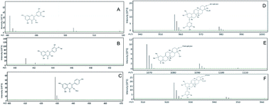
Black bean coats (Phaseolus vulgaris) contain bioactive compounds, including flavonoids and saponins, which have anti-fibrotic effects in which a standardized black bean extract (BBE) has been found to prevent liver fibrosis. Accordingly, the purpose of this study was to test whether BBE prevents remodeling in a murine model of non-ischemic cardiomyopathy. Saponins and flavonols were identified and quantified in BBE. Identification of flavonoids and saponins was confirmed by HPLC-MS-TOF. The cardiomyopathy model was produced by administering angiotensin and oral supplementation of L-NAME. Experimental animals received BBE in their diet at a dose equivalent to 40 mg per kg per day. The cardiomyopathy group (CMP) was characterized by severe maladaptive remodeling, left ventricular (LV) hypertrophy, decreased ejection fraction and increased LV end-diastolic volume. CMP mice treated with BBE had an improvement in ventricular function and reduction in LV mass. In addition, we found 65%, 85% and 83% reductions in interstitial fibrosis, brain natriuretic peptide (BNP) and transforming growth factor (TGFβ) expression, respectively. Consistent with those observations, collagen and TGFβ expression by isolated cardiac fibroblasts was reduced 82% and 70% following administration of BBE. BBE prevents adverse cardiac remodeling by reducing the extent of fibrosis and collagen deposition that occur in cardiomyopathy. These initial studies provide the basis for future research into the therapeutic potential of BBE in heart failure.
1. Introduction
Myocardial response to injury is characterized by myocyte hypertrophy and fibrosis. This process, also known as remodeling, is accompanied by a large number of molecular and cellular changes, but events central to this process are the signals responsible for cardiac myocyte enlargement and increased collagen deposition in the extra-cellular matrix.1
This response can be measured histologically and molecularly, but from a clinical perspective, it can be measured easily by imaging techniques looking for the characteristic features of increase in cardiac chamber size, increase in left ventricular mass, and changes in ventricular function.2 Notably, cardiac adverse remodeling is associated with worse prognosis and reverse remodeling with improved survival. Interventions that prevent adverse remodeling or reverse it, such as ACE inhibitors or beta-blockers, have changed the prognosis of patients with heart failure (HF) dramatically, regardless of the original insult that resulted in myocardial dysfunction.
Black bean (BB) is a legume widely consumed in Mexico and Latin America. The seed possesses relatively high protein and dietary-fiber contents and many proven nutraceutical properties.3 Using bioactivity screening of compounds associated with BB hulls, triterpenoids and flavonoids have been shown to exert anti-proliferative activity against colon, hepatic, and mammary cancer cells.4 In addition, López-Reyes et al. (2008) demonstrated that a methanolic BB extract (BBE) reduced expression of types I and IV collagen in a hepatic fibrosis murine model.5 Particularly, a previous study demonstrated that BB hulls had higher anti-inflammatory potential compared to counterparts obtained from pink or pinto beans.6 In addition, BB coat contains a number flavonoids and saponins, which are have been described as having immunomodulatory7 and anti-cytotoxic activities8 and, more recently, anti-fibrotic effects.9–11 Because of the high availability of black beans seeds, the significant role they play in nutrition in many areas of the world, and the potential anti-fibrotic effect they may have, we test whether use of BBE prevents adverse remodeling in a murine model of acute non-ischemic cardiomyopathy.
2. Experimental
2.1 Plant material
Black beans var. Otomí were planted and harvested at optimum maturity at Tecnológico de Monterrey's Experimental-Agricultural field located at Hualahuises, Nuevo León, México. The beans were immediately placed on 30 × 45 cm trays in preparation for dehydration in a convection oven set at 60 °C for 4 h. The seed coats were removed using an abrasive-disk mechanical sheller for 1.5 min and cotyledons were separated from seed coats using a set of sieves. This procedure was repeated 4 times.
2.2 Extraction of bioactive constituents of BBE
Phytochemicals associated to black beans seed coats were extracted with 80% methanol in a 1
:
10 (w/v) ratio using a shaking incubator (New Brunswick Scientific) at 25 °C and 140 rpm for 3 h. The extract was filtered, then concentrated with a rotary evaporator (Büchi) at 60 °C and finally freeze-dried (VirTis). The BBE was stored at −20 °C until use. The BBE was then provided to Teklad (Harlan Laboratories) for inclusion in standard mouse chow for animal experiments or dissolved directly in culture media for cell experiments.
2.3 Identification and quantification of saponins and flavonoids
Saponins and flavonoids were identified and quantified using an HPLC-DAD-ELSD (Agilent Technologies) system as previously reported.12 Briefly, peak identification of flavonoids was based on retention time and UV spectra and quantified using authentic standards of the corresponding aglycones: kaempferol, quercetin and myricetin. To confirm the detection of the DDMP-conjugated saponins their absorption maximum was obtained at 295 nm. The identification of saponins in BBE was confirmed by HPLC-MS-TOF (Agilent 1100). All saponins were quantified using evaporative light scattering detector (ELSD) with a standard curve obtained from soyasaponin I (Sigma-Aldrich).
2.4 Animals
All studies were approved by the Internal Committee for Care and Handling of Laboratory Animals of Tecnológico de Monterrey and the Methodist Hospital Research Institute. Twelve-week-old male C57BL/6 mice were used in all experiments. We used five animals for each treatment.
Non-ischemic cardiomyopathy mice model was obtained by a modified protocol of Oestreicher et al., (2003)13 to produces a severe malaptative remodeling with impaired ventricular function. Cardiomyopathy (CMP) mice were placed on water containing 0.1 mg mL−1 L-NAME and 1% NaCl. After 1 week acclimatization, mice were implanted subcutaneous with osmotic mini-pumps (Alzet model 1004) which delivered angiotensin II (AngII) (Sigma) at a rate of 0.7 mg per kg per day for 4 weeks. The mice receiving BBE were given a concentration of 400 mg kg−1 of BBE in their food pellets. The dose of BBE in the in vivo model was calculated according to the average mouse weight of 0.030 kg and average ingestion of 2.9 g per day; therefore, we calculated a total BBE intake of 40 mg per kg per day. All of the mice were euthanized at the end of the five-week protocol.
2.5 Mean arterial pressure measurements
Blood pressure measurements were taken when the animals were awake, three times each week throughout the protocol, using a CODA noninvasive tail cuff blood monitoring system (KENT Scientific). This system required ten cuff inflations and used internal software to calculate and record average MAP. We measured MAP at baseline and two, three, four, and five weeks, but not during the week of pump implants.
2.6 Magnetic resonance imaging analysis
Magnetic resonance imaging (MRI) was performed using a 9.4 T (89 mm bore) superconducting magnet with a Bruker AVANCE console that has live animal imaging capabilities. For mouse imaging, the system is equipped for anesthesia administration and cardiac gating, which is performed as a service by the imaging core at our facility (Methodist Hospital). Mice were anesthetized with inhaled isoflurane (1.5 vol% at 1 L min−1 oxygen flow) via a nose cone. The identities of all animals were blinded before imaging and uncoded following analysis. MRI analysis was performed at the completion of the 35 day of treatment.
2.7 Histological analysis
Mouse hearts were removed and sectioned mid-heart with apex portion used for PCR studies and base portion fixed in 2% paraformaldehyde, processed and embedded in paraffin and 5 micron sections cut. Sections were stained for 5 minutes in 0.1% Sirius red (Direct red 80) in 2% picric acid, followed by three 0.5% acetic acid washes. The slides were then cover slipped and analyzed at 20× magnification using an Olympus AX70 microscope. Images were taken of all regions of the left ventricle and analyzed for fibrosis using Image Pro Plus v4.0 analysis software (Media Cybernetics). Color cube-based selection criteria were used for positive staining and stained/unstained areas were measured. The results expressed are the average percent tissue area (pixels) stained by the dye.
2.8 PCR analysis
Total mRNA was isolated from cardiac tissues or cell culture using an RNA binding column kit (RNA4PCR, Ambion). RNA was immediately transcribed into cDNA using a kit employing random primers (iScript, BioRad) following manufacturer's directions. Real-time PCRs were performed at a final volume of 20 μL in 96-well plates analyzed in a MyIQ5 iCycler instrument (BioRad) which employs the use of SYBR green and contains software for analysis of signals. All samples were performed in quadruplicate and standardized to GAPDH run on the same plate. Mouse primers were chosen from the RTPrimerDB public primer database using the following sense and antisense primers for each molecule (5′ → 3′): brain natriuretic peptide (BNP) sense AGAACACGGCATCATTGCCTGG; BNP antisense, TGTGGCAAGTTTGTGCTCCAA; collagen I sense TGTTGGCCCATCTGGTAAAGA; collagen I antisense CAGGGAATCCGATGTTGCC; tumor necrosis factor (TNF-α) sense TTCTGTCTACTGAACTTCGGGGTGATCGGTCC; TNF-α antisense GTATGAGATAGCAAATCGGCTGACGGTGTGGG; GAPDH sense ACCCAGAAGACTGTGGATGG; GAPDH antisense ACACATTGGGGGTAGGAACA; transforming growth factor (TGFβ) sense CCGCAACAACGCCATCTATGA; TGFβ antisense GGGGGTCAGCAGCCGGTTAC. Reaction mixtures contained 11 μL H2O, 12.5 μL IQ SYBR Green Supermix (Bio-Rad), 1 μL primers and 1 μL DNA template. All plates were sealed, centrifuged at 500 × g for 60 s, and then amplified for 40 cycles of 10 s at 95 °C, 30 s at 55 °C, and 10 s at 72 °C. The expression levels were calculated by using (2Ct(gene)/2Ct(GAPDH)) × 1000 to standardize to GAPDH and expressed as relative transcript numbers for inter-assay comparisons. All GAPDH values were run on the same plate as the gene transcripts for each mouse/cell DNA assayed.
2.9 Cell culture
Primary cardiac fibroblasts were collected from adult mice and cultured to confluence. Briefly, excised tissue was minced into 1 to 2 mm pieces with a razor blade and digested in RPMI-1640 media (Sigma) containing 0.75 mg mL−1 collagenase B (Boehringer Mannheim) and placed in an Erlenmeyer flask on a shaking water bath maintained at 37 °C. Tissues were digested for 30 minutes and supernatant strained through 3 layers of cheesecloth and fresh media added to the tissue which was returned to the shaker. Three changes of supernatant were collected and pooled and centrifuged 5 min at 5000 × g. Cell pellets were resuspended in RPMI-1640 media containing 1% fetal bovine serum and penicillin/streptomycin (Sigma) and placed in culture plates and incubated in a CO2 incubator for 4 h. Plates were then washed twice to remove non-adherent cells and returned to the incubator with fresh media. Cells were grown for 2–3 weeks until confluent at which time assays were performed. For viability assays, media was replaced with media containing various concentrations of BBE and incubated for 3 h followed by addition of Trypan blue dye and cell viability counted and expressed as a percentage of viable cells. For PCR data, cells were incubated in various BBE concentrations for 3 h followed by removal of all media and isolation of mRNA as described under PCR heading.
2.10 Statistical analysis
Results are reported as the mean ± standard deviation. ANOVA test was used for comparisons among treatments, using Tukey post-test. A p ≤ 0.05 was considered statistically significant.
3. Results
3.1 Characterization of BBE
In accordance with Guajardo-Flores et al. (2012),14 the most abundant glycosylated flavonoid in BBE was quercetin-4-O-galactoside (Table 1). The saponins detected at 295 nm in BBE were group B soya-saponins conjugated to a 2,3-dihydro-2,5-dihydroxy-6-28-methyl-4H-pyran-4-one (DDMP) group. The soya-saponin αg was the most abundant, followed by soya-saponins βg and γg. Fig. 1 shows mass spectra identification.
Table 1 Concentration of main flavonoids and saponins found in black bean extract (BBE) for three determinations
Compound |
Concentration (mg/100 g) |
Myricetin-3-O-glucoside |
759.5 ± 36.6 |
Quercetin-4-O-galactoside |
4439.8 ± 224 |
Kaempferol-3-O-glucoside |
61 ± 5.1 |
Soyasaponin αg |
59.4 ± 3 |
Soyasaponin βg |
29.2 ± 0.6 |
Soyasaponin γg |
22.3 ± 0.3 |
 |
| Fig. 1 Major compounds and identities from BBE. Mass spectra of main flavonoids and saponins: myricetin-3-O-glucoside (A), quercetin-4-O-galactoside (B) and kaempferol-3-O-glucoside (C), soyasaponin αg (D), soyasaponin βg (E), soyasaponin γg (F). | |
3.2 Diet supplementation with BBE prevents adverse cardiac remodeling
Fig. 2 shows LV mass in control mice and CMP mice either untreated or treated with BBE (A), LV end-diastolic volume (B), and LV ejection fraction (C). As shown, the CMP group was characterized by an increase in LV mass, LV end-diastolic dimension, and a reduction in LV ejection fraction. LV mass was 62 ± 5 mm in controls, while in the CMP group, it was 78 ± 5 mm, and in the CMP + BBE group, it was 60 ± 2 mm (p-value < 0.001). LV end-diastolic volume was 0.072 ± 0.01 mL for controls, 0.09 ± 0.01 mL for the CMP group, and 0.07 ± 0.01 mL for the CMP + BBE group (p-value < 0.0001). Left ventricular ejection fraction (LVEF) was 65 ± 4% in controls, 42 ± 6% in the CMP group, and 51 ± 4% in the CMP + BBE group (p-value < 0.001). In contrast, the CMP group treated with BBE (CMP + BBE) did not show the same changes, and cardiac structure was similar to that in the untreated control group. As shown, the CMP group had increased LV mass and LV end-diastolic volume and decreased LVEF, and the CMP + BBE group showed improvement in all parameters measured.
 |
| Fig. 2 Effect of a black bean extract on myocardial function in a murine model of cardiomyopathy. Mice were grouped as control (n = 5) or treated to produce cardiomyopathy (CMP) (n = 5) and treated to produce CMP and simultaneously treated with BBE (CMP + BBE) (n = 5), as described in the method section. At the end of treatment MRI was performed on 5 mice in each group. The data shown is averaged ± SD. Representative sample images from one mouse in each group taken during systole and diastole at the mid-heart level are shown (A). The effect in left ventricle (LV) ejection fraction (EF) (B), end-diastolic volume (EDV) (C) and LV mass (LVM) is shown in (D). Both imaging and analysis was performed by a blinded operator to each mouse's experimental condition.* p-value < 0.05 vs. control.**p-value < 0.05 vs. CMP. | |
Fig. 2A shows a representative MRI example. Both imaging and analysis was performed by an operator blinded to each mouse experimental condition.
Fig. 3 shows changes in blood pressure over time during the experiment. Mean arterial pressure (MAP) was calculated by tail cuff measurement in the control, CMP, and CMP + BBE groups. The CMP and CMP + BBE mice had similar MAP values, but they were significantly higher compared to the control mice (p < 0.05). At five weeks, the MAP values were 98 ± 8 mmHg in the control group, 141 ± 11 mmHg in the CMP group, and 124 ± 13 mmHg in the CMP + BBE.
 |
| Fig. 3 Black bean supplementation did not reduce hypertensive response in the Angiotensin-II/L-Name model. Mean arterial pressure (MAP) was calculated by tail cuff measurements in control (n = 5), CMP (n = 5) and CMP + BBE (n = 5) groups. As shown an increase in MAP is seen as early as the third week of the experiment in the CMP and CMP + BBE groups. By the end of the experiment, 5 week, MAP were: control 98 ± 8, CMP 141 ± 11 and CMP + BBE 124 ± 13 mmHg.* p-value < 0.05 vs. control. | |
3.3 BBE prevents fibrosis as well as molecular changes of adverse remodeling
Fig. 4A shows the extent of fibrosis as measured by Sirius red in untreated controls, CMP, and CMP + BBE groups. The stained area in the control group was 3 ± 1%, in the CMP group it was 23 ± 3%, and in the CMP + BBE group it was 8 ± 2%. Fig. 4B shows a representative staining sections of control, CMP, and CMP + BBE mice with Sirius red. CMP showed a significant increase in stained area (in red-pink) in compared to the control or CMP + BBE group.
 |
| Fig. 4 Effect of black bean extract on myocardial fibrosis. (A) shows the extent of fibrosis as measured by Sirius red in untreated controls (n = 5), CMP (n = 5) and CMP + BBE (n = 5). The data shown is averaged ± SD. based of area stained of four separate experiments. (A) shows area stained in control was 3 ± 1%, CMP was 23 ± 3% and CMP + BBE was 8 ± 2%. (B) shows a representative experiment of Sirius red stained sections of a control mouse, CMP and CMP + BBE. As shown, a significant increased in area stained (in red-pink) is show in CMP compared to control or CMP + BBE. * p-value < 0.05 vs. control. **p-value < 0.05 vs. CMP. | |
Fig. 5 shows mRNA expression of TGFβ (A), BNP (B), TNF-α (C), and collagen I (D) in the control, CMP, and CMP + BBE groups. As shown, the CMP group had a significant (p < 0.001) increase in TGFβ, BNP, TNF-α, and collagen I: 0.28 ± 0.07, 1.60 ± 0.1, 1.32 ± 0.18, and 1.72 ± 0.2, respectively. The expressed levels of each of those genes in the CMP + BBE group were similar to those of the control group (TGFβ, 0.05 ± 0.004; BNP, 0.20 ± 0.03; TNF-α, 0.1 ± 0.04; and collagen I, 0.45 ± 0.07).
 |
| Fig. 5 Effect of black bean supplementation prevents expression of genes associated with cardiomyopathy. mRNA expression of TGFβ , BNP, TNF-α, and collagen I in the control (n = 5), CMP (n = 5), and CMP + BBE (n = 5) groups. As shown, the CMP group had a significant increase in TGFβ (A) BNP (B), TNF-α (C) and collagen I (D), whereas the CMP + BBE expressed levels of each of those genes similar to the control group. * indicates p < 0.05 vs. control group; ** indicates p < 0.05 vs. CMP group. The data is shown as average ± SD of five experiments. | |
3.4 BBE reduces collagen expression in murine fibroblasts
To explore the potential action mechanism of BBE on fibrosis and cardiac remodeling, we measured mRNA expression of collagen I after a 10% FBS stimulus on previous 1% FBS deprivation in murine cardiac fibroblasts (Fig. 6A). Expression of collagen I mRNA increased following stimulation with 10% FBS. The addition of increasing doses of BBE resulted in a powerful inhibition of collagen I expression; significant suppression of collagen I expression was observed at doses as low as 4 μg mL−1 of BBE. Fig. 6B shows fibroblast viability measured by Trypan blue uptake, in which fibroblast viability decreased to 75% at doses higher than 160 μg mL−1. Fibroblast viability was maintained at the doses at which BBE prevented collagen I expression. In addition, TGF-β expression decreased significantly following FBS stimulation at lower doses of BBE (Fig. 6C).
 |
| Fig. 6 Effect of black bean extract on collagen expression in murine fibroblasts. Murine cardiac fibroblasts were culture in control medium and control medium plus 10% FBS in the presence or absence of varying concentrations of BBE. (A) Expression of collagen I mRNA increased following stimulation with FBS. The addition of increasing doses of BBE resulted in powerful inhibition of collagen I expression; significant suppression of collagen I expression was observed at doses as low as 4 μg mL−1. (B) Viability of cardiac fibroblasts as measured by Trypan blue uptake. (C) Expression of TGF-β mRNA increased following stimulation with FBS. The addition of BBE (4 μg mL−1) decreased TGF-β expression. The figure shows that cardiac fibroblast viability decreased at doses higher than 200 μg mL−1. Fibroblast viability was intact at doses that prevented collagen I expression. *p < 0.05 vs. FBS. The data is shown as average ± SD (n = 5). | |
4. Discussion
Findings demonstrate that in a murine model of acute non-ischemic cardiomyopathy, oral supplementation of BBE prevents adverse cardiac remodeling in a powerful way. First, in vitro studies using murine cardiac fibroblasts stimulated with 10% FBS demonstrated a profound tendency to inhibit collagen expression by BBE at doses that were at least 40-fold lower than cytotoxic (Fig. 6). Second, in a murine model of CMP, increase in chamber size and LV mass and decrease in ejection fraction were prevented by oral administration of BBE. It is important to mention that the effect observed in BBE treatment likely also was the result of reduction in hypertensive response. Several studies have demonstrated that flavonoids possesses antihypertensive action by activating eNOS activity and inhibiting iNOS, endothelium-independent mechanisms, and Inter alia.15 Finally, we observed that these structural changes exerted by BBE treatment occurred in association with a significant reduction in fibrosis and a reduction in expression of BNP, TNF-α, TGFβ, and collagen I, genes that typify the HF phenotype.
4.1 BBE and the anti-fibrotic effect
Nowadays, medicinal herbs and daily intake foods are most likely taken into account for the pharmacological effects of particular herbs and to form the basis of their therapeutic efficacy on different pathologies. Several studies have indicated that both animals and humans are exposed significantly to some, but not all, compounds of an herbal medicine or a daily food constituent after dosing and that the pharmacokinetic profiles, like the pharmacological activities, can be used to scan the importance and usefulness of the individual BBE constituents. BBE is an extract of Phaseolus vulgaris L., a BB plant widely available, readily grown, and consumed in large quantities in many regions of the world. No major therapeutic aspects of BB consumption have been developed, but extensive literature is available regarding the effects of many BBE components. BB is a source of vitamin B6 and folic acid, and it contains enormous numbers of saponins and flavonoids, which exert diverse biological activities.16,17 Bioactive compounds with favorable properties contained in the BBE with adequate abundance, such as saponins and flavonoids, occur mainly in their glycoside forms. In general, saponins and flavonoids are two diverse groups of compounds present in many plants species, classified based on the biosynthesis and subsequent transformation of their carbon skeletons.18 As mentioned earlier, germinated BB seeds contain different amounts of saponins and flavonoids in the sprouts, seed coats, and cotyledons.12 Saponin and flavonoid compounds have been associated with several biological activities, such as antioxidant capacity and antiproliferative activities,14 as well as cholesterol micelle disruptor,19 antimicrobial,20 immunomodulatory,3 and anticytotoxic effects.8 It is important to mention that BBE was recently described as having antifibrotic effects, wherein the flavonoid quercetin provided a protective effect against pulmonary injury and fibrosis in a rat model.10 In addition, BBE has been shown to inhibit liver fibrosis.5 The findings of the present study were consistent with previous observations on the use of BBE, which demonstrated an antifibrotic effect in a murine model of liver cirrhosis. In that model, rats treated with CCl4 were either untreated or treated with BBE by intragastric administration. The use of BBE in doses similar to those used in the present study exhibited a dramatic reduction in liver fibrosis.5 The findings of the present study and the observations of the CMP mouse model are consistent, showing a common mechanism of protection whereby the accumulation of collagen I is prevented by BBE supplementation. Interest in these and other flavonoid-rich natural products has grown since the results of a double-blind, placebo-controlled study using flavonol-rich chocolate demonstrated beneficial effects in patients with heart failure.21
As mentioned previously, the BBE used in these experiments contains six major bioactive compounds that can be classified as flavonoids (quercetin-4-O-galactoside, myricetin-3-O-glucoside, and kaempferol-3-O-glucoside) and saponins (soyasaponin αg, soyasaponin βg, and soyasaponin).12 The six major bioactive compounds in the BBE used in these experiments represent only 5.4% of the extract; therefore, we calculated a real dose of 2.1 mg per kg per day of saponins and flavonoids ingested by the animals in our study.
In order to evaluate a possible mechanism of action of BBE, we evaluated TGFβ expression, as it is one of the most important molecules that play a pivotal role in heart failure as a consequence of the development of cardiac hypertrophy mediated by angiotensin networking. TGFβ has been well established as a key mediator of cardiac adaptation to hemodynamic overload, and it is critically involved in the pathogenesis of cardiac hypertrophy and failure. In summary, in cardiac myocytes and fibroblasts, activation of the AT1 receptor by AngII upregulates TGFβ expression. The induction of this cytokine is completely required for AngII-induced cardiac hypertrophy in vivo and in vitro.22 TGFβ induces the proliferation of cardiac fibroblasts and their differentiation to a myofibroblast phenotype. Myofibroblasts are in charge of producing ECM proteins such as collagen I, thus inducing structural remodeling of the ventricular wall in an autoparacrine way. One possible explanation for this mechanism has been shown in primary cardiac fibroblasts, wherein antioxidant treatment inhibited ECM production already converted to a myofibroblast phenotype (by prolonged TGFβ treatment), suggesting that continuous ROS production is necessary for maladaptive remodeling.23
4.2 BBE in the prevention of cardiomyopathy
The CMP model used in the present study represents a form of acute non-ischemic cardiomyopathy wherein the use of Angiotensin II (AngII) leads to hypertension and downstream effects that create the CMP phenotype. This model clearly is characterized by an increase in LV mass and LV end-diastolic dimension and a reduction in LV ejection fraction. In addition, significant histological and molecular changes accompany this phenotype. In the present study, we demonstrated a significant effect in cardiac fibrosis as measured by Sirius red and increase in BNP, TNF-α, and collagen I genes, clearly defining a CMP state. Interestingly, oral diet supplementation with BBE administered in pellets resulted in preventing adverse cardiac remodeling effects, as defined by MRI and histological studies. Those changes were supported further by the effect of BBE treatment on preventing up-regulation of BNP, TNF-α, and collagen I gene expression. Put together, these data are consistent in supporting a significant effect of BBE in preventing induction of CMP in this model. For instance, the flavonoid herperetin has been shown to exert beneficial effects against oxidative stress and fibrosis events related to cardiac hypertrophy, reducing remodeling markers such and exerting robust protection against ventricular dysfunction.24
This model is associated with a significant hypertensive response, as seen in the present study; at the end of five weeks of AngII treatment, the MAP of the CMP group was 141 mmHg compared to control animals, in which MAP was 98 mmHg. Interestingly, the CMP + BBE mice also developed significant hypertension. At the end of five weeks, their MAP was 124 mmHg, significantly higher than the controls, but somewhat lower than the MAP of the CMP group (p ≥ 0.05). This difference appears to be present during the early stages of the experiment and persists over time. Furthermore, the change in MAP in the CMP + BBE group was very significant compared to that in the control group, and it is unlikely that this effect was the result of the changes observed in cardiac structure and at the molecular level in this group, but that cannot be ruled out completely as a potential contributor. We suggest, however, that the findings are more consistent with BBE exerting a not significant hypertensive effect preventing the adverse remodeling.
4.3 Limitations
The model used to produce CMP is a form of acute non-ischemic cardiomyopathy induced by AngII, and it is possible that there is a specific interaction between AngII and BBE that may not be applicable to other forms of cardiac disease. However, increased AngII levels are characteristic of human HF, and therefore, these observations are significant. BBE was administered in the diet by creating pellets with a concentration that would predict consumption of 40 mg per kg per day. However, since we do not have a way to measure BBE concentration in the serum, we cannot establish a correlation between the effect and serum concentrations of BBE. Finally, BBE is an extract, and multiple active compounds are present in the preparation; dissecting individual components for further identification of the bioactive compounds is important and will be the focus of future research.
Disclosures
All authors have no conflicts to declare.
Abbreviations
AngII | Angiotensin II |
BB | Black bean |
BBE | Black bean extract |
BNP | Brain natriuretic peptide |
CMP | Cardiomyopathy |
DDMP | 2,3-Dihydro-2,5-dihydroxy-6-28-methyl-4H-pyran-4-one |
EDV | End-diastolic volume |
EF | Ejection fraction |
FBS | Fetal bovine serum |
HF | Heart failure |
LV | Left ventricular |
LVEF | Left ventricular ejection fraction |
LVM | LV mass |
MAP | Mean arterial pressures |
MRI | Magnetic resonance imaging |
TGF-β | Transforming growth factor beta |
TNF-α | Tumor necrosis factor alpha |
Acknowledgements
This work was partially supported by Cátedra de Cardiología-Tec de Monterrey (0020CAT131), Xignus Research Found (G. G.-R.) as well by grants from SEP-CONACYT: Projects 133591 (G. T.-A.), 131565 and 151136 (G. G.-R.) and BBE characterization was supported by FEMSA-Nutrigenomics Found.
References
- M. G. Sutton and N. Sharpe, Circulation, 2000, 101, 2981–2988 CrossRef CAS PubMed.
- G. D. Aquaro, G. Todiere, A. Barison, E. Strata, M. Marzilli, A. Pingitore and M. Lombardi, J. Card. Failure, 2011, 17, 384–391 CrossRef PubMed.
- Y. Papanikolaou and V. L. Fulgoni 3rd, J. Am. Coll. Nutr., 2008, 27, 569–576 CrossRef CAS PubMed.
- M. Dong, X. He and R. H. Liu, J. Agric. Food Chem., 2007, 55, 6044–6051 CrossRef CAS PubMed.
- A. G. López-Reyes, N. Arroyo-Curras, B. G. Cano, V. J. Lara-Diaz, G. E. Guajardo-Salinas, J. F. Islas, V. Morales-Oyarvide, L. A. Morales-Garza, F. J. Galvez-Gastelum, G. Grijalva and J. E. Moreno-Cuevas, Ann. Hepatol., 2008, 7, 130–135 Search PubMed.
- B. D. Oomah, A. Corbé and P. Balasubramanian, J. Agric. Food Chem., 2010, 58, 8225–8230 CrossRef CAS PubMed.
- A. S. Strimpakos and R. A. Sharma, Antioxid. Redox Signaling, 2008, 10, 511–545 CrossRef CAS PubMed.
- K. C. Choi, W. T. Chung, J. K. Kwon, J. Y. Yu, Y. S. Jang, S. M. Park, S. Y. Lee and J. C. Lee, Food Chem. Toxicol., 2010, 48, 2747–2753 CrossRef CAS PubMed.
- Q. Huang, R. Huang, S. Zhang, J. Lin, L. Wei, M. He, L. Zhuo and X. Lin, Toxicol. Lett., 2013, 217, 102–110 CrossRef CAS PubMed.
- H. K. Park, S. J. Kim, Y. Kwon do, J. H. Park and Y. C. Kim, Life Sci., 2010, 87, 181–186 CrossRef CAS PubMed.
- L. D. Hernández-Ortega, B. E. Alcántar-Díaz, L. A. Ruiz-Corro, A. Sandoval-Rodriguez, M. Bueno-Topete, J. Armendariz-Borunda and A. M. Salazar-Montes, J. Gastroenterol. Hepatol., 2012, 27, 1865–1872 CrossRef PubMed.
- D. Guajardo-Flores, M. García-Patiño, D. Serna-Guerrero, J. A. Gutiérrez-Uribe and S. O. Serna-Saldívar, Food Chem., 2012, 134, 1312–1319 CrossRef CAS PubMed.
- E. M. Oestreicher, D. Martinez-Vasquez, J. R. Stone, L. Jonasson, W. Roubsanthisuk, K. Mukasa and G. K. Adler, Circulation, 2003, 108, 2517–2523 CrossRef CAS PubMed.
- D. Guajardo-Flores, S. O. Serna-Saldívar and J. A. Gutiérrez-Uribe, Food Chem., 2013, 141, 1497–1503 CrossRef CAS PubMed.
- G. Siasos, D. Tousoulis, V. Tsigkou, E. Kokkou, E. Oikonomou, M. Vavuranakis, E. K. Basdra, A. G. Papavassiliou and C. Stefanadis, Curr. Med. Chem., 2013, 20, 2641–2660 CrossRef CAS.
- R. J. Nijveldt, E. van Nood, D. E. van Hoorn, P. G. Boelens, K. van Norren and P. A. van Leeuwen, Am. J. Clin. Nutr., 2001, 74, 418–425 CAS.
- H. Wiseman, in FLAVONOIDS Chemistry, biochemistry and applications, ed. Ø. M. Andersen and K. R. Markham, Informa, United States of America, 2006, pp. 371–398 Search PubMed.
- J. P. Vincken, L. Heng, A. de Groot and H. Gruppen, Phytochemistry, 2007, 68, 275–297 CrossRef CAS PubMed.
- R. A. Chávez-Santoscoy, J. A. Gutiérrez-Uribe and S. O. Serna-Saldívar, Plant Foods Hum. Nutr., 2013, 68, 416–423 CrossRef PubMed.
- V. J. Lara-Díaz, A. A. Gaytán-Ramos, A. J. Dávalos-Balderas, J. Santos-Guzmán, B. D. Mata-Cárdenas, J. Vargas-Villarreal, A. Barbosa-Quintana, M. Sanson, A. G. López-Reyes and J. E. Moreno-Cuevas, Basic Clin. Pharmacol. Toxicol., 2009, 104, 81–86 CrossRef PubMed.
- A. J. Flammer, I. Sudano, M. Wolfrum, R. Thomas, F. Enseleit, D. Periat, P. Kaiser, A. Hirt, M. Hermann, M. Serafini, A. Lévêques, T. F. Lüscher, F. Ruschitzka, G. Noll and R. Corti, Eur. Heart J., 2012, 33, 2172–2180 CrossRef CAS PubMed.
- M. Dobaczewski, W. Chen and N. G. Frangogiannis, J. Mol. Cell. Cardiol., 2011, 51, 600–606 CrossRef CAS PubMed.
- D. A. Siwik, P. J. Pagano and W. S. Colucci, Am. J. Physiol.: Cell Physiol., 2001, 280, C53–C60 CAS.
- W. Deng, D. Jiang, Y. Fang, H. Zhou, Z. Cheng, Y. Lin, R. Zhang, J. Zhang, P. Pu, Y. Liu, Z. Bian and Q. Tang, J. Mol. Histol., 2013, 44, 575–585 CrossRef CAS PubMed.
|
This journal is © The Royal Society of Chemistry 2015 |
Click here to see how this site uses Cookies. View our privacy policy here.