DOI:
10.1039/C5RA07700A
(Paper)
RSC Adv., 2015,
5, 46146-46157
Bio-inspired adhesion: fabrication and evaluation of molecularly imprinted nanocomposite membranes by developing a “bio-glue” imprinted methodology†
Received
28th April 2015
, Accepted 18th May 2015
First published on 18th May 2015
Abstract
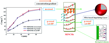
Nanocomposite membranes with specific recognition, durability and regeneration ability that can rapidly adsorb and separate target compounds have remarkable technological applications for areas ranging from solid-phase extraction devices to architecture. In this work, inspired by the highly bioadhesive performance of mussel protein, urgently desired molecularly imprinted nanocomposite membranes (MINCMs) were prepared by developing a simple “bio-glue” imprinted strategy. By simply immersing the “bio-glue” m-cresol-imprinted PDA@SiO2 into casting solution accompanied by persistently mechanically stirring, a highly bio-adhered and homo-dispersedly distributed structure could be generated into MINCMs during a phase inversion process, which directed the higher perm-selectivity and reusability. Additionally, due to the unique properties of PDA modified layers and SiO2 nanoparticles (high surface-to-volume ratio and large surface area), the as-prepared MINCMs not only exhibited rapid adsorption dynamics, but also possessed an excellent separation performance of template molecule (m-cresol in this work). The excellent separation (perm-selectivity factor is 3.477) and recognition behavior (imprinted factor is more than 3.0) along with the low preparation consumed and green, quick, facile synthesis conditions make the as-prepared MINCMs attractive in broad technological applications for areas ranging from drug delivery to bioseparation.
1. Introduction
Developing nanotechnology is a new research field which produces an enormous influence on broad kinds of research fields, such as materials, environment, and energy.1–6 In recent years, multifunctional nanocomposite membranes have attracted significantly important aspects in nanotechnology, which can keep both the strength–toughness of high polymer materials and the rigidity, special properties of nano materials.7–10 As a low-energy consuming, environmental friendly and efficient progress, nanocomposite membrane-based separation technique (NCMST) has caused worldwide concern over nanotechnology and membrane science during the past years.9–14 This technique could not only stand a chance to fit together the advantages of both polymer membranes (such as polysulfone/PSF and polyvinylidene fluoride/PVDF) and nanoparticles (SiO2, TiO2), but also increase the comprehensive performance of single membranes to meet the special requirements.10–14 As evidenced in the early studies, although the straightforward blending of nanoparticles into polymeric membranes can effectively improve the anti-fouling performance, mechanical strength, and hydrophilicity, several inherent drawbacks such as non-selectivity, aggregation of nanoparticles, and weak binding force will curb the development of nanocomposite membrane materials for further applications.15–17
It is generally known that molecularly imprinted technique (MIT) is a simple and well-established technique for creating recognition cavities complementary to the size, shape, and functionality of template molecules.18–22 Recently, molecularly imprinted polymers (MIPs) play central roles in bioseparation, medical diagnostics, drug delivery, and membrane separations because of their outstanding advantages such as easy preparation, low-cost, and chemical/mechanical stability.23–26 As the name implies, molecularly imprinted membranes (MIMs) are the membranes composed of MIPs or containing MIPs. Combination of MIPs into nanocomposite membranes could provide membrane-based specific recognition capacity for targets.27–30 In recent years, porous MIMs with highly diversified pore size, layered structure, and specific separation performance have attracted plenty of attention. At present, the most common approaches for synthesis of MIMs are casting or grafting imprinting layer at the surface of membranes. On the other side, although numerous reports have appeared, issues such as high diffusion barrier, low regenerability, poor perm-selectivity, and structure resistance still exist, and the widespread use of MIMs is still in its infancy.30 In particular, the major issue is the optimization of recognition sites and membrane permeability simultaneously. So, it is very valuable to find an effective and straightforward method to disperse the imprinting layer homogeneously and steadily into membrane materials, which directing both high recognition ability and perm-selectivity of specific compounds.
Inspired by the mussels' adhesive versatility, Messersmith and co-workers proposed that dopamine (DA) could be regarded as an excellent surface-adherent material for multifunctional coatings on the basis of DA self-polymerization.31 Because of the oxidation of catechol groups into quinone form in an alkaline DA solution, DA can polymerize onto all kinds of inorganic and organic surfaces to form a heterogeneous polydopamine (PDA) layer (Fig. S1†).32–34 Attributed to this breakthrough study, PDA coating layers can be regarded as a stabilized cross-linked structure which generate homogenous and stable imprinted cavities.18,19,30 Meanwhile, the PDA-based MIPs could be easily prepared by co-dissolving the target molecules with DA on many diverse surfaces. And the resultant PDA-modified monolayer could be regarded as a double-sided adhesive platform for secondary reactions. Considering the above-mentioned attractive properties of PDA-based MIPs and NCMST in the preparation of MIMs, a breakthrough strategy containing both the NCMST and DPA-based MIPs was engineered in this work.
Cresol, which is the mixture of m-cresol, p-cresol, o-cresol isomers, is mainly obtained from petroleum products. Recently, cresol and its downstream products herald vast potentials for development and considerable market prospects.35–39 m-Cresol has attracted increasing interests due to its wide array of applications, such as the synthesis of pharmaceuticals, dye, epoxy resin, and various crucial fine chemicals. And the related p-cresol and o-cresol isomers also play significant roles in modern material science, respectively.40–42 To date, because of the enormous market demand, numerous approaches such as chemical synthesis, fractional distillation, extractive crystallization have been utilized to separate and extract m-cresol from cresol mixtures. However, many drawbacks such as time-consuming and complex procedures, low specific separation ability and requirements of expensive apparatuses limit the further application and development of m-cresol.
Despite decades of intense research in this area, engineering and synthesis of MIMs with highly selective separation performance and enhanced comprehensive properties are still challenged. Therefore, development of practical and effective strategies capable of excellent adsorption capacity and highly specific separation properties is necessary. Inspired by the advantages of NCMST and PDA-based MIPs, for the first time, the design and engineering of novel molecularly imprinted nanocomposite membranes (MINCMs) capable of selectively recognizing and separating special compound (m-cresol in this work) were successfully achieved. Importantly, instead of directly constructing MIPs layer on the membrane surfaces or in situ synthesis of MIPs into membranes, we infiltrated m-cresol-imprinted PDA@SiO2 into polymeric materials (PVDF) during the formation of membranes by a phase inversion process. The PDA-based surfaces allowed for the homogeneous and steady distribution of imprinting layers onto membrane materials, which made it an effective strategy for selective separation and purification of template molecules. Furthermore, the approach reported here consists of several environment-friendly and straightforward techniques, which directs it as a promising candidate for large-scale applications.
2. Experimental section
2.1. Materials
PVDF powders, polyvinylpyrrolidone, HPLC grade water and acetonitrile were purchased from Sinopharm Chemical Reagent (Shanghai, China). m-Cresol (98%), p-cresol (98%), 2,4-dichlorophenol (2,4-DP, 98%) were obtained from Aladdin Reagent (Shanghai, China). Dopamine (DA, 98%, Aladdin), tetraethyl orthosilicate (TEOS, 98%, Aladdin), tris(hydroxymethyl)-aminomethane (Tris–HCl, 99%, Aladdin), 1-methyl-2-pyrrolidinone (NMP, 99%, Aladdin), ammonia solution (NH4OH, 28–30%, Aladdin) were used as received. Doubly distilled water was used in all cleaning processes and aqueous solutions.
2.2. Apparatus and characterization
Fourier transform infrared (FT-IR) spectra (4000–800 cm−1) for nanoparticles were recorded on a Nicolet 560 FTIR spectrophotometer (U.S.A.). The ATR-FTIR spectra (4000–800 cm−1) for different PVDF membranes were recorded on a FT-IR Nicolet 560 apparatus (U.S.A.), and ZnSe was used as the crystal plate. To test the surface chemical composition, X-ray photoelectron spectroscopy (XPS) measurement was carried out using polychromatic Mg Kα X-rays source with an Omicron ESCA probe spectrometer. The pressure in the analysis chamber was maintained at 5.0 × 10⁻⁸ Torr or lower during each measurement. Raman spectra recorded using 532 nm excitation laser was performed using a LabRAM HR Raman spectrometer (U.S.A.). High performance liquid chromatography (HPLC) (Agilent 1200 series, U.S.A.) was used for the determination of m-cresol, p-cresol, and 2,4-dichlorophenol. Detailed determined requirements were shown in ESI.† The static water contact angle of different modified membranes was measured by the contact angle detecting process. The deionized water droplet used in detection tests was placed on dry smooth membrane surfaces and the contact angle was obtained. The measured contact angle values were calculated by averaging over more than five contact angle values at different sites for insuring the accuracy.
2.3. Preparation of PDA-based molecularly imprinted silica nanoparticles self-polymerization of dopamine
Silica nanoparticles (SiO2) with different sizes were prepared through the hydrolysis of TEOS with NH4OH aqueous solution following the reported Stöber strategy. In a typical m-cresol-imprinted PDA@SiO2 synthesis, SiO2 (200 mg) were firstly dispersed in 50 mL Tris–HCl (pH = 8.5) aqueous solution by an ultrasonic process for 5 min, m-cresol (20 mg) was then added. The mixture was persistently shaken for 1.0 h. Subsequently, DA (100 mg) was added to the above mixture to start the self-polymerization procedure. The reaction system was under persistently mechanically stirred at room temperature for 3 h, which directed the formation of PDA imprinted layer on the surface of SiO2. Then the m-cresol-imprinted PDA@SiO2 were obtained after being rinsed with deionized water to wash out redundant PDA particles and unreacted DA. As the contrast, non-imprinted PDA@SiO2 were prepared without adding the template molecules and subjected to the same conditions.
2.4. Preparation of molecularly imprinted nanocomposite membranes (MINCMs) by blending with m-cresol-imprinted PDA@SiO2
The MINCMs were synthesized using an m-cresol-imprinted PDA@SiO2 infiltrated phase inversion process. In a typical MINCMs synthesis, the casting solution of MINCMs was prepared by adding 4.0 g PVDF powders, 10 mg polyvinylpyrrolidone and 300 mg m-cresol-imprinted PDA@SiO2 into 20 g NMP. The mixed solution was sealed under persistently mechanically stirring at 50 °C for 24 h, so that the m-cresol-imprinted PDA@SiO2 and PVDF powders fully mixed as to create the homo-disperse solution without air bubbles. After that, the as-prepared mixture was cast on a glass plate using a doctor knife. And then the casting solution on the glass substrate was instantly immersed into a coagulant bath containing pure water to undergo a phase inversion. Upon complete coagulation, the membranes were washed by deionized water after taken out from coagulation bath, and the formative MINCMs were obtained after being rinsed with a mixture of ethanol–acetic acid (95
:
5, v/v) to wash out template molecules. As the control, instead of adding m-cresol-imprinted PDA@SiO2, the non-imprinted PDA@SiO2 was used to prepare the non-imprinted nanocomposite membranes (NINCMs) by the same procedure.
2.5. Binding experiments
In this work, to optimize the synthesis conditions, the impacts of the concentrations of DA, DA self-polymerization time, amount of m-cresol-imprinted PDA@SiO2, and elution time were studied, and the best preparation condition was investigated in detail. In addition, in order to reduce errors and increase measurement precision, we employed the adsorption temperature of 30 °C and m-cresol concentration of 600 mg L−1 ethanol solution as the constant adsorption conditions in the whole optimized experiments.
To investigate the binding performance of the MINCMs and the control NINCMs, binding experiments were performed by batch mode operations. For the adsorption kinetics studies, one piece of MINCMs (25 mm in diameter) or NINCMs was added to 10 mL of m-cresol ethanol solution (600 mg L−1), the supernatant adsorbed solution was shaken in thermostatic water bath at 25 °C and then taken out at predetermined time intervals (0, 5.0, 10, 20, 30, 40, 50, 60 min). For the adsorption isotherm studies, one piece of MINCMs or control NINCMs was added to 10 mL of m-cresol ethanol solutions with different concentrations (80, 100, 200, 400, 800, 1000 mg L−1). And the experiments were carried out at 25 °C for 2.0 h. The HPLC apparatus was used for the determination of m-cresol at 280 nm. The adsorption capacities of MINCMs and NINCMs were calculated from the concentration differences of m-cresol after and before adsorption. The adsorption amount of m-cresol could be calculated as follow:
|
 | (1) |
|
 | (2) |
where
Ce and
C0 (mg mL
−1) represent the initial and equilibrium concentrations, respectively.
Ct (mg L
−1) is the concentration of
m-cresol at different time
t.
Qe and
Qt (mg mL
−1) represent the adsorption amount of
m-cresol.
V is the volume of adsorbed solution,
m is the molar mass of
m-cresol.
The selective adsorption experiments of MINCMs and NINCMs were investigated using m-cresol, p-cresol, and 2,4-DP as competitive molecules with the initial concentrations of 500 mg L−1. After selective binding processes, the binding solution including m-cresol, p-cresol, and 2,4-DP was detected by HPLC. The imprinting factor α was calculated by the following equation:
|
 | (3) |
where
Q values are the adsorption amounts of
m-cresol,
p-cresol, and 2,4-DP by MINCMs or NINCMs, respectively.
2.6. Selective permeation experiments
To investigate the perm-selectivity performance of MINCMs and NINCMs, the competitive permeation experiments were evaluated using the feeding ethanol solutions consisted of m-cresol, p-cresol, and 2,4-DP with different concentrations (ranging from 100 to 1000 mg L−1). Time-dependent perm-selectivity experiments (ranging from 0 to 100 min) were carried out towards m-cresol, p-cresol, and 2,4-DP to further confirm the existence of selective recognition and separation performance of MINCMs produced by the imprinting procedure. The permeability tests were all carried out in an H-model tube installation as shown in Fig. S2.† The concentrations of m-cresol, p-cresol, and 2,4-DP obtained from receiving phase which permeated through the MINCMs or NINCMs were measured by HPLC. In addition, the mixture solutions in both chambers were kept homogeneous by water bath thermostatic oscillator at 25 °C. The permeation flux J (mg cm−1 s−1), permeability coefficient P (cm2 s−1) and permeation factors βNINCMs/MINCMs and β2,4-DP/m-cresol are obtained as follows: |
 | (4) |
|
 | (5) |
|
 | (6) |
|
 | (7) |
where V, A, and d represent the volumes of feeding and receiving solution (mL), effective membrane area (cm2), and the membrane thickness, respectively. ΔCi/Δt is the change of concentration in the receiving solution. (CFi − CRi) is the concentration difference between feeding and receiving chambers.
3. Results and discussion
3.1. Covalent synthesis and characterization of molecularly imprinted nanocomposite membranes (MINCMs)
As schematically described in Fig. 1, the preparative method developed to infiltrate m-cresol-imprinted PDA@SiO2 into PVDF membranes involves two steps. Initially, inspired by a “bio-glue” imprinting strategy, through the straightforward immersion of SiO2 in DA buffered solution (pH = 8.5) in presence of m-cresol molecules at ambient temperature, DA can concurrently polymerize into PDA and deposit on the SiO2 surfaces. During the self-polymerization process of DA, the deposited PDA-based imprinted layers constructed the cross-linked structures which could generate stable three-dimensional imprinted cavities, the m-cresol molecules could be then trapped in the cross-linked polymeric network because of the imprinting effect.19,31,43 The reasonable explanation should be that the amino group in PDA layers could form hydrogen bonds with the hydroxyl group of m-cresol. Additionally, the m-cresol molecules keep the benzene rings in chemical structure. The structure similarity also led to the van der Waals interaction between m-cresol molecules and the PDA-based imprinting layers. That is to say, there were plenty of benzene rings in the PDA-based imprinting layers at the surface of SiO2, which maximized the spatial distribution between the imprinting cavities and m-cresol molecules. As a result, the specific sterically complementary imprinted cavities of template molecules were created in the synthesized m-cresol-imprinted PDA@SiO2.
 |
| Fig. 1 Possible reaction mechanism of preparation of MINCMs by developing a two-step surface polymerization strategy. | |
The morphological evolution of pristine SiO2 and m-cresol-imprinted PDA@SiO2 (with different self-polymerization time) was analyzed by SEM. Fig. 2 summarized the SEM results of various before and after DA self-polymerization. Compared with Fig. 2a (pristine SiO2), the smooth surfaces of pristine SiO2 became rough after the “bio-glue” imprinting procedure (DA self-polymerization in presence of m-cresol). Fig. 2b–e (m-cresol-imprinted PDA@SiO2 with different self-polymerization time) exhibited obviously rough surfaces, which also directed the successful construction of PDA-based imprinting layers on the SiO2 surface. As shown in Fig. 2b–e, with the DA self-polymerization time increasing, the m-cresol-imprinted PDA@SiO2 exhibited the thicker and rougher surface morphologies. Due to more and more PDA particles were bound on the surface of SiO2, the morphologies of the PDA-based polymer layers became much denser and apparent. The surface spectroscopies of pristine SiO2 and m-cresol-imprinted PDA@SiO2 were further investigated by FTIR and XPS. The FTIR of pristine SiO2 and m-cresol-imprinted PDA@SiO2 were presented in Fig. 3A. As shown, after the DA self-polymerization procedure in presence of m-cresol, there came out several new absorption signals. The emerging peaks at 1625 cm−1 and 3700 cm−1 were because of the C
C and phenolic groups (Ar–OH) resonance vibrations in the aromatic rings, respectively.44 These results evidentially illustrate the successful formation of PDA-based imprinting layers which are further confirmed by XPS. The XPS spectrum was also used to analyze the differences of the surface chemical compositions for pristine SiO2 and m-cresol-imprinted PDA@SiO2. Fig. 3B and C illustrated the XPS wide scans and narrow scan for N1s peaks of m-cresol-imprinted PDA@SiO2. According to XPS wide spectra (Fig. 3B), the new emerge of the N1s peak, together with the narrow scan for N1s peaks of m-cresol-imprinted PDA@SiO2, demonstratively indicated the formation of PDA-based imprinting layers.32,33
 |
| Fig. 2 SEM images of m-cresol-imprinted PDA@SiO2 with different self-polymerization time of DA. | |
 |
| Fig. 3 (A) FTIR spectra of pristine SiO2 and m-cresol-imprinted PDA@SiO2. X-ray photoelectron spectroscopy wide scan (B) and narrow scan (C) for N1s peak of m-cresol-imprinted PDA@SiO2. | |
Secondly, the m-cresol-imprinted PDA@SiO2 infiltrated phase inversion process was applied for the synthesis of MINCMs. The mixture of PVDF powders, NMP, and m-cresol-imprinted PDA@SiO2 was stored at 50 °C and under persistently mechanically stirring to obtain the evenly dispersed solution. The constant stirring speed could make a homogeneous distribution of m-cresol-imprinted PDA@SiO2 in the cast solution. Furthermore, due to the “bio-glue” imprinting layers on the surface of SiO2, the m-cresol-imprinted PDA@SiO2 could also be homo-distributed onto the as-prepared membranes with highly adhesive attraction. Finally, after the removal of m-cresol molecules, the specific sterically complementary imprinted cavities of m-cresol were created in the synthesized MINCMs.
After the phase inversion process, the synthesized m-cresol-imprinted PDA@SiO2 nanoparticles could be adhered onto the MINCMs (Fig. 4a–c). From the cross-section views depicted in Fig. 4d and e, the homo-distributed and adhesive m-cresol-imprinted PDA@SiO2 nanoparticles onto MINCMs can be obviously observed. One can reasonably speculate that the persistently mechanically stirring of casting solution and the “bio-glue” property of the PDA-based m-cresol-imprinted PDA@SiO2 can form highly adhesive attractions with polymeric membranes. Fig. 5 illustrated the ATR-FTIR spectra of pristine PVDF membrane and MINCMs, at the side of pristine PVDF membrane, MINCMs exhibited several new absorption peaks at 1640 cm−1 and 1515 cm−1, which are corresponded to the C
C resonance vibrations and N–H bending vibrations, respectively.44,45 Meanwhile, the m-cresol-imprinted PDA@SiO2 inserted structure of MINCMs was recorded by XPS to further confirm the surface chemical composition. Fig. 6 presents the wide scan and narrow scans for Si2p, N1s, O1s of MINCMs. According to Fig. 6 describing XPS wide spectra, the Si2s, Si2p, O1s, and N1s perks emerge, which may attribute to the existence of m-cresol-imprinted PDA@SiO2. In addition, Fig. 6b and c summary the curve-fitting results of N1s and O1s, the peak of N1s located at 399.8 eV and another peak of O1s located at 532.3 eV correspond to the N–H groups and Si–O–Si groups, respectively. These findings, together with the results from ATR-FTIR, evidently indicate the successful formation and adhesion of m-cresol-imprinted PDA@SiO2 on the surface of MINCMs.
 |
| Fig. 4 Top view (a–c) and cross-section (d–f) SEM images of the MINCMs prepared by a “bio-glue” imprinted phase inversion process. | |
 |
| Fig. 5 ATR-FTIR spectra of the pristine PVDF membrane, and the MINCMs. | |
 |
| Fig. 6 X-ray photoelectron spectroscopy wide scan and narrow scans for Si2p (a), N1s (b), and O1s (c) peaks of MINCMs. | |
3.2. Optimization of preparation conditions
To evaluate the influence of DA, 200 mg SiO2, 20 mg m-cresol, and different amounts of DA (60, 80, 100, 120, and 140 mg) were selected as the preparation conditions of MINCMs. Fig. 7A presented the adsorption capacities of MINCMs with various amount of DA. As shown, the adsorption capacities of MINCMs for m-cresol increased as amount of monomer DA increased until 100 mg. One can reasonably speculate that the increasing concentration of DA could increase the thickness of PDA-based imprinting layer at the surface of SiO2, which could create much more recognition cavities during the imprinting procedure and increase the binding capacity of as-prepared MINCMs. However, a little reduction in the adsorption capacity of MINCMs could be observed. It may lie in the overthickness of the PDA-based imprinting layer, which blocked the site accessibility and led to the decrease in the binding amount of m-cresol. Therefore, the optimized DA amount of 100 mg was obtained.
 |
| Fig. 7 Effect of (A) the amount of DA, (B) DA self-polymerization time, (C) amount of m-cresol-imprinted PDA@SiO2, and (D) washing time on the adsorption capacity of MINCMs. | |
To create more recognition cavities and to get rapid response, the thickness of PDA-based imprinting layer on the surface of m-cresol-imprinted PDA@SiO2 was controlled by adjusting reaction time. The influence of DA self-polymerization time was studied by ranging the reaction time from 2.0 to 10 h, and the results are given in Fig. 7B. The adsorption capacities of MINCMs for m-cresol significantly increased with the increasing self-polymerization time and reached maximum at 6 h. The adsorption capacity was then decreasing with the polymerization time extended, which could be because that the longer reaction time would increase the thickness of the PDA-based imprinting layer on the SiO2 surface, and m-cresol molecules are perhaps buried deep within overthickness of PDA-based imprinting layer and are difficult to extract to form effective recognition sites.
Similarly, to evaluate the influence of the m-cresol-imprinted PDA@SiO2 concentration, the amount of m-cresol-imprinted PDA@SiO2 was ranging from 100 to 500 mg. As shown in Fig. 7C, the binding capacities of MINCMs obviously increased as the amount of m-cresol-imprinted PDA@SiO2 was below 300 mg. However, a little decrease in the adsorption amount was observed with m-cresol-imprinted PDA@SiO2 amounts above 300 mg, which may be because excess of m-cresol-imprinted PDA@SiO2 flocked together and blocked the channels of the as-prepared MINCMs.
Elution process is significantly important for the formation of MIPs, because it directly influences the selectivity and rebinding capacity of the as-prepared MIPs to templates. In this work, a mixture of methanol and acetic acid (95
:
5, v/v) was used as eluant to extract m-cresol from the PDA-based imprinting layers in MINCMs. With the extraction going on, more and more templates could be extracted from the PDA-based imprinting layers in MINCMs, leaving an increasing number of imprinted cavities of m-cresol. Fig. 7D presented the adsorption capacities of MINCMs as the extraction time was going on. As seen, the rebinding capacity of MINCMs reached the maximum value at the washing time of 8 h. With extending the washing time, the recognition sites in the PDA-based imprinting layers may be partially destroyed. Finally, the optimized synthesis conditions were obtained as DA content of 100 mg, reaction time of 6 h, m-cresol-imprinted PDA@SiO2 amount of 200 mg, and wash time of 8 h.
3.3. Binding analysis of MINCMs
To obtain a deep realization into the origin of the imprinted effects, the binding isotherms of MINCMs and NINCMs were investigated in detail. According to Fig. 8A showing the equilibrium binding experiments, the m-cresol molecules bound by both MINCMs and NINCMs increased with the increase of initial concentration, and the MINCMs exhibited much higher adsorption capacities of m-cresol than that of NINCMs, which strongly illustrated the creation of abundant empty and high affinity recognition sites of m-cresol in the PDA-based imprinting layers. Especially, the MINCMs reached the saturated binding capacity at 29 mg g−1 as the feeding concentration of artemisinin was set at 600 mg L−1, which was much higher than that of NINCMs (more than three times). In addition, the maximum adsorption capacity (29 mg g−1) of the synthesized MINCMs in this research was superior to those of most MIMs as reported previously.46–50 This significantly high adsorption capacity may also originate from the combination of PDA-based nanoparticles and polymeric membranes, directing the large area of imprinting layers. Therefore, it could be demonstrated that due to the formation of “bio-glue” like nanocomposite structure into the as-prepared MINCMs, a large number of recognition sites were created onto the imprinting surfaces.
 |
| Fig. 8 (A) Adsorption isotherms of m-cresol Langmuir model on MINCMs and control NINCMs. (B) Kinetic data and modeling for the adsorption of m-cresol on MINCMs and control NINCMs. (C) Adsorption selectivity of MINCMs and control NINCMs toward different targets. Inset: structures of the different targets. | |
To further investigate the adsorption mechanism of MINCMs and NINCMs, Langmuir isotherm adsorption model was used for fitting the experimental data.51 The Langmuir equation is listed as follow:
|
 | (8) |
where
Qe (mg g
−1) and
Qm (mg g
−1) represent the equilibrium and maximum adsorption capacity of template molecules, respectively.
Ce (mg L
−1) is the equilibrium concentration of template molecule,
KL (L mg
−1) is the Langmuir constant. The linear regression values fitting with Langmuir model and the data of isothermal adsorption experiments are listed in
Table 1. As shown in
Fig. 8A and
Table 1, it could be seen that the Langmuir isotherm model fitted well with experimental data, indicating the excellent imprinting factor of MINCMs. In addition, the linear Langmuir plot and the appropriate fitting curve (
KL, 0.003945 L mg
−1;
R2, 0.9937) might also originate from the homogeneous distribution of PDA-based imprinting layers in MINCMs. Finally, the obtained remarkably higher adsorption capacity than that of other works in the preparation of MIMs could be attributed to the combination of the large surface area of polymeric membranes and appropriate PDA-based imprinting layers on the surface of SiO
2 nanoparticles.
Table 1 Langmuir data for the adsorption of m-cresol onto MINCMs and NINCMs at 25 °C
Membranes |
Qe,expa (mg g−1) |
Qe,cb (mg g−1) |
KL (mg−1) |
R2 |
Qe,exp is the experimental value of Qe (mg g−1). Qe,c is the calculated value of Qe (mg g−1) by Langmuir adsorption model. |
MINCMs |
29.435 |
29.964 |
0.0128 |
0.9983 |
NINCMs |
9.637 |
16.3290 |
0.0063 |
0.9974 |
Similarly, to further comprehend the dynamic performance of MINCMs such as binding and rate-controlling mechanism, the adsorption kinetics was studied by bath model experiments. A series of contact times ranging from 5.0 to 150 min were applied in this case, as shown in Fig. 8B, the whole dynamic adsorption process could be divided into two parts. In the first 30 min, the MINCMs exhibited a remarkable rapid adsorption dynamics and reached nearly 80% of the equilibrium amount. Then the adsorption equilibrium was slowly reached within 120–150 min. As the control, it could be easily observed that NINCMs exhibited a much slower adsorption rate and lower equilibrium adsorption amount than that of MINCMs. Compared with other studies, the high binding capacity together with the fast adsorption kinetics of the synthesized MINCMs in our work may originate from the abundant and uniform distribution of m-cresol-imprinted PDA@SiO2 into polymeric membranes, which led the formation of the large surface-to-volume ratio and high ratio of surface-imprinted sites.47–50,52
The pseudo-first-order53 and pseudo-second-order54 rate equations were applied to further investigate the kinetic properties of MINCMs and NINCMs. The pseudo-first-order (9) and pseudo-two-order eqn (10) are listed as follows:
|
Qt = Qe − Qe e−k1t
| (9) |
|
 | (10) |
where
Qe and
Qt (mg g
−1) are the adsorption amounts of MINCMs and NINCMs at equilibrium and at different contact time
t.
k1 (min
−1) and
k2 (g mg
−1 min
−1) are constants of first-order model and second-order model, respectively. The pseudo-two-order fitting curves and binding data from kinetic adsorption experiments of MINCMs and NINCMs are described in
Fig. 8B. And the results such as linear regression values and adsorption rate constants from two models are listed in
Table 2.
R2 values calculated from second-order model (more than 0.99) were found to be significantly higher than that of the first-order model, the
Qe values calculated from second-order model were also simultaneously close to the experimental data. As described in the end, the results from pseudo-two-order model fitted better than that of pseudo-second-order model, suggesting the significant influence of chemical reactions in the adsorption process and the specific recognition forces between MINCMs and template molecules.
Table 2 Kinetics constants for the pseudo-first-order and pseudo-second-order rate equations
Membranes |
Qe,expa (mg g−1) |
Pseudo-first-order model |
Pseudo-second-order model |
Qe,calb (mg g−1) |
k1c (min−1) |
R2 |
Qe,cal (mg g−1) |
k2d (g mg−1 min−1) |
R2 |
Qe,exp is the experimental value of Qe (mg g−1). Qe,cal is the calculated value of Qe (mg g−1). k1 is the rate constant of pseudo-first-order model. k2 is the rate constant of pseudo-second-order model. |
MINCMs |
30.637 |
29.687 |
0.0144 |
0.8704 |
30.844 |
0.00226 |
0.9994 |
NINCMs |
10.174 |
10.022 |
0.0097 |
0.8844 |
10.397 |
0.00285 |
0.9957 |
3.4. Selectivity analysis and recognition mechanism
Fig. 8C depicted the adsorption capacities of MINCMs and NINCMs for different targets, the structures of these similarities (m-cresol, p-cresol, 2,4-DP) are shown in Fig. 8C (inset). As evidenced in Fig. 8C, it could be obviously seen that MINCMs exhibited a much higher binding amount of m-cresol than that of other similarities, indicating the better rebinding ability toward template molecules. The binding amount of MINCMs for different targets presented the following order, m-cresol > p-cresol > 2,4-DP. In addition, the differences in the adsorption capacity of MINCMs and NINCMs are 18.8, 4.3, and 0.618 mg g−1 for m-cresol, p-cresol, 2,4-DP, respectively. These results together with the higher specific adsorption capacity obviously prove that the as-prepared MINCMs are specific to m-cresol but non-specific to non-template molecules (p-cresol, 2,4-DP), because both the MINCMs and NINCMs possess the similar rebinding amount for p-cresol and 2,4-DP.
In addition, although the similar hydrogen bonds and π–π stacking interactions could be formed between the target molecules and functional monomers during the imprinting procedure, different imprinted effects may also be based on the structure, functional groups, and distinct sizes of the targets. Therefore, the recognition sites formed in the PDA-based imprinting layers were spatially oriented and sterically complementary to m-cresol according to size and shape. Although the complete targets (p-cresol, 2,4-DP) present the similar weight with template molecules, the steric complementarity and microenvironment of imprinting cavities are not suitable for these compounds. Therefore, we can confirm that attribute to the abundant existence of specific recognition cavities on the surfaces of m-cresol-imprinted PDA@SiO2, the as-prepared MINCMs could possess a high adsorption selectivity and specificity toward template molecules.
3.5. Transport performance and perm-selectivity analysis of MINCMs
It is significantly important to study the transport performance of MIMs, because it can provide a deeper insight of the relationship between the form of recognition sites and the arrangement of the functional monomer DA in these cavities. Fig. 9A described the transport properties of MINCMs and NINCMs toward template molecules m-cresol with various concentrations. As seen, the transport flux of m-cresol through the MINCMs was much higher than that of NINCMs. One can reasonably speculate that due to the imprinted effects produced by DA self-polymerization in presence of m-cresol, leading the formation of imprinting cavities on the m-cresol-imprinted PDA@SiO2 surfaces in MINCMs. Furthermore, as shown in Fig. 9A, the permeation factors βNINCMs/MINCMs were all more than 4.5 in the whole permeation experiments. The main reason should be the non-formation of imprinted cavities in NINCMs, and non-imprinting effects could be achieved in the permeation experiments of NINCMs.
 |
| Fig. 9 (A) Permeation performances of MINCMs and control NINCMs toward m-cresol. Time-dependent perm-selectivity curves of MINCMs (B) and NINCMs (C) toward m-cresol, p-cresol, and 2,4-DP. (D) Possible perm-selectivity mechanism of the as-prepared MINCMs. | |
The time-dependent perm-selectivity curves of MINCMs and NINCMs were shown in Fig. 9B and C. The permeability coefficients of m-cresol, p-cresol, and 2,4-DP through MINCMs and NINCMs were summarized in Table 3. On the one hand, as depicted in Fig. 9B, MINCMs processed a much lower transport flux of m-cresol than that of m-cresol and 2,4-DP, which should be mainly attributed to the presence of sterically complementary imprinting cavities of m-cresol molecules that hindered the transport of m-cresol via binding/desorption onto recognition sites in MINCMs. While other substances (p-cresol and 2,4-DP) presented no specific interactions with MINCMs, thus facilitating the transport by convection or diffusion. On the other hand, by comparing Fig. 9B with Fig. 9C, a contrary phenomenon could be observed in NINCMs, nearly the same transport performance for m-cresol, p-cresol, and 2,4-DP was obtained, which also suggested the inexistence of imprinting cavities in NINCMs. Meanwhile, the random arrangement of the functional groups of DA in NINCMs resulted in no imprinting effects. Therefore, attributing to the bio-adhered and homo-distributed m-cresol-imprinted PDA@SiO2 in MINCMs, together with the β values of MINCMs (more than 3.0) calculated by eqn (7), we can envision that the as-prepared MINCMs exhibited excellent selective separation property of template molecules (m-cresol in this work).
Table 3 Time-permeation results of MINCMs and NINCMs for m-cresol, p-cresol, and 2,4-DP (the data are the mean of at least three independent experiments)
Membranes |
Substrates |
J (mg cm−2 h−1) |
P (cm2 h−1) |
βp-cresol/m-cresol |
β2,4-DP/m-cresol |
MINCMs |
m-Cresol |
1.503 |
15.367 |
3.07 |
3.477 |
p-Cresol |
2.559 |
47.196 |
2,4-DP |
2.664 |
53.438 |
NINCMs |
m-Cresol |
3.310 |
143.151 |
1.04 |
0.937 |
p-Cresol |
3.332 |
149.908 |
2,4-DP |
3.2788 |
134.199 |
As to the selective separation mechanism of MIMs, besides sieving, two diametrically opposite mechanisms can be summarized to retarded permeation and facilitated permeation.30 (i) Facilitated permeation driven by preferential sorption of the template together with the better diffusion path availability (no or slower transport of non-specific solutes); (ii) retarded permeation due to affinity binding (no or slower transport of the template). Transport of templates are retarded either by binding or binding/desorption to imprinting cavities in MIMs, while another substrate which has no specific interactions with the recognition sites in imprinted membranes will be transported by diffusion or convection (membrane adsorber). In this work, as depicted in Fig. 9D, the second mechanism (retarded permeation) played the main role in selective separation of m-cresol. The m-cresol molecules were firstly approached the surface of the MINCMs and then absorbed onto the imprinted cavities. While the p-cresol and 2,4-DP molecules could transport straightforwardly through the MINCMs with less resistance. Finally, the perm-selectivity results together with the transport performance of MINCMs evidently suggested that the as-prepared MINCMs with highly enhanced perm-selectivity and recognition performance could be used as a high-efficiency separation method for selective separation of target compound.
3.6. Regeneration analysis of MINCMs
Regeneration performance is also significantly important for the further applications of molecularly imprinted materials. In this study, an adsorption–desorption cycle was repeated for five times to study the regenerability and stability of the as-prepared MINCMs. As depicted in Fig. 10, after adsorption–desorption cycles in five times, the MINCMs could still possessed effective rebinding capacities with only 8.6% decrease of the maximum adsorption capacity. It significantly reflected the high stability and regeneration performance of MINCMs for persistently separation of m-cresol. The reasonable cause for the reduction in adsorption capacity of MINCMs might be the partly deformation of the recognition sites in imprinting layers during the adsorption–desorption cycles, and thus, the non-specific sites (no longer match the templates) were left.
 |
| Fig. 10 Adsorption stability and regeneration performances of MINCMs after several adsorption–desorption cycles. | |
4. Conclusion
To summarize, inspired by a simple “bio-glue” imprinted strategy, for the first time, the novel MINCMs with efficiently selective separation performance for special compound (m-cresol in this case) were engineered, mathematically modeled and synthesized. Importantly an interestingly, the synthesized homodisperse nanocomposite structure is created by infiltrating m-cresol-imprinted PDA@SiO2 (with “bio-glue” imprinted layers) into the cast solution to undergo the phase inversion process simultaneously. After the formation of MINCMs, the m-cresol-imprinted PDA@SiO2 could be bio-adhered and distributed onto the polymeric chains in MINCMs. Additionally, the obtained bio-inspired structure could not only largely improve the comprehensive performance of membranes, but also enhance the adsorption capacity, perm-selectivity performance (β2,4-DP/m-cresol = 3.477, βp-cresol/m-cresol = 3.07), and reinforce the regeneration performance (durability) of MINCMs. We believe that this versatile method is promising in constructing the next generation of MIMs with both high perm-selectivity and recognition performance.
Importantly in this work, by changing the synthesis conditions, the optimized thickness of the thin adherent PDA-based imprinting layer was obtained. And thus a great deal of efficient imprinted cavities could be eventually generated on SiO2 surfaces, which could evidently demonstrate the remarkably high binding capacity of MINCMs. More importantly, according to the “bio-glue” property of PDA-based polymeric films and the persistently mechanically stirring of casting solution, the synthesized m-cresol-imprinted PDA@SiO2 could be highly bio-adhered and homo-dispersedly distributed into the MINCMs during the phase inversion process. It can reasonably explain the achieved highly selective separation and regeneration performance of the as-prepared MICNMs. Finally, coupling of the highly selective separation performance with excellent durability evidence, we envision that the as-produced strategy could be used for broad technological applications for areas ranging from drug delivery to bioseparation.
Acknowledgements
This work was financially supported by the National Natural Science Foundation of China (nos 21446015, 21406085), Ph.D. Programs Foundation of Ministry of Education of China (no. 20123227120015) and Natural Science Foundation of Jiangsu Province (BK20140580), and Special Financial Grant from the China Postdoctoral Science Foundation (2014T70488).
References
- O. C. Farokhzad and R. Langer, ACS Nano, 2009, 3, 16 CrossRef CAS PubMed.
- P. S. Weiss and P. A. Lewis, ACS Nano, 2010, 4, 1249 CrossRef CAS PubMed.
- N. C. Seeman, C. D. Mao and H. Yan, Acc. Chem. Res., 2014, 47, 1643 CrossRef CAS PubMed.
- H. D. Chen, J. S. Seiber and M. Hotze, J. Agric. Food Chem., 2014, 62, 1209 CrossRef CAS PubMed.
- H. H. Yang, S. Q. Zhang, F. Tan, Z. X. Zhuang and X. R. Wang, J. Am. Chem. Soc., 2005, 127, 1378 CrossRef CAS PubMed.
- I. S. Chronakis, B. Milosevic, A. Frenot and L. Ye, Macromolecules, 2006, 39, 357 CrossRef CAS.
- S. S. Ray, K. Yamada, M. Okamoto and K. Ueda, Nano Lett., 2002, 2, 1093 CrossRef CAS.
- A. J. Webb, D. Bloor, M. Szablewski and D. Atkinson, ACS Appl. Mater. Interfaces, 2014, 6, 12573 CAS.
- R. C. Pangule, S. J. Brooks, C. Z. Dinu, S. S. Bale, S. L. Salmon, G. Y. Zhu, D. W. Metzger, R. S. Kane and J. S. Dordick, ACS Nano, 2010, 4, 3993 CrossRef CAS PubMed.
- H. X. Che, S. P. Yeap, M. S. Osman, A. L. Ahmad and J. K. Lim, ACS Appl. Mater. Interfaces, 2014, 5, 16508 Search PubMed.
- X. Hou, F. Yang, L. Li, Y. L. Song, L. Jiang and D. B. Zhu, J. Am. Chem. Soc., 2010, 132, 11736 CrossRef CAS PubMed.
- W. Guo, H. W. Xia, L. X. Cao, F. Xia, S. T. Wang, G. Z. Zhang, Y. L. Song, Y. G. Wang, L. Jiang and D. B. Zhu, Adv. Funct. Mater., 2010, 20, 3561 CrossRef CAS PubMed.
- X. Hou, H. C. Zhang and L. Jiang, Angew. Chem., Int. Ed., 2012, 51, 5296 CrossRef CAS PubMed.
- X. Hou, W. Guo and L. Jiang, Chem. Soc. Rev., 2011, 40, 2385 RSC.
- F. Liu, N. A. Hashim, Y. Liu, M. R. M. Abed and K. Li, J. Membr. Sci., 2011, 375, 1 CrossRef CAS PubMed.
- T. H. Bea and T. M. Tak, J. Membr. Sci., 2005, 249, 1 CrossRef PubMed.
- S. K. Kumar and J. D. Hong, Langmuir, 2008, 24, 4190 CrossRef CAS PubMed.
- J. Luo, S. S. Jiang and X. Y. Liu, J. Phys. Chem. C, 2013, 117, 18448 CAS.
- G. H. Yao, R. P. Liang, C. F. Huang, Y. Wang and J. D. Qiu, Anal. Chem., 2013, 85, 11944 CrossRef CAS PubMed.
- J. K. Awino and Y. Zhao, J. Am. Chem. Soc., 2013, 135, 12552 CrossRef CAS PubMed.
- J. M. Pan, L. Z. Li, H. Hang, R. R. Wu, X. H. Dai, W. D. Shi and Y. S. Yan, Langmuir, 2013, 29, 8170 CrossRef CAS PubMed.
- G. Z. Kyzas, D. N. Bikiaris and N. K. Lazaridis, Chem. Eng. J., 2009, 149, 263 CrossRef CAS PubMed.
- Z. Zhang, J. H. Li, X. Y. Wang, D. Z. Shen and L. X. Chen, ACS Appl. Mater. Interfaces, 2015, 7, 9118 CAS.
- J. Gauczinski, Z. H. Liu, X. Zhang and M. Schönhoff, Langmuir, 2012, 28, 4267 CrossRef CAS PubMed.
- K. Haupt and K. Mosbach, Chem. Rev., 2000, 100, 2495 CrossRef CAS PubMed.
- D. M. Gao, Z. P. Zhang, M. H. Wu, C. G. Xie, G. J. Guan and D. P. Wang, J. Am. Chem. Soc., 2007, 129, 7859 CrossRef CAS PubMed.
- J. Yao, K. Wang, M. Ren, J. Z. Liu and H. J. Wang, J. Membr. Sci., 2012, 421, 8 CrossRef PubMed.
- B. Wang and Z. J. Lai, J. Membr. Sci., 2012, 405, 275 Search PubMed.
- N. A. Hashim, F. Liu, M. R. M. Abed and K. Li, J. Membr. Sci., 2012, 415, 399 CrossRef PubMed.
- M. Ulbricht, J. Chromatogr. B: Anal. Technol. Biomed. Life Sci., 2004, 804, 113 CrossRef CAS PubMed.
- H. Lee, S. M. Dellatore, W. M. Miller and P. B. Messersmith, Science, 2007, 318, 426 CrossRef CAS PubMed.
- H. Lee, Y. Lee, A. R. Statz, J. Rho, T. G. Park and P. B. Messersmith, Adv. Mater., 2008, 20, 1619 CrossRef CAS PubMed.
- Q. Liu, N. Y. Wang, J. Caro and A. S. Huang, J. Am. Chem. Soc., 2013, 135, 17679 CrossRef CAS PubMed.
- M. Zhang, X. Zhang, X. He, L. Chen and Y. Zhang, Nanoscale, 2012, 4, 3141 RSC.
- D. V. Suriapparao, N. Pradeep and R. Vinu, Energy Fuels, 2015, 29, 2571 CrossRef CAS.
- D. Rajkumar and K. Palanivelu, Ind. Eng. Chem. Res., 2003, 42, 1833 CrossRef CAS.
- L. S. Richard, C. E. S. Bernardes, H. P. Diogo, J. P. Leal and M. E. Minas da Piedade, J. Phys. Chem. A, 2007, 111, 8741 CrossRef CAS PubMed.
- W. H. Hatcher and F. W. Skirrow, J. Phys. Chem. A, 1917, 39, 1939 CAS.
- H. P. Messner and J. F. E. French, J. Am. Chem. Soc., 1952, 74, 1000 CrossRef.
- J. A. Yong, J. Chem. Educ., 2006, 83, 1753 CrossRef.
- R. C. Huston and P. S. Chen, J. Am. Chem. Soc., 1933, 55, 4214 CrossRef CAS.
- S. L. Whittenburg, S. J. Mckinnon, V. K. Jain and R. F. Evilia, J. Phys. Chem., 1988, 92, 4236 CrossRef CAS.
- J. Luo, S. S. Jiang and X. Y. Liu, J. Phys. Chem. C, 2013, 117, 18448 CAS.
- B. A. Zangmeister, T. A. Morris and M. J. Tarlov, Langmuir, 2013, 29, 8619 CrossRef PubMed.
- L. Shao, Z. X. Wang, Y. L. Zhang, Z. X. Jiang and Y. Y. Liu, J. Membr. Sci., 2014, 461, 10 CrossRef CAS PubMed.
- X. Kou, Q. Li, J. D. Lei, L. Y. Geng, H. Q. Deng, G. F. Zhang, G. H. Ma, Z. G. Su and Q. Y. Jiang, J. Membr. Sci., 2012, 417, 87 CrossRef PubMed.
- X. J. Wang, Z. L. Xu, J. L. Feng, N. C. Bing and Z. G. Yang, J. Membr. Sci., 2008, 313, 97 CrossRef CAS PubMed.
- Z. Y. Jiang, Y. X. Yu and H. Wu, J. Membr. Sci., 2006, 280, 867 CrossRef PubMed.
- R. R. Chen, L. Qin, M. Jia, X. W. He and W. Y. Li, J. Membr. Sci., 2010, 363, 212 CrossRef CAS PubMed.
- H. J. Wang, W. H. Zhou, X. F. Yin, Z. X. Zhuang, H. H. Yang and X. R. Wang, J. Am. Chem. Soc., 2006, 128, 15954 CrossRef CAS PubMed.
- I. Langmuir, J. Am. Chem. Soc., 1918, 40, 1361 CrossRef CAS.
- Y. L. Wu, M. Yan, Y. S. Yan, X. L. Liu, M. J. Meng, P. Lv, J. M. Pan, P. W. Huo and C. X. Li, Langmuir, 2014, 30, 14789 CrossRef CAS PubMed.
- Y. S. Ho and G. McKay, Water Res., 1999, 33, 578 CrossRef CAS.
- Y. S. Ho and G. McKay, Process Biochem., 1999, 34, 451 CrossRef CAS.
Footnote |
† Electronic supplementary information (ESI) available. See DOI: 10.1039/c5ra07700a |
|
This journal is © The Royal Society of Chemistry 2015 |
Click here to see how this site uses Cookies. View our privacy policy here.