Isolation of doxorubicin from a bacterial culture using immobilised metal ion affinity chromatography

I. Nakano, C. Z. Soe and R. Codd*
School of Medical Sciences (Pharmacology), The University of Sydney, New South Wales 2006, Australia. E-mail: rachel.codd@sydney.edu.au

Received 27th April 2015 , Accepted 19th May 2015

First published on 19th May 2015


Abstract

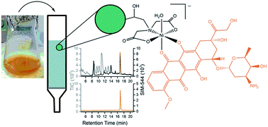
The restricted supply of the anticancer agent doxorubicin (DXR) prompted the development of a simple method to isolate DXR from a Streptomyces peucetius var. caesius liquid culture that could facilitate its local production. The metal-coordinating ability of DXR enabled its purification using immobilised metal ion affinity chromatography (IMAC). The entirety of a DXR standard (94 nmol) bound to 1 mL of Ni(II)-charged iminodiacetic acid (IDA) IMAC resin at pH 7.5 and was eluted as a free ligand at pH 5.5. The DXR binding capacity of the Ni(II)-charged IDA resin was about 4.7 μmol mL−1. An aliquot of the crude S. peucetius liquid culture containing native DXR (∼2 nmol) was retained on 1 mL of the Ni(II)-charged IDA resin in 38%. The reduced binding capacity was likely due to competing ligands in the bacteriological medium. Re-application of a sub-sample of the unbound DXR on a second column gave 81% DXR retention. The utility of IMAC to purify DXR on an analytical scale warrants further evaluation of industrial-scale IMAC processing as a potential route to this essential medicine.
1. Introduction

The supply of the anticancer agent doxorubicin (DXR) became restricted in 2011, due to operating losses at the major US manufacturer.1,2 As one of the most successful drugs for the treatment of leukaemia, Hodgkin's lymphoma, soft tissue sarcoma and bladder and breast cancer,3–9 its shortage caused widespread concern for clinicians and patients, and highlighted the vulnerability of drug supply chains.10,11 This situation prompted us to examine the development of a simple DXR purification protocol that could support its local production and increase the security of DXR supply. The annual requirement for DXR is about 225 kg.12 DXR is a natural anthracycline product of Streptomyces peucetius var. caesius (Fig. 1) and is accessed from fermentation mixtures using multiple extraction steps with organic solvents (acetone, chloroform, methanol), filtration and chromatography.13,14 The biosynthetic precursor of DXR, daunorubicin (Fig. 1, DNR), produced by several strains of Streptomyces peucetius, was first used in chemotherapy, but has been superseded by DXR, which has a higher therapeutic index.
image file: c5ra07639k-f1.tif
Fig. 1 Anthracyclines: doxorubicin (DXR, 1) and daunorubicin (DNR, 2).

Our group has developed immobilised metal ion affinity chromatography (IMAC) as a platform for isolating non-protein clinical agents and drug leads from fermentation mixtures.15–19 Secondary metabolites that have metal coordinating ability as an inherent and/or functional property are viable targets. The principle underlying the use of IMAC for isolating non-protein secondary metabolites is identical to that of its intended and more common use for the purification of recombinant histidine-tagged proteins. The target binds to the vacant sites of a hemi-saturated Ni(II)–iminodiacetic acid (IDA) complex that is covalently attached to an insoluble resin matrix. After washing unbound components from the resin, the target is displaced upon washing with an imidazole buffer or a low-pH solution.

The coordination chemistry of anthracyclines and substituted anthraquinones has been established in solution, with a limited number of complexes characterised by X-ray crystallography.26–30 Complexes between DXR and Cu(II), Co(II), Ni(II), Fe(III) or Pd(II), have been characterised by electronic absorption spectroscopy, circular dichroism spectroscopy, electron paramagnetic resonance spectroscopy, resonance Raman spectroscopy and electrochemistry.20–23,31–37 The affinity constants of complexes formed between different IMAC-compatible metal ions and DXR, DNR or IDA (Table 1), were within a range that supported the potential of IMAC-based DXR purification.18 This work describes the optimisation of an IMAC procedure for DXR purification using a DXR standard solution and the utility of the system as applied to a native DXR-containing S. peucetius fermentation mixture.

Table 1 log[thin space (1/6-em)]K values of complexes of defined metal–ligand stoichiometry formed between selected metal ions and DXR, DNR or IDA
  log[thin space (1/6-em)]K
Ni(II) Cu(II) Fe(III) Zn(II)
1[thin space (1/6-em)]:[thin space (1/6-em)]1 1[thin space (1/6-em)]:[thin space (1/6-em)]2 1[thin space (1/6-em)]:[thin space (1/6-em)]1 1[thin space (1/6-em)]:[thin space (1/6-em)]2 1[thin space (1/6-em)]:[thin space (1/6-em)]1 1[thin space (1/6-em)]:[thin space (1/6-em)]3 1[thin space (1/6-em)]:[thin space (1/6-em)]1 1[thin space (1/6-em)]:[thin space (1/6-em)]2
a N/A, not available.b From ref. 20.c From ref. 21.d A value of 19.3 has been reported in ref. 22.e From ref. 23.f A value of 33.4 has been reported in ref. 22.g From ref. 24.h From ref. 25.
DXR N/Aa 12.0b 12.3c 16.7c,d N/Aa 28.3e,f N/Aa N/Aa
DNR N/Aa N/Aa N/Aa N/Aa 11.0g N/Aa 4.5g N/Aa
IDA 8.1h 14.1h 10.6h 16.5h 10.7h N/Aa 7.2h 12.5h


2. Experimental

2.1. Materials

Chemicals were obtained from Sigma-Aldrich: K2HPO4·3H2O, NaH2PO4, NaCl, NaOH, HCl, imidazole, glucose, biotin, folic acid, pyridoxine hydrochloride, thiamine hydrochloride, 4-aminobenzoic acid, (−)-riboflavin, nicotinic acid, D-pantothenic acid hemicalcium salt, vitamin B12, (±)-α-lipoic acid, Na2B4O7·10H2O, (NH4)6Mo7O24·4H2O; Merck: Na2HPO4, CuCl2·2H2O, MnCl2·2H2O, NiCl2; Ajax: MgSO4·7H2O, ZnCl2; BD/Difco: asparagine; Amresco: HEPES, Na2EDTA·2H2O; APS: FeCl3·6H2O; or Euroasia: doxorubicin·HCl (DXR). IMAC experiments were conducted using His GraviTrap™ columns (GE Healthcare Life Sciences) prepacked with 1 mL of Sepharose™ 6 Fast Flow resin, charged with Ni(II). Milli-Q water was used for all experiments.

2.2. Instrumentation

The pH measurements were made using a Microprocessor pH meter (model: pH 211) with a 5 mm glass pH electrode (model: HI 1330) from Hanna Instruments. UV/visible spectra (350–850 nm) were obtained from solutions in 96-well BD Falcon tissue-culture treated microplates using a BMG Labtech POLARstar Omega plate reader. High performance liquid chromatography coupled with mass spectrometry (HPLC-MS) was performed using an Eclipse XDB-C18 reverse-phase (RP) column (Agilent; particle size: 5 μm; column dimensions: 150 mm × 4.6 mm internal diameter) with a gradient of 0–100% B over 30 min (A: H2O; B: acetonitrile (ACN), both containing 0.1% formic acid), at a flow rate of 0.4 mL min−1 and an injection volume of 10 μL. The RP HPLC-MS was performed using an Agilent Technologies System, made up of an autosampler (100 μL loop), Agilent 1260 Infinity degasser, a quaternary pump, a 1200 series diode array detector (DAD) and 6120B series single Quadrupole electrospray ionisation mass spectrometer (ESI-MS). The DAD was set to 220 nm and 490 nm, and the capillary voltage of the ESI-MS was set to 4000 V in positive ion mode. Agilent Chemstation software was used to process mass chromatograms in both the total ion current (TIC) and the selected ion monitoring (SIM) detection modes.

2.3. Preparation of solutions

The following solutions were prepared. 0.5 M NaCl in 20 mM NaH2PO4/Na2HPO4, pH 8.0; 0.3 M NaCl and 100 mM EDTA in 20 mM NaH2PO4/Na2HPO4, pH 7.2; 0.5 M NaCl and 0.5 M imidazole in 20 mM NaH2PO4/Na2HPO4, pH 8.0; 0.2 M NaCl in 10 mM HEPES at pH 6.0, 7.0, 7.5, 8.0, 9.0 (binding buffer, BB) or pH 5.5 (elution buffer, EB); aqueous solutions (0.1 M) of Ni(II), Cu(II), Zn(II) or Fe(III); aqueous DXR stock solution of 7.5 mM, with additional DXR solutions (0.4 μM, 47 μM or 750 μM) in the relevant buffer system. Adjustments of pH values were made with 0.1 M HCl or 0.1 M NaOH. Caution DXR is a known carcinogen (Category 1B) and should be handled with the required personal protective equipment (latex gloves, safety goggles and lab coat). All waste and contaminated materials should be disposed of in cytotoxic bins.

2.4. Immobilised metal ion affinity chromatography (IMAC)

IMAC columns were conditioned prior to sample loading with 10 column volumes (CV) of water and equilibrated with 10 CV BB. For each IMAC run, after loading 1 CV of sample, the column was washed with approximately 5 CV of BB. Elution of DXR was achieved by washing with EB (pH 5.5) until all DXR had been removed from the resin. The resin was stripped of Ni(II) upon washing with 10 CV of EDTA solution, 10 CV of phosphate buffer and 10 CV of water and was recharged with 0.5 CV Ni(II) solution, or other metal ion (Cu(II), Zn(II), Fe(III)) followed by 10 CV of phosphate buffer and 10 CV of water. Unbound Ni(II) (or other metal ion) was removed upon washing with 1 CV imidazole buffer, 10 CV of phosphate buffer and 10 CV of water. Columns were stored at 4 °C in 20% v/v aqueous ethanol. Fractions were analysed using absorbance at 490 nm or by LC-MS.

2.5. Bacterial cultures

Streptomyces peucetius var. caesius ATCC 27952 (Risk Group 1) was obtained from the American Type Culture Collection. The permanent stock was kept in 10% DMSO at −80 °C. S. peucetius was cultured in liquid medium according to literature methods.38 For the pre-culture, S. peucetius cells were inoculated into a 250 mL flask containing 100 mL of medium and a steel coil. The culture was incubated aerobically at 40 °C for 26 h at 210 rpm, before incubating for a further 5 days at 29 °C at 210 rpm. Approximately 4 mL of cells from the preculture were inoculated into a 250 mL flask containing 70 mL medium, which was incubated at 29 °C at 210 rpm for 2.5 days before harvesting. Culture supernatant was collected following centrifugation (3900 rpm for 20 min) and was concentrated to dryness. The pellet was dissolved in 600 μL water and 600 μL BB (pH 7.5) (×2 concentration) and the pH value was measured as pH 7.46. The solution was centrifuged (15[thin space (1/6-em)]000 rpm for 10 min). A 200 μL aliquot was used for LC-MS analysis and the remaining 1 mL was processed using IMAC.

3. Results and discussion

3.1. Optimisation of DXR binding conditions

The retention of 0.94 nmol DXR on a bed of Ni(II)-charged IDA resin was examined at pH 6.0, 7.0, 7.5, 8.0 and 9.0 (Fig. S1). At pH 6.0, no DXR bound to the resin, with about 90% of the sample retained at pH 7.0. The degradation of DXR was a potential issue under high pH conditions. Separate experiments showed that after 24 h, 15% or 80% DXR was degraded at pH 8.0 or pH 9.0, respectively (Fig. S2). At pH 7.5, the retention of DXR on the resin was close to 100% and pH-induced degradation over the 2 h time course of an IMAC experiment was negligible. All subsequent experiments were conducted at pH 7.5, as the optimal condition for DXR binding, with elution at pH 5.5. A typical binding curve of DXR using Ni(II)-based IMAC showed the sample was retained on the resin at pH 7.5, and was eluted at pH 5.5 (Fig. 2a). The retention of DXR on the resin showed the relative magnitudes of the 1[thin space (1/6-em)]:[thin space (1/6-em)]1 affinity constants of Ni(II)–IDA and Ni(II)–DXR complexes (Table 1) were favourably positioned to form a stable IDA–Ni(II)–DXR ternary complex.
image file: c5ra07639k-f2.tif
Fig. 2 (a) Profile of DXR (0.94 nmol) as processed on a Ni(II)-charged IMAC resin; and characterization of eluted product using (b) liquid chromatography, (c) mass spectrometry, or (d) electronic absorption spectroscopy (mid gray), with overlaid spectra from solutions of standard DXR (black) and Ni(II)–DXR (light gray).

In some instances, the immobilised metal ion can be leached from the resin by the ligand, resulting in the elution of the metal–ligand complex. This can occur in cases where the affinity between the target ligand and the immobilised metal ion is significantly greater than the 1[thin space (1/6-em)]:[thin space (1/6-em)]1 metal–IDA affinity constant.18 This phenomenon of metal-ion-transfer occurred in the use of Cu(II)-based IMAC for the purification of bleomycin.17,19 This is a potential disadvantage of IMAC, since additional purification steps would be needed to access the metal free drug for clinical use.

Analysis of the DXR eluted from the resin showed that it eluted as a free ligand. A single signal was observed in the liquid chromatogram (Fig. 2b), which gave three signals in the mass spectrum (Fig. 2c) consistent with DXR present as the aglycone form ([M]+ m/zcalc 397.1, m/zobs 397.1), or as a protonated adduct of a DXR monomer ([M + H]+ m/zcalc 544.2, m/zobs 544.2) or dimer ([2M + H]+ m/zcalc 1087.4, m/zobs 1087). The electronic absorption spectrum of the bound fraction eluted from the resin was coincident with the spectrum from a DXR standard solution, and was distinct from that of a Ni(II)–DXR complex, which had absorbance maxima at 510, 544 and 584 nm (Fig. 2d), similar to literature values.20 The mass spectrometry and electronic absorption spectroscopy data corroborated that DXR was isolated from the IMAC resin as a metal free complex.

The isolation of DXR using IDA resin charged with other IMAC-compatible metal ions was examined, in addition to non-specific binding. Non-specific binding of DXR on metal-free IMAC resin was negligible (Fig. 3a). The elution peak of DXR from a Ni(II)-charged IDA resin was Gaussian (Fig. 3b). DXR was retained on Cu(II)-charged IDA resin, but required a greater volume of elution buffer to recover the sample (Fig. 3c). The electronic absorption spectrum from the bound DXR fraction eluted from the Cu(II)–IDA resin was coincident with the spectrum from standard DXR and distinct from a Cu(II)–DXR solution. This showed that the DXR target was isolated as a free ligand, with no evidence of metal-ion transfer. Neither Zn(II)- (Fig. 3d) nor Fe(III)-charged (Fig. 3e) IDA IMAC resins retained DXR. The Zn(II)–DXR affinity was likely to be minimal, based upon data from Zn(II)–DNR complexes (log[thin space (1/6-em)]β110 4.5) (Table 1).24 DXR appeared to form a complex on the Fe(III)-charged resin that was unable to be removed. The precipitation of Fe(OH)3 from solutions of Fe(III) and DXR has been noted.37 This series of experiments supported that Ni(II) was the optimal metal ion for IMAC-based capture of DXR. Although Cu(II) showed some potential, there were disadvantages in the requirement for greater volumes of elution buffer, thereby increasing processing times, and the precipitation of aged Cu(II)–DXR solutions on the resin.


image file: c5ra07639k-f3.tif
Fig. 3 Profiles of DXR (column at left, DXR loading as bracketed) as processed on IDA IMAC resin (1 mL) charged with: (a) no metal (1.5 μmol), (b) Ni(II) (0.94 μmol), (c) Cu(II) (1.0 μmol), (d) Zn(II) (1.0 μmol), or (e) Fe(III) (0.94 μmol); or DXR (column at right) on Ni(II)-charged IDA IMAC resin (1 mL) at a loading of: (f) 0.47 μmol, (g) 0.94 μmol, (h) 1.4 μmol, (i) 2.8 μmol, or (j) 4.7 μmol.

The capacity for DXR binding on the Ni(II)-charged IDA resin was conservatively estimated as 4.7 μmol mL−1 (Fig. 3f–j). At this loading, the entire DXR sample was retained on the resin, indicating that the resin was not fully saturated. The recovery of DXR required the use of a larger volume of elution buffer and resulted in a bigaussian elution peak. This operational reason was used as the criterion for capacity. The nominal binding capacity of Ni(II)-charged IDA resin for DXR (4.7 μmol mL−1) is about 1.5 times greater than the binding capacity for the hydroxamic acid siderophore desferrioxamine B (3 μmol mL−1).15

3.2. Isolation of native DXR from Streptomyces peucetius var. caesius culture

Streptomyces peucetius var. caesius was grown in liquid cultures and on agar plates.38 The production of anthracycline metabolites was evident from the orange-red pigmented cells and supernatant. DXR and DNR were detected using SIM mode in LC-MS from cells scraped from the agar that were lysed in water. LC-MS analysis of the S. peucetius liquid culture using TIC detection showed multiple species present, ascribable to bacterial secondary metabolites and medium components (Fig. 4a).
image file: c5ra07639k-f4.tif
Fig. 4 LC-MS from a culture of S. peucetius var. caesius detected by (a) TIC; or (b) SIM (m/z 544), in the absence (black) or presence (gray) of authentic DXR. The gradient in (a) was the same in (b).

A signal at tR 16.76 min detected using SIM at m/z 544 was ascribed to native DXR ([M + H]+ m/zcalc 544.2), based on the selective increase in the intensity of the signal upon reanalysing the solution augmented with a defined concentration of authentic DXR (Fig. 4b). From standard addition analysis, the concentration of native DXR in the culture was estimated as 12 μg L−1, which was typical of low DXR yields in native culture.39 There was no detectable DNR in the liquid culture. The culture was taken to dryness and the pellet was re-dissolved in a minimal amount of binding buffer. An aliquot containing about 0.7 μg DXR was processed on a 1 mL resin bed of Ni(II)-charged IDA IMAC resin. Each 1 mL fraction collected during the binding event and the elution event was analysed using LC-MS with simultaneous detection modes of TIC and SIM 544 (Fig. S3).

Analysis of the areas of the LC peaks attributable to DXR showed that 62% DXR was eluted as an unbound species, with 38% DXR retained on the resin (Fig. 5a). The reduced retention of DXR when present in a complex mixture was attributed to high concentrations of ligands in the bacteriological medium, including glucose, asparagine, pyridoxine, thiamine, pantothenic acid, riboflavin and folic acid (Table S1), that could compete for resin binding sites.


image file: c5ra07639k-f5.tif
Fig. 5 Capacity of a 1 mL bed of Ni(II)-charged IDA resin towards binding: (a) native DXR in a crude sample of S. peucetius var. caesius culture; or (b) a sub-sample of the first-round IMAC-processed unbound fraction from trace ((a) gray symbol).

One DXR-containing fraction that eluted with other unbound species was re-adsorbed onto a second IMAC column and reprocessed. This sample comprised a similarly complex mixture of species with a reduced DXR concentration. The LC-MS trace from the solution applied to the resin showed the presence of multiple species, as detected using TIC (Fig. 6a). Simultaneous detection using SIM at 544 showed a strong signal at tR 16.82 min attributable to DXR, in addition to others species with lower retention times. Analysis of each 1 mL fraction using LC-MS showed a significant number of species were removed in the step upon washing with BB (Fig. 6b–e). Upon elution, the dominant species evident using both TIC and SIM detection modes was DXR (Fig. 6f–j). The signal at tR 16.87 min (Fig. 6g, black) was confirmed as DXR from spiking the sample with an authentic sample of DXR (Fig. 6g, dotted). The IMAC-based capture capacity for the second-round native DXR sub-sample was 81% (Fig. 5b). In these experiments, the first-round IMAC column effectively served as a pre-column to remove a proportion of competing ligands. This could also be achieved using a more conventional resin, such as XAD-2, to remove polar medium components.


image file: c5ra07639k-f6.tif
Fig. 6 LC-MS traces (detection: SIM 544, black; TIC, gray) from a sub-sample of first-round IMAC-processed native DXR-containing S. peucetius var. caesius culture ((a) fraction marked with * in Fig. 5a); and the fractions collected during the binding (b–e) or elution (f–j) of the sample during second-round processing on a Ni(II)-charged IDA IMAC resin. The gradient in (a) was the same in (b–j). The sample in (g) was reacquired upon the addition of authentic DXR (dotted).

4. Conclusions

DXR standard solutions were bound to a Ni(II)-charged IDA resin at pH 7.5, with DXR eluted from the resin as a free ligand at pH 5.5. The nominal DXR binding capacity of this IMAC resin format was 4.7 μmol mL−1. Low levels of native DXR produced in S. peucetius var. caesius culture were isolable using two purification rounds with the optimised IMAC format. Significant sample loss in the first-round IMAC processing was attributed to the presence of competing ligands in the bacteriological medium. Removing these ligands using a macrorecticular XAD-type pre-column could improve DXR capture by IMAC. The absence of metal-ion transfer in this IMAC-based DXR isolation method, together with its aqueous compatibility and simple operation, could provide impetus for studies to establish the viability of scale-up for industry production. The method would be applicable to the isolation of DXR and to DNR as the precursor of DXR. A guaranteed supply of DXR through a combination of improved isolation methods and the development of overproducing strains,40 would enable optimal chemotherapy regimens are maintained for cancer patients.

Acknowledgements

The University of Sydney is acknowledged for funding. Ms J. Gu is acknowledged for technical advice early in the project.

Notes and references

  1. J. L. Berger, A. Smith, K. K. Zorn, P. Sukumvanich, A. B. Olawaiye, J. Kelley and T. C. Krivak, OncoTargets Ther., 2014, 7, 1409–1413 CAS.
  2. M. L. Gatesman and T. J. Smith, N. Engl. J. Med., 2011, 365, 1653–1655 CrossRef CAS PubMed.
  3. R. H. Blum and S. K. Carter, Ann. Intern. Med., 1974, 80, 249–259 CrossRef CAS PubMed.
  4. R. B. Weiss, Semin. Oncol., 1992, 19, 670–686 CAS.
  5. J. W. Lown, Pharmacol. Ther., 1993, 60, 185–214 CrossRef CAS.
  6. G. Minotti, P. Menna, E. Salvatorelli, G. Cairo and L. Gianni, Pharmacol. Rev., 2004, 56, 185–229 CrossRef CAS PubMed.
  7. Y. Pommier, E. Leo, H.-L. Zhang and C. Marchard, Chem. Biol., 2010, 17, 421–433 CrossRef CAS PubMed.
  8. P. Ma and R. J. Mumper, Nano Today, 2013, 8, 313–331 CrossRef CAS PubMed.
  9. F. Yang, S. S. Teves, C. J. Kemp and S. Henikoff, Biochim. Biophys. Acta, 2014, 1845, 84–89 CAS.
  10. A. McBride, L. M. Holle, C. Westendorf, M. Sidebottom, N. Griffith, R. J. Muller and J. M. Hoffman, Am. J. Health-Syst. Pharm., 2013, 70, 609–617 CrossRef PubMed.
  11. M. Mitka, J. Am. Med. Assoc., 2011, 306, 1069–1070 CrossRef CAS PubMed.
  12. C. R. Hutchinson and A. L. Colombo, J. Ind. Microbiol. Biotechnol., 1999, 23, 647–652 CrossRef CAS PubMed.
  13. F. Arcamone, G. Cassinelli, G. Fantini, A. Grein, P. Orezzi, C. Pol and C. Spalla, Biotechnol. Bioeng., 1969, 11, 1101–1110 CrossRef CAS PubMed.
  14. F. Arcamone, F. Animati, G. Capranico, P. Lombardi, G. Pratesi, S. Manzini, R. Supino and F. Zunino, Pharmacol. Ther., 1997, 76, 117–124 CrossRef CAS.
  15. N. Braich and R. Codd, Analyst, 2008, 133, 877–880 RSC.
  16. N. Ejje, E. Lacey and R. Codd, RSC Adv., 2012, 2, 333–337 RSC.
  17. J. Gu and R. Codd, J. Inorg. Biochem., 2012, 115, 198–203 CrossRef CAS PubMed.
  18. R. Codd, J. Gu, N. Ejje and T. Lifa, in Inorganic Chemical Biology: Principles, Techniques and Applications, ed. G. Gasser, John Wiley & Sons, Ltd, Chichester, UK, 2014, pp. 1–35 Search PubMed.
  19. J. Gu and R. Codd, RSC Adv., 2015, 5, 3443–3453 RSC.
  20. P. S. Guin, P. C. Mandal and S. Das, J. Coord. Chem., 2012, 65, 705–721 CrossRef CAS PubMed.
  21. H. Beraldo, A. Garnier-Suillerot and L. Tosi, Inorg. Chem., 1983, 22, 4117–4124 CrossRef CAS.
  22. P. M. May, G. K. Williams and D. R. Williams, Eur. J. Cancer, 1980, 16, 1275–1276 CrossRef CAS.
  23. H. Beraldo, A. Garnier-Suillerot, L. Tosi and F. Lavelle, Biochemistry, 1985, 24, 284–289 CrossRef CAS.
  24. R. Kiraly and R. B. Martin, Inorg. Chim. Acta, 1982, 67, 13–18 CrossRef CAS.
  25. A. E. Martell and R. M. Smith, Critical Stability Constants, Plenum Press, New York, 1974 Search PubMed.
  26. M. J. Maroney, R. O. Day, T. Psyris, L. M. Fleury and J. P. Whitehead, Inorg. Chem., 1989, 28, 173–175 CrossRef CAS.
  27. S. Das, A. Saha and P. C. Mandal, Talanta, 1996, 43, 95–102 CrossRef CAS.
  28. E. E. Langdon-Jones and S. J. A. Pope, Coord. Chem. Rev., 2014, 269, 32–53 CrossRef CAS PubMed.
  29. P. Das, C. K. Jain, S. K. Dey, R. Saha, A. D. Chowdhury, S. Roychoudhury, S. Kumar, H. K. Majumder and S. Das, RSC Adv., 2014, 4, 59344–59357 RSC.
  30. S. Roy, P. Mondal, P. S. Sengupta, D. Dhak, R. C. Santra, S. Das and P. S. Guin, Dalton Trans., 2015, 44, 5428–5440 RSC.
  31. M. M. L. Fiallo and A. Garnier-Suillerot, Biochemistry, 1986, 25, 924–930 CrossRef CAS.
  32. M. Tachibana, M. Iwaizumi and S. Tero-Kubota, J. Inorg. Biochem., 1987, 30, 133–140 CrossRef CAS.
  33. P. K. Dutta and J. A. Hutt, Biochemistry, 1986, 25, 691–695 CrossRef CAS.
  34. M. M. L. Fiallo, A. Garnier-Suillerot, B. Matzanke and H. Kozlowski, J. Inorg. Biochem., 1999, 75, 105–115 CrossRef CAS.
  35. K. Nawara, H. Beeckman, P. Krysinski and G. J. Blanchard, J. Phys. Chem. B, 2013, 117, 6868–6873 CrossRef CAS PubMed.
  36. K. Nawara, J. L. McCracken, P. Krysinski and G. J. Blanchard, J. Phys. Chem. B, 2013, 117, 6859–6867 CrossRef CAS PubMed.
  37. K. D. Mjos, J. F. Cawthray, G. Jamieson, J. A. Fox and C. Orvig, Dalton Trans., 2015, 44, 2348–2358 RSC.
  38. M. L. Dekleva, J. A. Titus and W. R. Strohl, Can. J. Microbiol., 1985, 31, 287–294 CrossRef CAS.
  39. S. Malla, N. P. Niraula, B. Singh, K. Liou and J. K. Sohng, Microbiol. Res., 2010, 165, 427–435 CrossRef CAS PubMed.
  40. N. Lomovskaya, S. L. Otten, Y. Doi-Katayama, L. Fonstein, X.-C. Liu, T. Takatsu, A. Inventi-Solari, S. Filippini, F. Torti, A. L. Colombo and C. R. Hutchinson, J. Bacteriol., 1999, 181, 305–318 CAS.

Footnote

Electronic supplementary information (ESI) available: Optimisation of DXR isolation/stability with pH; first-round IMAC DXR processing; bacteriological medium components. See DOI: 10.1039/c5ra07639k

This journal is © The Royal Society of Chemistry 2015
Click here to see how this site uses Cookies. View our privacy policy here.