Yuchuan Guoa,
Shuihong Caib,
Guofeng Gua,
Zhongwu Guo*ab and
Zhongzhu Long*b
aNational Glycoengineering Research Center, Shandong University, Jinan, China. E-mail: zwguo@sdu.edu.cn
bQidong Dongyue Pharmaceutical Company, Qidong, Jiangsu Province, China. E-mail: longzhongzhu@dybiochem.com
First published on 19th May 2015
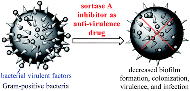
Sortase A (SrtA) is a membrane-associated enzyme responsible for the covalent anchoring of many virulent factors of Gram-positive bacteria onto the cell wall. It has been shown that SrtA plays a pivotal role in the pathogenic processes of bacterial infection. Additionally, SrtA is not essential for microbial growth and viability, and its inhibition does not therefore place pressure on bacteria to develop drug-resistant mechanism. As an extracellular membrane enzyme, it can more readily be targeted by drugs relevant to intracellular enzymes. SrtA is thus an excellent target for the design and development of novel anti-virulence drugs against the drug-resistant Gram-positive bacteria that have become a major worldwide health problem. A number of SrtA inhibitors have so far been identified by techniques such as the rational design of substrate mimetic inhibitors based on the structure of the enzyme and enzyme substrates, identification of novel inhibitors among natural products, the discovery and development of SrtA inhibitors via high-throughput, and in silico screening of small molecule libraries followed by structural optimization. The present article reviews the progress made recently in the development of SrtA inhibitors as new antibacterial agents using similar techniques.
Owing to increasing difficulty in the discovery and development of new antibiotics, there is an urgent need to explore novel antibacterial drug targets and strategies.2 The development of anti-virulence agents for the treatment of bacterial infections has, for example, recently received increasing attention.2 Unlike conventional antibiotics, which inhibit bacterial growth or kill bacteria directly, these agents reduce bacterial virulence and make the pathogens more susceptible to the host immune system. As a result, they impose little selective pressure on pathogens to develop drug resistance. Molecules associated with bacterial virulence, such as bacterial sortase (Srt), have therefore gained widespread interest as new targets for antibacterial drug design.
Srt is a type of membrane-bound cysteine transpeptidase responsible for the covalent anchoring of surface proteins to the Gram-positive bacterial cell wall.3–5 Phylogenetic analysis of 61 Srt genes encoded in 22 Gram-positive bacterial genomes has divided Srt into four separate groups, known as A, B, C and D.6 SrtA is present in almost all low GC% Gram-positive bacterial strains. The primary SrtA-mediated protein anchoring process has been elucidated.7–9 Firstly, proteins destined for cell wall anchorage are secreted in association with the extracellular membrane. These proteins contain a number of epitopes located at their carboxyl terminus, including a sorting signal consisting of a leucine, proline, X, threonine, and glycine (LPXTG), where X can be any amino acid motif, a membrane-binding hydrophobic region, or a tail with charged residues which direct the secretion process. Next, the LPXTG motif is recognized by SrtA and is cleaved at Thr and Gly to form a reactive thioester between the acyl group of Thr and the Cys thiol group at the active center of SrtA. Finally, the amino terminus of the pentaglycine moiety of the cell wall precursor lipid II attacks the reactive thioester to form an amide linkage with the carboxyl terminus of the protein. In this process surface proteins are linked to lipid II, and the products are eventually transformed into mature peptidoglycans via crosslinking reactions catalyzed by penicillin-binding proteins.
Recent studies have revealed that many virulent bacterial factors are anchored onto the cell surface by SrtA.10–13 These factors play an important role in the pathological process of bacterial infection, such as bacterial adhesion and invasion to the host cell, biofilm formation, colonization, bacterial evasion of the host immune system, and so on (Table 1).14 As a result, inhibiting SrtA activity in bacteria causes their failure to properly display virulent factors, and hence the significant reduction of virulence and infection.12,15–17 For example, it has been demonstrated that a SrtA gene knockout mutant of S. aureus, which was defective of various LPXTG motif-containing cell surface proteins, was unable to establish renal abscess and acute infection in mice.10,11 These results were similarly confirmed in a rat endocarditis model. Moreover, the SrtA knockout bacterial mutant was found to be more susceptible to killing by macrophage.18
Name | Abb. | Function | Pathogenic process | Signal sequence |
---|---|---|---|---|
a The table is adapted from ref. 14. | ||||
Collagen-binding adhesion | Cna | Adhesion for collagen (type I and IV) | Adhesion | LPKTG |
Clumping factor A | ClfA | Platelet adhesion (fibrin-mediated); binds complement regulator factor I | Adhesion; colonization; evasion of innate immune defense | LPDTG |
Clumping factor B | ClfB | Platelet adhesion (fibrin-mediated); binds cytokeratin 10 | Adhesion; colonization; evasion of innate immune defense | LPETG |
Fibronectin-binding protein homolog | FnbA | Adhesion for fibrinogen, fibronectin and elastin | Adhesion; colonization; biofilm formation | LPETG |
Fibronectin-binding protein homolog | FnbB | Adhesion for fibronectin and elastin | Adhesion; colonization; biofilm formation | LPETG |
Serin-aspartate repeat protein C | SdrC | Adhesion | Adhesion; colonization | LPETG |
Serin-aspartate repeat protein D | SdrD | Adhesion | Adhesion; colonization | LPETG |
Serin-aspartate repeat protein E | SdrE | Adhesion | Adhesion; colonization | LPETG |
Protein A | Spa | Binds Fc domain for immunoglobulins; binds complement protein C3 | Interference with innate and adaptive immune response | LPETG |
S. aureus surface protein C | SasC | Binds extracellular matrix | Adhesion; biofilm formation | LPNTG |
S. aureus surface protein G | SasG | Binds extracellular matrix | Adhesion; biofilm formation | LPDTG |
Iron-regulated surface determinant A | IsdA | Adhesion factor for fibronectin, fibrinogen, transferrin, hemoglobin (expressed in iron-restricted environment) | Adhesion; colonization | LPKTG |
Plasmin sensitive protein | Pls | Methicillin-resistant surface protein | Resistance | LPDTG |
Knocking out the SrtA gene in other Gram-positive pathogens, such as S. pneumoniae,13 S. suis,19 and Listeria monocytogenes,12 has also led to failure in their display of cell surface proteins, reduction of biofilm formation, and attenuation of virulence, and the mutant bacteria become more susceptible to macrophage-mediated killing.5
Despite the fact that SrtA is responsible for anchoring various virulent factors onto the bacterial cell surface and plays a critical role in bacterial infection and virulence, it is not essential for bacterial growth and viability. SrtA inhibition does not therefore place pressure on bacteria to develop drug-resistance mechanisms. Moreover, SrtA is an extracellular membrane enzyme that can easily be targeted by drugs, and no eukaryotic SrtA homologs have currently been identified, which renders drugs specific in targeting SrtA. SrtA is thus a promising target for the design and development of novel anti-virulence drugs.14,20
The crystal structure of SrtA has been described by a number of workers. For example, Ilangovan et al. first reported the crystal structure of a variant of S. aureus SrtA, in which the 59 amino acids at the N-terminus of the enzyme were truncated.21 As shown in Fig. 1, SrtA possesses an eight-stranded β-barrel fold, and includes two short helices and several loops. Strands β7 and β8 form the floor of a hydrophobic depression, with walls composed of amino acids located in the loops connecting strands β3–β4, β2–β3, β6–β7, and β7–β8. In all the sortases Cys184 and His120 are absolutely conserved and are essential for SrtA catalysis. While Cys184 is anchored at β7, His120 is located within a helical region connecting β2 and β3, with its imidazole group in the vicinity of the thiol group of Cys184, Arg197 which is anchored in β8, is located in close proximity and parallel to the Cys184 active site and acts as a hydrogen donor interacting with the carbonyl groups on the LP backbone of the LPXTG substrate in the inactive form of SrtA, and interacts with the TG backbone carbonyls in the active form, which may be important for holding the substrate in position and during the catalytic process.
![]() | ||
Fig. 1 Crystal structure of SrtA (C184A) bound to the LPETG peptide. The structure was generated from atomic coordinates deposited in the Protein Data Bank, PDB ID: 1T2W.22 |
High resolution X-ray analysis of SrtA bound to the LPETG peptide has provided additional insights into the molecular interaction between SrtA and its substrate.22 The substrate binding site resides in a concave plane molded by the β7 and β8 strands, while the scissile peptide bond between Thr and Gly is positioned between the side-chains of Cys184 and Arg197. The Leu and Pro residues of the LPETG motif are bound to the C-terminal region of β7, surrounded by several highly hydrophobic amino acids. Residues perturbed after ligand binding have also been mapped to the C-terminal region of the β7 strand (Thr180 and Ile182) and to the vicinity of the loop connecting strands β3 and β4. Importantly, Thr180 and Ala118 are absolutely conserved, and Ile182 partially conserved, among sortases. Mutation of these residues significantly impairs Srt activity in vitro.23
Elucidation of the SrtA structure and its catalytic mechanism has not only facilitated the wide application of SrtA in organic synthesis and chemical biology,24–29 but has also provided the molecular basis for the design and development of SrtA inhibitors as anti-virulence agents. To date a number of SrtA inhibitors have been discovered through rational design and modification of analogs of the SrtA substrate. Novel inhibitors have also been discovered among natural products. In addition, modern technologies, including fluorescence resonance energy transfer-based high-throughput, and in silico virtual screening, have also been successfully employed in the design and discovery of SrtA inhibitors.
The present article reviews recent progress in the development of SrtA inhibitors by a variety of techniques.
In 2002 Scott et al. reported the synthesis, kinetic analysis, and biological evaluation of the first type of SrtA inhibitors designed on the basis of the structure of its native substrate.30 In this study peptidyl-diazomethane and -chloromethane analogs of the LPXTG motif, benzyloxycarbonyl(Cbz)-LPAT-CHN2 and Cbz-LPAT-CH2Cl, respectively, were found to show time-dependent irreversible inhibition on recombinant SrtA. The diazoketone or chloromethyl ketone group was chosen as a replacement for the scissile amide linkage between T and G, due to their ability to alkylate the thiol group of Cys184 at the active site of the enzyme. The inhibitory constants for the peptidyl-diazomethane and peptidyl-chloromethane analogs were 0.22 and 0.21 μM, respectively. The functional mechanisms of these inhibitors were believed to involve covalent binding with SrtA to form a Michaelis complex (Fig. 2) and inactivation of the enzyme. Their SrtA inhibitory activities were assessed by fluorescence resonance energy transfer (FRET) technology using a synthetic self-quenched fluorescent probe, i.e., 4-([4-(dimethyl-amino)-phenyl]azo)benzoyl (Dabcyl, the fluorescent donor)-Gln-Ala-Leu-Pro-Thr-Gly-Glu-Glu-[5-[(2-aminoethyl)amino]naphthalene-L-sulphonic acid] (Edans, the fluorescence quencher), which is also a SrtA substrate. The Km and Kcat values for the SrtA-catalyzed cleavage of this self-quenched substrate were calculated by fitting the data points into the Michaelis–Menten equation for substrate hydrolysis (Fig. 2) using GraFit2 software.30 It was also found that the peptidyl-chloromethane analog, with a specificity constant of 5.3 × 10⁴ M−1 min−1, was ca. twice as potent as the peptidyl-diazomethane analog as SrtA inhibitors. Connolly et al. designed and synthesized a different irreversible SrtA inhibitor by replacing the scissile T–G moiety in the SrtA recognition motif LPXTG with a vinyl sulfone (CC–SO2Ph), which could covalently modify the active-site thiol group of SrtA.31 Since the vinyl sulfone group has lower electrophilicity than chloromethane and diazomethane ketones, the inhibitor constant for the vinyl sulfone analog was significantly increased (Ki = 9 μM). By analyzing the pH dependence of SrtA inhibition and NMR studies, they excluded the thiolate–imidazolium ion pair mechanism for the transpeptidation reaction and proposed a general base catalysis mechanism, namely that Cys184 as a nucleophile is neutral at physiological pH and His120 functions as a general base.
![]() | ||
Fig. 2 The mechanism for SrtA inhibition by substrate-mimetic inhibitors Cbz-LPAT-CHN2 and Cbz-LPAT-CH2Cl. |
Subsequently, Jung et al. prepared two tetrapeptide analogs of the sorting signal motifs of SrtA and SrtB, in which (2R,3S)-3-amino-4-mercapto-2-butanol was used to replace L-Thr.32 These analogs were shown to inhibit SrtA and SrtB via the reaction of their thiol group with the Cys residue at the Srt active site to generate a disulfide bond. In addition, Kruger et al. prepared a unhydrolyzable phosphinic peptidomimetic of the LPXTG motif, NH2-YALPE-AlaØ(PO2H–CH2)G-EE-NH2, where Ø indicates that the –C(O)NH– moiety between two amino acids had been replaced by the given functional group, in which a phosphinic isostere was utilized to replace the scissile T–G bond, as an analog of the transition state of the SrtA-catalyzed reaction.33 It was shown to be a reversibly competitive inhibitor of SrtA, but its inhibitory activity was relatively low (IC50 = 10 mM).
Kim et al. reported the first attempt to find SrtA inhibitors from natural sources. After screening 80 medicinal plants, they found that Cocculus trilobus, Fritillaria verticillata, Rhus verniciflua, and Liriope platyphylla had relatively strong SrtA inhibitory activities,34 among which the ethyl acetate fraction extracted from the rhizomes of C. trilobus had the strongest (IC50 = 1.52 μg ml−1). Later, glucosylsterol β-sitosterol-3-O-glucopyranoside (Fig. 3) was isolated from the bulbs of F. verticillata, and identified as the first natural product with confirmed SrtA inhibitory activity (IC50 = 18.3 μg ml−1 or 31.72 μM).35 However, this natural product exhibited antibacterial activities against S. aureus, Bacillus subtilis, and Micrococcus leuteus with MIC values of 346.71, 86.68, and 693.42 μM, respectively. In addition, after deglycosylation, the resultant aglycon, sitosterol, showed no SrtA inhibition or bacterial cell growth inhibitory activity, confirming the importance of the glucopyranosyl residue.
Flavonols are a further class of natural products able to inhibit SrtA. Kang et al. obtained nine flavonols from Rhus verniciflua showing SrtA inhibitory activity, among which morin, myricetin, and quercetin (Fig. 3) were the strongest, with IC50 values of 11.29, 13.99, and 15.91 μg ml−1 (37.35, 43.96 and 52.64 μM), respectively.36 None of these compounds showed obvious inhibitory effect on growth of S. aureus Newman, whereas they reduced bacterial clumping to fibrinogen in a dose-dependent manner. Huang et al. demonstrated that morin derived from S. mutans had inhibitory activity to SrtA UA159 (IC50 = 8.21 μg ml−1 or 27.2 μM).37 At a concentration of 9 μg ml−1 (30 μM), it significantly reduced the biofilm formation of S. mutans, while the bacterial viability was not affected.
Oh et al. isolated a series of flavonoids from the roots of Sophora flavescens.38 Evaluation of their activity in inhibiting SrtA and microbial growth demonstrated that Kurarinol (Fig. 3) was the most potent SrtA inhibitor, with an IC50 value of 48.8 μg ml−1 (107 μM), and had antibacterial activity against S. aureus, with a MIC of 99 μg ml−1 (219 μM).
Park et al. found that curcumin (Fig. 3) extracted from Curcuma longa L. rhizome was a potent SrtA inhibitor, with an IC50 value of 13.8 ± 0.7 μg ml−1 or 37.5 μM, without inhibition of bacterial cell growth (MIC value greater than 200 μg ml−1 or 542.9 μM).39 Moreover, similar to SrtA gene knockout, curcumin treatment could reduce S. aureus cell adhesion to fibronectin in a dose-dependent manner (2.5–20 μg ml−1). This highlighted its potential for the treatment of S. aureus infections through inhibition of Srt activity.
Hu et al. evaluated in vitro the inhibitory activity of curcumin to purified SrtA derived from S. mutans UA159, and its IC50 value was 13.8 μg ml−1 (10.2 μM), which was lower than the MIC value of 61.7 μg ml−1 (175 μM) and MBC value of 123.5 μg ml−1 (350 μM).40 Subsequently, Hu et al. found that a low dose of curcumin (5.29 μg ml−1 or 15 μM) induced the release of Pac proteins to the supernatant and significantly reduced the biofilm formation of S. mutant, but this was not caused by the decrease in bacterial growth.41
Kim et al. isolated several isoquinoline alkaloids from the Coptis chinenesis rhizome and evaluated their inhibitory activities against SrtA, among which berberine chloride (Fig. 3) was the most potent (IC50 = 8.7 μg ml−1 or 23.4 μM) and showed moderate antibacterial activity against Gram-positive bacteria (MIC range of 50–400 μg ml−1 or 134.5–1075.8 μM).42 Recently, Lee et al. isolated three new lignans, together with eight known lignans and phenyl propanoids, from the dry roots of Pulsatilla koreana and found that these compounds could significantly inhibit SrtA derived from S. mutans OMZ65. (−)-Rosmarinic acid and coffeic acid (Fig. 3) were also shown to be potent SrtA inhibitors, with IC50 values of 7.2 and 3.6 μg ml−1 (20.0 and 20.0 μM), respectively.43
Oh et al. isolated several bis(indole) alkaloids, which have two indole moieties connected by a heterocyclic unit, from the marine sponge Spongosorites sp. and evaluated their SrtA inhibitory activities.44 Deoxytopsentin (Fig. 3) was identified as the most potent SrtA inhibitor (IC50 = 15.67 μg ml−1 or 48.02 μM), but it had antibacterial activity (MIC = 6.25 μg ml−1 or 19.15 μM). Interestingly, 4,5-dihydrogenation of the imidazole ring led to total loss of SrtA inhibitory activity (IC50 > 100 μg ml−1), suggesting the significant influence of the imidazole ring substituents on the SrtA inhibitory activity of topsentins. Structure–activity relationship analysis suggested that the imidazole and pyrazinone skeletons were important features of their activity.
Bromodeoxytopsentin (Fig. 3), which had potent SrtA inhibitory activity (IC50 = 19.4 μg ml−1 or 47.99 μM) and moderate antibacterial activity (MIC = 100 μg ml−1 or 247.4 μM), was evaluated by fibronectin-binding assay and was shown to reduce the capacity of bacteria to adhere to fibronectin-coated surfaces dose-dependently, in the range 0–40 μg ml−1 (0–99.0 μM). Jang et al. isolated four aaptamines, 1H-benzo[de][1,6]-naphthyridine alkaloids, from the marine sponge Aaptos aaptos and evaluated their SrtA inhibitory activity.45 Isoaaptamine (Fig. 3) was found to be a potent inhibitor of SrtA (IC50 = 3.7 ± 0.2 μg ml−1 or 16.2 μM) and could reduce cell adhesion to fibronectin-coated surface in a dose-dependent manner (0–16 μg ml−1 or 0–70.0 μM). Structure–activity relationship analysis revealed that the methyl group at the isoaaptamine N-1 position was important for their SrtA inhibitory activity.
Jeon et al. isolated a number of pyrroloiminoquinone alkaloids of the discorhabdin class from the sponge Sceptrella sp.46 Biological studies revealed that (−)-discorhabdin Z (Fig. 3), which contained an unusual hemiaminal group, was a potent SrtA inhibitor (IC50 = 2.19 μg ml−1 or 6.14 μM), which did not affect microbial viability (MIC > 100 μg ml−1 or 280.4 μM) but was cytotoxic to the K562 cell line (IC50 = 0.74 μg ml−1 or 2.08 μM). Bae et al. isolated eight sesterterpenes and related pentaprenyl hydroquinones (halisulfates and suvanine) from the sponge Coscinoderma sp.,47 and found that halisulfate 1 (Fig. 3) was the most potent SrtA inhibitor (IC50 = 21.34 μg ml−1 or 38.33 μM).
In contrast to other SrtA inhibitors, halisulfates were active against both Gram-positive and Gram-negative bacteria, the mechanism of which has not yet been clarified. In addition, Won et al. isolated several furarines and beta-carboline alkaloids from Synoicum sp.,48,49 such as cadiolide E and eudistomin Y3 (Fig. 3), that had moderate SrtA inhibitory activity (IC50 = 78.25 and 145 μM, respectively). However, these compounds had some antibacterial activity and cytotoxicity.
The natural origins, enzyme inhibitory activities, fibronectin-binding inhibitory activities, antibacterial activities, and related references of all the natural products mentioned as SrtA inhibitors are summarized in Table 2.
Name | Source | SrtA IC50 (μM) | Fibronectin binding inhibition (μM) | MIC (μM) | Ref. |
---|---|---|---|---|---|
β-Sitosterol-3-O-glucopyranoside | F. verticillata | 31.72 (S. aureus) | N.D. | 346.7 (S. aureus) | 35 |
Morin | R. vernicifua | 37.35 (S. aureus) | Dose dependent | >2977 (S. aureus) | 36 and 37 |
27.2 (S. mutans) | |||||
Myricetin | R. vernicifua | 43.96 (S. aureus) | Dose dependent | >2828 (S. aureus) | 36 |
Quercetin | R. vernicifua | 52.64 (S. aureus) | Dose dependent | >2977 (S. aureus) | 36 |
Kurarinol | S. flavescens | 107 (S. aureus) | N.D. | 219 (S. aureus) | 38 |
Curcumin | C. longa L. | 37.5 (S. aureus) | Dose dependent 6.8–54.3 | >542.9 (S. aureus) | 39 |
10.2 (S. mutans) | N.D. | 175 (S. mutans) | 40 and 41 | ||
Berberine chloride | C. chinensis | 23.4 (S. aureus) | N.D. | 269 (S. aureus) | 42 |
(−)-Rosmarinic acid | P. koreana | 20.0 (S. mutans) | N.D. | N.D. | 43 |
Coffeic acid | P. koreana | 20.0 (S. mutans) | N.D. | N.D. | 43 |
Deoxytopsentin | Spongosorities sp. | 48.02 (S. aureus) | N.D. | 19.15 (S. aureus) | 44 |
Bromodeoxytop-sentin | Spongosorities sp. | 47.99 (S. aureus) | Dose dependent 0–99.0 | 247.4 (S. aureus) | 44 |
Isoaaptamin | Aaptos aaptos | 16.2 (S. aureus) | Dose dependent 0–70.0 | 219.2 (S. aureus) | 45 |
(−)-Discorhabdin Z | Sceptrella sp. | 6.14 (S. aureus) | N.D. | >280.4 (S. aureus) | 46 |
Halisulfate 1 | Coscinoderma sp. | 38.33 (S. aureus) | N.D. | 2.8–44.9 (S. aureus) | 47 |
Cadiolide E | Synoicum sp. | 78.25 (S. aureus) | N.D. | 3.9 (S. aureus) | 48 |
Eudistomin Y3 | Synoicum sp. | 145 (S. aureus) | N.D. | 14.0 (S. aureus) | 49 |
Via high-throughput screening, Oh et al. identified compound 1 (IC50 = 231 μM) (Fig. 4) as a promising lead for SrtA inhibitors.50 They synthesized a series of its derivatives and studied their structure–activity relationships, which suggested that the positioning of the two phenyl groups and the introduction of a nitrile group in the side-chain were pivotal for SrtA inhibition. Compound 2 (Fig. 4), which had an IC50 value of 9.2 μM, was the most active inhibitor among all the synthetic derivatives, and was almost 25 times more potent than compound 1 for the inhibition of SrtA. Kinetic studies disclosed that compound 2 was a competitive inhibitor, with a Ki value of 6.81 μM.
![]() | ||
Fig. 4 Structure of some synthetic small molecule SrtA inhibitors and their S. aureus SrtA inhibition activity. |
Molecular modeling was also performed to study the relationship between the structure of a compound and its inhibitory activity. Although compound 2 does not interact with the side-chain of Cys184, its nitrile group forms hydrogen bonds with the two guanidyl NH groups of Arg197, and its phenyl rings have a relatively strong lipophilic interaction with the large hydrophobic binding pocket at the SrtA active site.
Subsequently, Oh et al. evaluated the in vivo biological activity of 2 in Balb/c mice.51 After inoculation with 107 CFU of S. aureus Newman, all the mice without treatment died within two weeks, but the mice receiving intraperitoneal injection of 2 at doses of 100, 20, and 4 mg kg−1 had survival rates of 75%, 100%, and 97%, respectively. Moreover, 2 reduced joint and kidney infections mediated by SrtA. However, it was observed that the animal survival rate was lower in the higher dose (100 mg kg−1) group than for the lower dose (20 mg kg−1) group, suggesting possible toxic and side-effects of 2. Another study further revealed that like some natural SrtA inhibitors, such as β-sitosterol-3-O-glucopyranoside, berberine chloride and psammaplin A1, 2 could inhibit the adhesion of S. aureus cells to fibronectin.52
Kudryavrsev et al. prepared some cis-5-phenyl prolinates with electrophilic substituents at the 4-position of the pyrrolidine ring via 1,3-dipolar cycloaddition reactions of arylimino esters with divinyl sulfone or acrylonitrile.53 They found that 4-vinylsulfinyl 5-phenyl prolinates inhibited S. aureus SrtA irreversibly through modification of the enzyme Cys184 residue, but they were relatively weak inhibitors (IC50 of the order of mM). Nonetheless, they can be used as leads for the development of new antibacterial and anti-virulence agents.
After screening a library of 135625 structures, Maresso et al. found that aryl β-aminoethyl ketones (AAEK) had SrtA inhibitory activity. AAEK1 3 and AAEK2 4 (Fig. 4) had IC50 values of 47 and 15 μM for S. aureus SrtA and IC50 values of 4.8 and 5.6 μM for Bacillus anthracis SrtA, respectively.54 The proposed Srt inhibition mechanism was that, under the influence of SrtA, they could transform into reactive olefins via β-elimination and that the olefins could covalently modify thiol groups. These molecules thus inhibited SrtA through irreversible, covalent modification of the Cys residue at the enzyme active sites.
Chenna et al. identified new S. aureus SrtA inhibitors through in silico virtual screening of commercial compound libraries by means of SYBYL software.55 Preliminary structure–activity studies on the lead compound resulted in the development of compounds with improved activity, such as 5 (Fig. 4), which had an IC50 value of 58 μM. Subsequently they systematically analyzed the structure–activity relationship of compound 5 and found that the stereochemistry of the double bond was an important factor in their bioactivity.56 In most cases, changing the E double bond to the Z isomer, or to a rigid triple bond, reduced the enzyme inhibitory activity. Reducing the double bond to a C–C single bond resulted in complete loss of activity. The amide carbonyl group, the NH group, and morpholine ring oxygen were also important for SrtA inhibitory activity.
After high-throughput screening of 30000 compounds, Suree et al. found three classes of novel small molecule SrtA inhibitors, rhodanines, pyridazinones, and pyrazolethiones, which inhibited SrtA in a reversible manner, with IC50 values in the sub-micromolar range.57 The most active SrtA inhibitors were rhodanine 6, pyridazinone 7, and pyrazolethione 8 (Fig. 4), with IC50 values of 3.7, 0.20, and 0.30 μM, respectively. Pyridazinone is the most potent SrtA inhibitor known to date, and its inhibition of SrtA was suggested to take place via a thiol–disulfide exchange reaction with SrtA Cys184. Structure–activity relationship studies highlighted the significant impact of the location and nature of substituents on the pyridazinone ring on its SrtA inhibitory activity. On the other hand, cell-based assays showed that these compounds had no impact on S. aureus and B. anthracis viability, suggesting that they are SrtA-specific inhibitors. This supports their potential for further development as anti-virulence drugs.
On the basis of these encouraging results, as well as the results for other derivatives of 6, 7, and 8,58 Uddin et al. created a pharmacophore model and investigated the three-dimensional quantitative structure–activity relationships. The model was verified through comparative molecular field analysis and comparative molecular similarity indices analysis, and could be applied to allow better understanding and explanation of the correlation between the structural features of the compounds and their biological activity.
Based on the discovery that indole-containing natural products had inhibitory activities against S. aureus SrtA and isocitrate lyase from Candida albicans,59,60 Lee et al. prepared a series of structural analogs of the natural products,61 among which six compounds exhibited higher SrtA inhibitory activities than the positive control, p-hydroxymercuribenzoic acid. In particular, compound 9 (Fig. 4) showed the highest SrtA inhibition, with an IC50 value of 25 μM, and had no influence on bacterial viability (MIC > 200 μg ml−1 against S. aureus).
Zhang et al. used virtual screening technology to identify new structures that could bind to the active site of SrtA, and proved that 3,6-disubsitituted triazolothiadiazole compounds were SrtA inhibitors in both in vitro and in vivo studies.62 Compound 10 (Fig. 4) was the most active, with IC50 values of 9.3 μM and 0.8 μM against SrtAs from S. aureus and S. pyogenes, respectively, whereas it had no influence on staphylococcal growth in vitro (MIC > 40 mM). A BALB/c mouse model was used to evaluate the in vivo activities of 10, and it was found to be efficacious in preventing lethal bacteremia and infections induced by S. aureus.
Kahlon et al. studied tetralene and indene compounds that had shown inhibitory activity against human pathogen, Mycobacterium tuberculosis, as SrtA inhibitors, by in silico analysis followed by biological assays.63 Indeed, compounds 11 and 12 (Fig. 4) showed some SrtA inhibitory activity (IC50 values of 117 and <89.8 μM, respectively). However, 11–13 also exhibited antibacterial activities against S. aureus, with MIC values of 14.67, 14.04, and 224.5 μM, respectively, which suggested that these compounds might not be SrtA-specific and could hit other targets as well, reducing their potential as ideal drug candidates. Nevertheless, these compounds did not exhibit significant cytotoxicity to Vero and WRL-68 cell lines.
Zhulenkovs et al. screened a library of 50240 compounds against SrtA and identified 14 (Fig. 4) as a SrtA inhibitor, with an IC50 value of 6.11 μM, whereas its MIC value against S. aureus was 2.92 μM and its IC50 value against NIH3T3 cell was 1.27 μM.64 In order to improve the SrtA inhibitory potency and reduce cytotoxicity, structural optimization was performed, which resulted in compound 15 (Fig. 4), which demonstrated a higher MIC value and a ten-fold decrease in cytotoxicity. Its IC50 value against SrtA, MIC and IC50 values against NIH3T3 cell were 3.8, 41.51, and 14.39 μM, respectively.
Currently, SrtA inhibitors are mainly discovered or developed using three approaches. One of these is the design of SrtA inhibitors by mimicking its substrate structure. This type of SrtA inhibitor is able to bind specifically to the enzyme, in either a reversible or irreversible manner, to block its enzymatic activity, and thereby to be potent while remaining nontoxic. Although some progress has been made in this area, no particularly strong SrtA inhibitors have yet been developed, possibly due to the susceptibility of the synthetic peptide mimics to proteases. Nevertheless, we believe that this is a promising direction, and the key topics for future research will be to further improve the binding affinity between the enzyme and the substrate analog and to improve the stability of inhibitors, for example by replacing the peptide bonds with bonds more stable to proteases.
The other two approaches are to identify new inhibitors among natural products, or by high-throughput in silico screening of small molecule libraries. The key advantage for identifying SrtA inhibitors among natural products is that structurally unique inhibitors may arise, and these can be used as lead compounds for further structural optimization and development. However, this approach is limited by the natural resources and manpower available. The small molecule library approach can take advantage of the convenience and power of high-throughput and in silico virtual screening technologies to probe a broad range of structures and thus improve search efficiency, and the inhibitors identified can be further optimized through rational design.
To date the most potent SrtA inhibitors have been discovered through the screening of small molecule libraries. With the rapid growth in molecular libraries and databases, we believe that more potent SrtA inhibitors may be identified using this approach. Potential problems associated with small molecule drugs are that they often may interact with multiple molecular targets and thus give rise to side-effects. As each approach has its own advantages and disadvantages, we believe that a combination of different approaches is a promising strategy for the future discovery and development of SrtA inhibitors. For example, new and improved SrtA inhibitors may be designed by combining the promising specificity and targeting ability of substrate-based inhibitors with the concept of small molecule pharmacophores.
Another important research topic in the development of SrtA-based anti-virulence drugs is the methods employed to evaluate SrtA inhibitors. Currently, SrtA inhibitory activity is assessed by means of FRET technology utilizing a synthetic self-quenching fluorescent probe, which is also a cleavable SrtA substrate. However, in common with other anti-virulence drugs, an ideal Srt inhibitor-based antibacterial agent may not have a significant influence on bacterial growth and viability. Consequently, to evaluate the therapeutic potential of a specific SrtA inhibitor and determine whether that inhibitor deserves further development, it must also be assessed by other methods, such as evaluation of its impact on bacterial biofilm formation, its influence on bacterial adhesion to the host cell by means of fibronectin-binding assay, its capacity to effect macrophage-mediated killing of bacteria, and by in vivo anti-infectious assays. These processes are time-consuming, and this affects the efficiency of drug screening.
In addition, the screening models for these studies are based on drug-resistant S. aureus, and it remains to be verified whether the SrtA inhibitors are effective against other bacteria. Consequently, the establishment of a more effective and convenient method for the high-throughput evaluation of the anti-virulence activities of SrtA inhibitors, or other similar antibacterial agents, is an important topic, and any progress in this direction will have a great impact.
Finally, unlike conventional antibiotics, anti-virulence agents do not directly kill bacteria but merely attenuate their virulence, interrupt their adhesion and invasion of the host cell, and make them more susceptible to the human immune system. For these antibacterial agents to work well, the immune system of the host must therefore function properly in order to help in removing the pathogens. As a result, they are not suitable for immuno-compromised patients. To treat these patients and to further improve the overall therapeutic efficacies of SrtA inhibitors as anti-virulence drugs, a future research direction may be the combined use of anti-virulence drugs and immuno-stimulants.
This journal is © The Royal Society of Chemistry 2015 |