Self-assembled core–shell nanospheres and dendritic nanostructure of novel tetra-(3-phenyprop-2-allyloxy) phthalocyanine in different solvents

Yinghui Bian, Jinshe Chen, Shaotang Xu, Lijun Zhu, Yulu Zhou, Yuzhi Xiang and Daohong Xia*
State Key Laboratory of Heavy Oil Processing, China University of Petroleum (East China), Qingdao 266580, China. E-mail: xiadh@upc.edu.cn; Fax: +86 532 86981787; Tel: +86 532 86981869

Received 22nd April 2015 , Accepted 29th April 2015

First published on 29th April 2015


Abstract

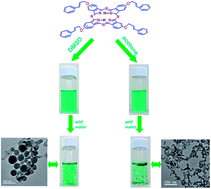
Novel metal-free tetra-(3-phenyprop-2-allyloxy) phthalocyanine (TPAO-Pc) was synthesized and characterized by nuclear magnetic resonance spectrum (1H NMR), MALDI-TOF MS, Fourier-transform infrared (FT-IR) and UV-Visible absorption (UV-Vis) spectra. The effects of concentrations and solvents on the properties of self-assembly were investigated via absorption and fluorescence spectra, transmission electron microscopy (TEM). TPAO-Pc was stable in most organic solvents, such as toluene, acetone and dimethylsulfoxide (DMSO). However, the TPAO-Pc exhibited the tendency of forming “face-to-face” stacking mode self-assemblies (H-aggregate) in DMSO–water and acetone–water mixed solutions. Self-assembly of the TPAO-Pc was also demonstrated by fluorescence spectra. The TEM images displayed the core–shell nanospheres and dendritic nanostructure of TPAO-Pc formed in the DMSO–water and acetone–water mixed solutions, respectively. The mechanism of the self-assemblies growth was proposed on the basis of the experimental results. The morphology of the TPAO-Pc self-assemblies was related with the selected solvent conditions and the aggregation time. H-bond, π–π interaction may be the main driving force for the formation of core–shell nanospheres and dendritic nanostructure.


Introduction

The aromatic macrocyclic phthalocyanines (Pc) containing an extended aromatic 18-π electron system are well known synthetic porphyrin analogues that have received considerable attention in recent years.1,2 The phthalocyanine structure with smart modification has been shown to improve its properties of photochemistry and photophysics, and usually enhance their solubility effectively. Because of their excellent chemical and thermal stability, phthalocyanine compounds have been used in various potential applications such as chemical sensors,3 electrocatalyst, data storage systems, nonlinear optics, liquid crystals4 and photosensitizers for photodynamic therapy.5 These advanced technological applications often require the aggregates of phthalocyanine for functional materials because the supramolecular assemblies of phthalocyanine possess more advantageous electrical, chemical, physical and optical properties than dispersed monomer molecular.6–9

Molecular materials and machines, based on supramolecular self-assembly, perform some special functions such as electrical conductivity, chemical sensing,10 and mechanical movement.11 The supramolecular self-assembly of phthalocyanines can be achieved through the cooperation of intermolecular interactions such as metal coordination, donor–acceptor, hydrogen-bond, acid–base interactions, and π–π stacking.12–15 In general, great progress has been made in the self-assembly of phthalocyanines that forms various morphology nanostructures.16 Recently, Zhu and coworkers17 have reported the design and synthesis of two phthalocyanine compounds with optical activity. And their self-assembly properties was comparatively investigated. The metal-free and Zn phthalocyanine self-assembled to nanosheets and helical nanofibers, respectively. Kim et al.18 have reported the phthalocyanine nanospheres with a very thin shell and hollow interior via rational design and synthesis, without using any templates or emulsifiers. In general, many developments have been made in the design and synthesis of phthalocyanine derivatives. However, these studies were focused on the formation of aggregates and aggregated structures, few studies have been reported the effect of solvent on the structure and morphology of aggregates.

In the present paper, we report the synthesis and an unexpected solvent effect of DMSO–water and acetone–water on the self-assembly of novel tetra-(3-phenyprop-2-allyloxy) phthalocyanine (TPAO-Pc) (Scheme 1). TPAO-Pc forms core–shell nanospheres in a mixture of DMSO–water (1/4, v/v), whereas in acetone–water (1/4, v/v) it self-assembles into dendritic nanostructure without using any templates or emulsifiers. The process of core–shell nanospheres and dendritic structure growth was also studied by TEM. For TPAO-Pc, π–π stacking in cooperation with hydrogen bonding results in the formation of self-assembly in DMSO–water and acetone–water.


image file: c5ra07308a-s1.tif
Scheme 1 Synthetic route of novel tetra-(3-phenyprop-2-allyloxy) phthalocyanine (TPAO-Pc).

Experimental

Materials

Cinnamic alcohol (compound 1) and 1,8-diazabicyclo[5.4.0]undec-7-ene (DBU) were purchased from Aladdin. All solvents were dried and purified as described by Perrin and Armarego.19 All reactions were carried out under nitrogen atmosphere. The progress of the reactions was monitored by TLC (SiO2). Column chromatography was carried out on silica gel (Haiyang, Kieselgel 60, 200–300 mesh) with the indicated eluents. 4-Nitrophthalonitrile (compound 2) was prepared according to the literature procedures.20

Synthesis of 4-(3-phenyprop-2-allyloxy) phthalonitrile (compound 3)

4-Nitrophthalonitrile (compound 2) (0.5 g, 2.888 mmol) and cinnamic alcohol (compound 1) (0.775 g, 5.78 mmol) were added to dry DMF (20 mL) successively with stirring and the mixture was stirred under N2 stream at 55 °C. Then the powdered anhydrous potassium carbonate (K2CO3) (0.8 g, 5.80 mmol) was added in eight equal portions for two hours and the mixture was stirred vigorously at 55 °C for 24 h. Then, the crude product was precipitated by adding 150 mL ice-water. The precipitate was collected by filtration. Then, the precipitate was washed successively with water and methanol several times. Finally, the crude product was crystallized repeatedly from ethanol to give light brown crystalline powder. Yield: 0.462 g, 61.5%, MA = 260.3 g mol−1, m.p. = 136 °C. IR νmax/(cm−1): 2924–2880 ν(aliph. CH), 3091 ν(Ar-H), 2229 ν(C[triple bond, length as m-dash]N), 1588–1479 ν(Ar C[double bond, length as m-dash]C), 1095–1010 ν(C–O–C), 995 ν(CH). 1H NMR (600 MHz, CDCl3): (δ: ppm): 7.71 (d, J = 8.8 Hz, 1H, Ar-H), 7.41 (d, J = 7.4 Hz, 2H, Ar-H), 7.35 (t, J = 7.5 Hz, 2H, Ar-H), 7.30 (dd, J = 14.0, 2H, Ar-H), 7.27–7.21 (m, 1H, Ar-H), 6.75 (d, J = 16.0 Hz, 1H, [double bond, length as m-dash]CH), 6.35 (dt, J = 16.0, 1H, CH[double bond, length as m-dash]), 4.80 (dd, J = 5.9, 0.9 Hz, 2H, –CH2). MS (ESI), (m/z): 260.52 [M]+. For C17H12N2O anal. calc.: C, 78.44; H, 4.62; N, 10.76. Found: C, 78.28; H, 4.63; N, 10.74.

Synthesis of tetra-(3-phenyprop-2-allyloxy) phthalocyanine (TPAO-Pc) (compound 4)

4-(3-Phenyprop-2-allyloxy) phthalonitrile (compound 3) (0.205 g, 0.79 mmol) was dissolved in dry n-pentanol (5 mL). The reaction mixture was heated at 90 °C under N2, and then 0.44 mL DBU (0.80 mmol) was added to the mixture. The reaction mixture was refluxed for over night under N2. After cooling down to room temperature, n-hexane (30 mL) was added to the mixed solution. The resulting precipitate was filtrated and washed successively with methyl alcohol, alcohol and ether, and then the product was dried. The raw product was purified with column chromatography on silica gel using CH2Cl2–CH3OH (20/1, v/v) mixtures for elution to give dark green solid. Yield: 0.24 g, 29.1%, m.p. > 300 °C. IR νmax/(cm−1): 3475–3280 (–NH), 3090 ν(Ar-CH), 2965–2868 ν(aliph. CH), 1721–1610 ν(C[double bond, length as m-dash]C), 1602 δ(–NH), 1132 δ(C–N), 1093–1001 δ(C–O–C), 985 δ(CH), 758 δ(Ar-H). UV–Vis (dichloromethane): λmax nm (log[thin space (1/6-em)]ε) 730 (3.93), 693 (3.81). 1H NMR (600 MHz, CDCl3) (δ: ppm): 7.74–7.08 (m, 32H, Ar-H), 6.89–6.67 (m, 4H, [double bond, length as m-dash]CH), 6.36 (dt, 4H, CH[double bond, length as m-dash]), 5.03–4.81 (m, 8H, –CH2). MALDI-TOF MS: calculated: 1043.22 [M]; found: 1044.58 [M + H]+. For C68H50N8O4 anal. calc.: C, 78.29; H, 4.83; N, 10.74. Found: C, 78.56; H, 4.81; N, 10.78.

Characterization

The 1H NMR (600 MHz) spectra were performed in CDCl3 on a Bruker AVANCE III 600. MALDI-TOF mass spectrum was taken on a Bruker BIFLEX III with ultra-high resolution. Elemental analysis was recorded on an Elementar Vario EL III instrument. The FT-IR spectra were recorded with a Nicolet 6700 Fourier transform infrared (FT-IR, Thermo Scientific) instrument using potassium bromide pellets. The UV-Vis spectra were measured on Hitachi U-3900H spectrophotometer. Fluorescence emission spectra were recorded on a Horiban Jobin Yvon FluoroMax-4 spectrofluorometer. TEM images of the aggregates were performed on a JEOL 1200EX electron microscopy (Japan). The size measurements of nanoaggregates were made by dynamics light scattering (DLS) (nano ZS) technique.

Results and discussion

Synthesis and characterization of TPAO-Pc

The synthetic route of novel tetra-(3-phenyprop-2-allyloxy) phthalocyanine is depicted in Scheme 1. Nucleophilic aromatic nitro displacement reaction of 4-nitrophthalonitrile in dry DMF in the presence of K2CO3 as base catalyst under N2 atmosphere at about 60 °C for 24 h has been carried out by many scientists.20,21 This is accomplished by nucleophilic substitution of 4-nitrophthalonitrile in the presence of base as catalyst in dipolar aprotic solvent, such as DMF or DMSO.21,22 In this respect, initial 4-(3-phenyprop-2-allyloxy) phthalonitrile (compound 3) was obtained from reaction between cinnamic alcohol (compound 1) and 4-nitrophthalonitrile (compound 2) in dry DMF at 55 °C. TPAO-Pc was synthesized based on typical procedures by processing the 4-(3-phenyprop-2-allyloxy) phthalonitrile in the existence of DBU in dry n-pentanol under N2 atmosphere at 138 °C for 24 h. In each case, the product was obtained as a mixture of four different possible isomers and purified by extraction and column chromatography.23 The pure metal-free TPAO-Pc was obtained in a good yield. The MALDI-TOF mass spectra of TPAO-Pc exhibited intense signal corresponding to the molecular ion [M + H]+. The characterization of the compounds was also carried out by some other methods, including 1H NMR, UV-Vis and FT-IR spectra. The proposed structures of the compounds were confirmed by the results of these analyses.24 The TPAO-Pc is soluble in lots of organic solvents, such as dimethylformamide (DMF), tetrahydrofuran (THF), chloroform, acetone, ethanol, dichloromethane, DMSO, pyridine, toluene and ethyl acetate.

UV-Vis spectra of the TPAO-Pc

Phthalocyanines display typical two distinct regions in absorption spectra. One of them is called as the “B” or Soret band at about 300 nm and the other one is called as the “Q” band at 600–750 nm.25,26 The Q band of the metal-free phthalocyanine was observed as splitted two bands due to D2h symmetry, as Qx and Qy bands.27,28 Fig. 1 depicts the absorption spectra of metal-free TPAO-Pc in acetone, dichloromethane, THF, DMF, DMSO and toluene at the concentration of 24 μM. The TPAO-Pc in these organic solvent exhibits splitting Q-bands with absorption maxima (λmax) at about 690 and 730 nm which is characteristic for metal-free phthalocyanines. The B or Soret absorption band for TPAO-Pc at near UV region is observed at λmax 342 nm. All these absorption spectra indicate that the monomeric phthalocyanines present in the above solution.
image file: c5ra07308a-f1.tif
Fig. 1 Absorption spectra of TPAO-Pc in DMF, THF, acetone, dichloromethane, DMSO and toluene. The measured concentration is 24 μM.

Aggregation is usually described as a progress of the compounds with coplanar association of rings from monomer to dimer and higher order complexes.29 It depends on the property of the substituents, solvents and concentration, environmental temperature and type of coordinate metal ions.30–32 The aggregation or self-assembly behavior of phthalocyanines in solution indicates the presence of interactions between the aromatic macrocycles and substituents. The formation of Pc aggregation could be investigated by the electronic absorption spectrum effectively.33 For the formation of phthalocyanine aggregates, the maximum absorption peak exhibits a blue-shift or red-shift of the Q band with some decrease in intensity.34 In this study, the self-assembly behavior of the TPAO-Pc was firstly investigated at different concentrations in dichloromethane, acetone, DMSO, ethanol, toluene and ethyl acetate. For example, with the concentration range of 2.4 × 10⁻⁶–21.6 × 10⁻⁶ M of TPAO-Pc in DMSO, acetone and toluene (Fig. 2a–c), the intensity of the Q band increased with the increasing of the concentration and no new bands appeared due to the aggregates for the studied TPAO-Pc.30,33 This indicated that the TPAO-Pc did not show aggregation in DMSO, acetone and toluene in this concentration range. Beer–Lambert law was obeyed for TPAO-Pc in the concentrations range of 2.4 × 10⁻⁶–21.6 × 10⁻⁶ M as shown in the inserts of Fig. 2. In the other organic solvents, Beer–Lambert law was also obeyed for TPAO-Pc in the measured concentration range (Fig. S3). It indicated that the presence of TPAO-Pc is mainly a monomeric form in these solvents.35


image file: c5ra07308a-f2.tif
Fig. 2 Absorption spectra changes of TPAO-Pc in DMSO (a), acetone (b) and toluene (c) at different concentrations: 2.4 × 10⁻⁶ (A), 3.6 × 10⁻⁶ (B), 4.8 × 10⁻⁶ (C), 6.0 × 10⁻⁶ (D), 7.2 × 10⁻⁶ (E), 8.4 × 10⁻⁶ (F), 9.6 × 10⁻⁶ (G), 12 × 10−6 (H), 14.4 × 10⁻⁶ (I), 16.8 × 10⁻⁶ (J), 19.2 × 10⁻⁶ (K), 21.6 × 10⁻⁶ M (L). Inset: plot of absorbance versus concentration in Q-band.

The aggregation behavior of TPAO-Pc was subsequently studied by mixing the DMSO or acetone solutions of TPAO-Pc with water in various volume ratios (10/0–1/9 (v/v)). As a result of solvent effect, the electronic spectra of the TPAO-Pc in mixed solutions showed some differences with those in DMSO or acetone. As shown in Fig. 3a, the absorption intensity at 728 nm decreased with an increase in water volume percent in DMSO–water mixed solutions (concentration of the solutions was kept constant at 19.2 × 10⁻⁶ M for all tested ratios) and a blue-shift of the Q-band. However, in acetone–water mixed solutions, the absorption intensity at 723 nm decreased with increasing the water percentage from 0 to 70% and increased when the water percentage was larger than 70% (Fig. 3b). The Q band of TPAO-Pc in acetone–water mixed solutions also showed a slight blue-shift. In DMSO–water and acetone–water mixed solutions, the well-defined Q bands of TPAO-Pc at about 705 and 725 nm disappeared, while a broad absorption band appeared,17 which was promoted by the addition of water.36 On the basis of Kasha's rule,37 a blue-shift in the Q bands of TPAO-Pc upon aggregation, which is a sign of the effective intermolecular interaction of TPAO-Pc, typically. This indicates the formation of the H-aggregate and reveals the face-to-face molecular self-assembly in DMSO–water and acetone–water.


image file: c5ra07308a-f3.tif
Fig. 3 Absorption spectra of TPAO-Pc in DMSO–water (a) and acetone–water (b) mixed solvent with different volume ratios (10/0–1/9 (v/v)). Concentration of the sample was kept at 19.2 × 10⁻⁶ M. Inset: dependence of absorption intensity in Q-band on the percentage of water.

Fluorescence spectroscopy of the TPAO-Pc

Fluorescence spectroscopy can also be used as a method to investigate the formation of aggregates in phthalocyanine systems, since the formation of aggregate results in significant quenching of fluorescence.38,39 The fluorescence emission spectrum of TPAO-Pc was monitored as increasing the percentage of water in DMSO–water and acetone–water mixtures. The fluorescence spectra of TPAO-Pc in DMSO–water (a) and acetone–water (b) obtained at an excitation wavelength of 660 nm and 658 nm are displayed in Fig. 4. Concentration of the sample was kept at 19.2 × 10⁻⁶ M. The fluorescence band with a maximum at 735 nm (a) and 737 nm (b) is observed. And the intensity decreases down to baseline without the appearance of a new emission band,36 which suggests that the new species formed is non-fluorescent. The emission bands of TPAO-Pc in DMSO–water (v/v, 1/9) and acetone–water (v/v, 1/9) are broadened and blue-shifted about 10 nm compared to those from TPAO-Pc in DMSO and acetone (DMSO–water and acetone–water, v/v, 1/0). Fluorescence quantum yields (Φf) are determined by using zinc phthalocyanine (Φf = 0.3) in DMF as a standard.40 Φf values for TPAO-Pc decrease dramatically to 0.0015, 0.0012 in DMSO–water (v/v, 1/9), acetone–water (v/v, 1/9) from 0.20, 0.25 in DMSO and acetone, respectively. The significant fluorescence quenching and emission bands blue-shift is observed for TPAO-Pc in DMSO–water and acetone–water, indicating the formation of H-aggregates with strong intermolecular interactions of the phthalocyanine molecules. These observations are consistent with the investigation of absorption spectra on the H-aggregates upon addition of water to DMSO and acetone.
image file: c5ra07308a-f4.tif
Fig. 4 Changes in the emission spectrum of TPAO-Pc (19.2 × 10⁻⁶ M) upon the addition of different content of water in DMSO (a) and acetone (b) (the ratio of DMSO–water and acetone–water is from 10/0 to 1/9 (v/v)); excitation at 660 nm (a) and 658 nm (b). Inset: dependence of fluorescence intensity on the content of water in the mixed solutions.

Morphology of self-assembled TPAO-Pc in solution

The absorption and fluorescence spectra of TPAO-Pc in DMSO–water and acetone–water mixed solvents are indicative of its strong tendency of aggregation. It is interesting to know whether these microscopic intermolecular interaction patterns that result in the aggregation of TPAO-Pc are really reflected by the macroscopic superstructures.41 Using Transmission Electron Microscopy (TEM) and Scanning Electron Microscopy (SEM) could investigate directly the morphology of self-assembled TPAO-Pc in solution. As seen from the TEM image (Fig. 5C), the TPAO-Pc molecules assemble into core–shell nanospheres with a diameter of ca. 50 ± 10 nm in DMSO–water after 12 h of aggregation, and the SEM image in Fig. S4a shows that the TPAO-Pc molecules assemble into non-solid nanospheres. When the solvent is changed to acetone–water, further change in the morphology of the nanostructure of self-assembled is observed, revealing the solvent effect on the formation of self-assembled nanoscale aggregates. As shown in TEM and SEM image (Fig. 5F and S4b), after 12 h of aggregation, molecules of TPAO-Pc self-assembled into nanostructures with dendritic morphology with a length from 600 to 200 nm and a width from 40 to 20 nm. To investigate the process of self-assemble nanostructure growth in more detail, the TPAO-Pc self-assemblies formed in DMSO–water (from Fig. 5A to C) and acetone–water (from Fig. 5D to F) with different aggregation time was studied by TEM. As shown in Fig. 5A, there are no distinct nano-aggregates formed in DMSO–water and acetone–water after five minutes of aggregation. As the self-assembly time was increased to 1 h, the spherical-like nano-aggregates were formed in DMSO–water with an average diameter of 40 ± 10 nm (Fig. 5B). And the small belt-like nano-aggregates was formed in acetone–water (Fig. 5E) with about 20–30 nm in length and 10–20 nm in width. The π–π stacking and H-bond promotes the growth of aggregates. Along with aggregation time prolonged to 12 h, the core–shell nano-spheral and dendritic structure was formed in DMSO–water and acetone–water, respectively. DLS was also used for characterization of the diameter sizes of the TPAO-Pc nano-aggregates formed in DMSO–water and acetone–water. The spectra were analyzed for particle size distribution using the CONTIN algorithm.42 The corresponding size intensity distribution was shown in the insets of Fig. 5B, C, E and F. The distribution of nano-aggregates formed in DMSO–water showed the diameter in the range of 19–55 nm and 35–55 nm with the center at 30 and 40 nm (Fig. 5B and C). And the distribution of nano-aggregates formed in acetone–water showed the diameter in the range of 8–22 nm and 30–55 nm with the center at 12 and 45 nm (Fig. 5E and F). These results were consistent with those by TEM measurements. Accordingly, the morphology and size of the TPAO-Pc core–shell nanospheral and dendritic self-assemblies was related with the selected solvent conditions and the aggregation time.
image file: c5ra07308a-f5.tif
Fig. 5 TEM images of the morphologies self-assembled from DMSO–water after (A) 5 min, (B) 1 h, (C) 12 h, and acetone–water after (D) 5 min, (E) 1 h, (F) 12 h. The sample was prepared by dripping a drop of DMSO–water and acetone–water mixed solution on copper grid, and then the solvent was quickly evaporated under vacuum. Inset: DLS results for TPAO-Pc in DMSO–water and acetone–water (B, C, E and F).

FT-IR spectra of TPAO-Pc and self-assemblies

The TPAO-Pc and its self-assembled nanostructures formed in DMSO–water and acetone–water were also studied by FT-IR spectra.41 As shown in Fig. S5, most vibration bands observed in the FT-IR spectra of the nanoscale aggregates can be found in the spectrum of TPAO-Pc, which unambiguously confirms the formation of corresponding aggregates. In the FT-IR spectrum of monomeric TPAO-Pc (Fig. S5A in the ESI), two absorptions at approximately 1134 and 1032 cm−1 are assigned to δ(C–N) and δ(C–O–C) bending vibration, respectively. Similar absorption peaks at 1131, 1132 cm−1 and 1044, 1025 cm−1 are also observed in the FT-IR spectra of self-assemblies fabricated from TPAO-Pc in DMSO–water and acetone–water mixed solutions (see Fig. S5B and C in the ESI). It is worth noting that the broad absorption peak appeared at about 3475 cm−1 for TPAO-Pc (Fig. S5A) due to the –NH stretching vibration, which is clearly broadened and shifted lower frequency (3372 and 3385 cm−1) in the self-assembled nanostructures formed in acetone–water and DMSO–water.43–45 This indicates the formation of H-bonds between the –NH in one TPAO-Pc molecule and [double bond, length as m-dash]N– in other one TPAO-Pc molecule in the nanostructure. In particular, the FT-IR spectrum of TPAO-Pc self-assemblies formed in DMSO–water has some difference with self-assemblies in acetone–water. It is also revealed that the solvents have a great influence on the formation of TPAO-Pc self-assembled nanostructures and these results are consistent with the results of electronic spectra and TEM.

Mechanism of self-assemblies

Based on the results of UV-Vis, fluorescence and TEM described above, the formation process of the core–shell nanospheres in DMSO–water and dendritic nanostructure in acetone–water was illustrated (Fig. 6). The UV-Vis spectra depended on the concentration of TPAO-Pc did not show the self-assemblies formed in DMSO and acetone, which was possibly caused by the formation of intermolecular hydrogen bonding between the molecules of TPAO-Pc and DMSO, acetone. Each TPAO-Pc molecule was possibly bound with two DMSO or acetone molecules via hydrogen bonding, leading to a relatively large intermolecular distance for the formation of self-assemblies. Therefore, TPAO-Pc was stable in DMSO and acetone. After mixing the DMSO and acetone solutions of TPAO-Pc with water, more stable hydrogen bonding would be formed between the molecules of water and DMSO, acetone, thus resulting in the disruption of the hydrogen bonding existing in the TPAO-Pc and DMSO, acetone molecules. As a result, TPAO-Pc molecules connect adjacent TPAO-Pc molecules into a face-to-face stacking supramolecular structure (H-aggregates) depending on the H-bond and π–π interaction. After 1 h aggregation, the intermolecular interaction and the accumulation of surface free energy promoted to the growth of the small nano-aggregates. With increasing of the aggregation time, adjacent small nano-aggregates attached together gradually. Finally, the small nano-aggregates would construct well-defined core–shell nanospheres and dendritic nanostructure after 12 h, which were the most stable morphology as shown in Fig. 5C and F. Accompanied by the information deduced from the UV-Vis, fluorescence and IR spectra, the formation of nanoscale self-assemblies morphological transitions were mainly due to the influence of hydrogen bonding and π–π stacking formed in neighboring phthalocyanine molecules. The formation process of TPAO-Pc core–shell nanospheres in DMSO–water was the same as dendritic nanostructure in acetone–water, while the morphologies of the synthesized phthalocyanine nanoaggregates were affected by the selected solvent and/or the aggregating time.
image file: c5ra07308a-f6.tif
Fig. 6 Schematic illustration for the formation process of core–shell nanospheres and dendritic nanostructure of TPAO-Pc self-assembles in DMSO–water and acetone–water.

Conclusions

In summary, the novel metal-free tetra-(3-phenyprop-2-allyloxy) phthalocyanine (TPAO-Pc) was successfully synthesized and the self-assembly properties was also investigated. TPAO-Pc exhibits excellent solubility in most organic solvents without aggregation. But TPAO-Pc shows aggregation tendency in DMSO–water and acetone–water mixed solutions in the concentration range of about 10−6 M, indicating the formation of “face-to-face” stacking mode self-assemblies. Further comparative investigation on the self-assembly behavior of TPAO-Pc reveals the formation of core–shell nanospheres self-assemblies in DMSO–water. However, TPAO-Pc self-assembles into dendritic nanostructure in acetone–water. These results imply the solvent effect on the morphology of nanoscale self-assemblies. The driving force for the formation of nano-aggregates is proposed to be the intermolecular H-bond in cooperation with π–π interaction between TPAO-Pc molecules. In addition, the core–shell nanospheres and dendritic nanostructure described in this work may be used to design nanostructured materials with excellent catalytic, electric and organic properties.

Acknowledgements

The authors thank the National Natural Science Foundation of China (Grant 21376265) and the Fundamental Research Funds for the Central Universities (no. 14CX02008A) for financial support.

Notes and references

  1. (a) K. M. Kadish, K. M. Smith and R. Guillard, The Porphyrin Handbook, Academic, San Diego, 2003, vol. 15–20 Search PubMed; (b) C. C. Leznoff and A. B. P. Lever, Phthalocyanines: Properties and Applications, VCH, Weinheim, 1989, vol. 1–4 Search PubMed.
  2. A. M. Sevim, S. Arikan, A. Koca and A. Gül, Dyes Pigm., 2012, 92, 1114–1121 CrossRef CAS PubMed.
  3. (a) G. de la Torre, P. Vázquez, F. Agulló-López and T. Torres, J. Mater. Chem., 1998, 8, 1671–1683 RSC; (b) G. de la Torre, P. Vázquez, F. Agulló-López and T. Torres, Chem. Rev., 2004, 104, 3723–3750 CrossRef CAS PubMed.
  4. (a) J. Fabian, H. Nakazumi and M. Matsuoka, Chem. Rev., 1992, 92, 1197–1226 CrossRef CAS; (b) H. W. Schinidt, Adv. Mater., 1989, 1, 218–224 CrossRef PubMed.
  5. (a) R. Bonnett, Chem. Soc. Rev., 1995, 24, 19–33 RSC; (b) L. Y. Xue, S. M. Chiu and N. L. Oleinick, Oncogene, 2001, 20, 3420–3427 CrossRef CAS PubMed.
  6. J. Y. Liu, P. C. Lo and K. P. Ng, Struct. Bonding, 2010, 35, 169–210 CrossRef.
  7. N. R. Armstrong, W. Wang, D. M. Alloway, D. Placencia, E. Ratcliff and M. Brumbach, Macromol. Rapid Commun., 2009, 30, 717–731 CrossRef CAS PubMed.
  8. M. V. Martinez-Diaz and G. Bottari, J. Porphyrins Phthalocyanines, 2009, 13, 471–480 CrossRef.
  9. K. R. A. Nishida, B. Wiggins, K. W. Hipps and U. Mazur, J. Phys. Chem. C, 2011, 115, 16305–16314 CAS.
  10. (a) C. F. van Nostrum and R. J. M. Nolte, Chem. Commun., 1996, 2385–2392 RSC; (b) D. C. Tully and J. M. Fréchet, Chem. Commun., 2001, 1229–1239 RSC.
  11. (a) A. R. Pease, J. O. Jeppesen, J. F. Stoddart, Y. Luo, C. P. Collier and J. R. Heath, Acc. Chem. Res., 2001, 34, 433–444 CrossRef CAS PubMed; (b) R. Ballardini, V. Balzani, A. Credi, M. T. Gandolfi and M. Venturi, Acc. Chem. Res., 2001, 34, 445–455 CrossRef CAS PubMed.
  12. Z. Y. Yang, L. H. Gan, S. B. Lei, L. J. Wan, C. Wang and J. Z. Jiang, J. Phys. Chem. B, 2005, 109, 19859–19865 CrossRef CAS PubMed.
  13. (a) W. J. Schutte, M. Sluyters-Rehbach and J. H. Sluyters, J. Phys. Chem., 1993, 97, 6069–6073 CrossRef CAS; (b) E. J. Osburn, L.-K. Chau, S. Y. Chen, N. Collins, D. F. O'Brien and N. R. Armstrong, Langmuir, 1996, 12, 4784–4796 CrossRef CAS.
  14. M. V. Martinez-Diaz, M. S. Rodriguez-Morgade, M. C. Feiters, P. J. M. van Kan, R. J. M. Nolte, J. F. Stoddart and T. Torres, Org. Lett., 2000, 2, 1057–1060 CrossRef CAS.
  15. A. Escosura, M. V. Martínez-Díaz, P. Thordarson, A. E. Rowan, R. J. M. Nolte and T. Torres, J. Am. Chem. Soc., 2003, 125, 12300–12308 CrossRef PubMed.
  16. (a) W. J. Feast, P. W. Lövenich, H. Puschmann and C. Taliani, Chem. Commun., 2001, 505–506 RSC; (b) H. Yao, M. Omizo and N. Kitamura, Chem. Commun., 2000, 739–740 RSC.
  17. C. C. Zhang, L. Jing, X. M. Zhang and P. H. Zhu, ChemPhysChem, 2013, 14, 3827–3833 CrossRef CAS PubMed.
  18. R. Hota, K. Baek, G. Yun, W. S. Ahne and K. Kim, Chem. Sci., 2013, 4, 339–344 RSC.
  19. D. D. Perrin and W. L. F. Armarego, Purification of Laboratory Chemicals, Pergamon, Oxford, 1989 Search PubMed.
  20. G. J. Young and W. Onyebuagu, J. Org. Chem., 1990, 55, 2155–2159 CrossRef.
  21. E. T. Saka, M. Durmus and H. Kantekin, J. Organomet. Chem., 2011, 696, 913–924 CrossRef CAS PubMed.
  22. K. Serbest, Y. Ünver, M. Er, C. Kantar and K. Sancak, J. Organomet. Chem., 2007, 692, 5646–5654 CrossRef CAS PubMed.
  23. X. Huang, F. Q. Zhao, Z. Y. Li, Y. W. Tang, F. S. Zhang and C. H. Tung, Langmuir, 2007, 23, 5167–5172 CrossRef CAS PubMed.
  24. M. S. Ağırtaşa, M. Çelebia, S. Gümüşa, S. Özdemirb and V. Okumuş, Dyes Pigm., 2013, 99, 423–431 CrossRef PubMed.
  25. M. Koçak, A. Cihan, A. Ì. I. Okur, A. Gül and Ö. Bekaroglu, Dyes Pigm., 2000, 45, 9–14 CrossRef.
  26. Z. Bıyıklıoğlu and H. Kantekin, Transition Met. Chem., 2007, 32, 851–856 CrossRef.
  27. B. Akkurt and E. Hamuryudan, Dyes Pigm., 2008, 79, 153–158 CrossRef CAS PubMed.
  28. H. Y. Yenilmez, A. I. Okur and A. Gül, J. Organomet. Chem., 2007, 692, 940–945 CrossRef PubMed.
  29. R. Bayraka, H. T. Akçay and M. Durmus, J. Organomet. Chem., 2011, 696, 3807–3815 CrossRef PubMed.
  30. H. Yaník, D. Aydín, M. Durmus and V. Ahsen, J. Photochem. Photobiol., A, 2009, 206, 18–26 CrossRef PubMed.
  31. M. Durmus, M. M. Ayhan, A. G. Gürek and V. Ahsen, Dyes Pigm., 2008, 77, 570–577 CrossRef CAS PubMed.
  32. H. Engelkamp and R. J. M. Nolte, J. Porphyrins Phthalocyanines, 2000, 4, 454–459 CrossRef CAS.
  33. A. W. Snow, in The Porphyrin Handbook, ed. K. M. Kadish, K. M. Smith and R. Guillard, Academic, San Diego, 2003, vol. 17, pp. 129–139 Search PubMed.
  34. G. Pawlowski and M. Hanack, Synthesis, 1980, 4, 287–288 CrossRef.
  35. C. F. Choi, P. T. Tsang, J. D. Huang and D. K. P. Ng, Chem. Commun., 2004, 2236–2237 RSC.
  36. X. F. Zhang, Q. Xi and J. M. Zhao, J. Mater. Chem., 2010, 20, 6726–6733 RSC.
  37. M. Kasha, H. Rawls and R. M. A. EI-Bayoumi, Pure Appl. Chem., 1965, 11, 371–392 CrossRef CAS.
  38. A. Lyubimtsev, Z. Iqbala, G. Cruciusa, S. Syrbub and E. S. Taraymovich, J. Porphyrins Phthalocyanines, 2011, 15, 39–46 CrossRef CAS.
  39. R. D. George, A. W. Snow, J. S. Shirk and W. R. Barger, J. Porphyrins Phthalocyanines, 1998, 2, 1–7 CrossRef CAS.
  40. D. J. Darwent, P. Douglas, A. Harriman, G. Porter and M. C. Richoux, Coord. Chem. Rev., 1982, 44, 83–126 CrossRef.
  41. X. Zhang, D. M. Gao, J. Gao, P. H. Zhu, M. Bouvet and Y. L. Chen, RSC Adv., 2014, 4, 14807–14814 RSC.
  42. G. Verma, V. K. Aswal, G. Fritz-Popovski, C. P. Shah, M. Kumar and P. A. Hassan, J. Colloid Interface Sci., 2011, 359, 163–170 CrossRef CAS PubMed.
  43. J. A. Duro, G. de la Torre, J. Barbera, J. L. Serrano and T. Torres, Chem. Mater., 1996, 8, 1061–1066 CrossRef CAS.
  44. Z. J. Hao, X. C. Wu, R. R. Sun, C. Q. Ma and X. M. Zhang, ChemPhysChem, 2012, 13, 267–273 CrossRef CAS PubMed.
  45. G. F. Lu, X. M. Zhang, X. Cai and J. Z. Jiang, J. Mater. Chem., 2009, 19, 2417–2424 RSC.

Footnote

Electronic supplementary information (ESI) available. See DOI: 10.1039/c5ra07308a

This journal is © The Royal Society of Chemistry 2015
Click here to see how this site uses Cookies. View our privacy policy here.