Charge heterogeneity induced binding and phase stability in β-lacto-globulin–gelatin B gels and coacervates at their common pI

Jyotsana Pathaka, Kamla Rawat*bc and H. B. Bohidar*ab
aPolymer and Biophysics Laboratory, School of Physical Sciences, Jawaharlal Nehru University, New Delhi 110067, India. E-mail: bohi0700@mail.jnu.ac.in; Fax: +91 11 2674 1837; Tel: +91 11 2670 4699
bSpecial Center for Nanosciences, Jawaharlal Nehru University, New Delhi 110067, India. E-mail: kamla.jnu@gmail.com
cInter University Accelerator Centre (IUAC), New Delhi 110067, India. E-mail: kamla.jnu@gmail.com

Received 21st April 2015 , Accepted 28th July 2015

First published on 29th July 2015


Abstract

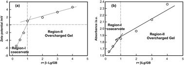
An understanding of the interactions between gelatin B (GB) and β-lacto-globulin (β-Lg) mainly arising from surface selective patch binding occurring at their common pI (≈5.0 ± 0.5) in the absence of added salt. Heterogeneous surface charge distribution on β-Lg facilitated such interaction at different mixing ratio ([GB]: [β-Lg] = r) and the GB–β-Lg complexes carried distinctive surface charge (seen through their zeta potential, ζ). For r < 1[thin space (1/6-em)]:[thin space (1/6-em)]1 (partial charge neutralization, ζ ≈ 0) a turbid solution was formed which gives the indication of formation of intermolecular soluble complexes. For r > 1[thin space (1/6-em)]:[thin space (1/6-em)]1 (overcharged regime, ζ > 0) the dispersion remained transparent and homogeneous which gives no phase separation, but the dispersion formed a gel on waiting. The overcharged gels were homogeneous, more rigid and higher melting temperature in comparison to coacervate. In the coacervate phase, the intensity of the scattered light Is, and its time-correlation function [g2(t) − 1] did not evolve with time. In contrast, the gel phase displayed considerable change with aging time tw. For gels, as tw → ∞ the system moved from an ergodic to non-ergodic state. At tw = 0, the correlation function exhibited one relaxation mode due to the system residing deeply inside the ergodic phase and purely mirroring Brownian dynamics. After a characteristic waiting time, tw an additional mode (slow relaxation) appeared which was attributed to inter-chain interaction induced reorganization of entanglements. This characteristic time was the time required for the system to get dynamically arrested, similar observation was made from rheology measurements too. A comprehensive phase diagram depicting the stability of the dispersion in various charged soft matter states of the complex under various temperature conditions was established.


1. Introduction

Protein–protein interaction is very strongly dependent on reaction environment like, the pH, ionic strength, temperature, protein surface charge distribution, protein structure etc. The heterogeneous surface charge distribution on near globular proteins1 like bovine serum albumin (BSA), β-lacto globulin (β-Lg), human serum albumin (HSA) etc. make their interaction with polyelectrolytes in general and polyampholytic proteins like gelatin, in particular quite interesting.2–4 The inter-protein interaction at their common pI is very revealing because such interactions are sometimes governed by a lesser known interaction called surface patch binding (SPB).2 Unlike electrostatic interactions where associations are dominated by net charge on oppositely charged protein molecules, SPB interactions can prevail even when the net charge on both proteins is the same. It has been realized in many biological assemblies that SPB type interactions cause intermolecular association leading to soluble complex formation.3–11 The heterogeneous surface charge distribution on one of the protein molecules, and the small persistence length of its binding partner offer adequate possibility for the surface selective binding to take place where the chain segment of the polyampholyte molecule intelligently chooses the oppositely charged patch on the other protein overcoming the repulsion between the similarly charged chain segment and surface patch of the colloidal protein molecule.12 Therefore, for SPB interactions to be relevant, it is imperative to have a pair of proteins where one is a colloidal protein molecule (like BSA, HSA, β-Lg etc.) and the other is preferably a low persistence length polyampholyte (like gelatin A and gelatin B). In general, there can be another scenario where a pair of biopolymers is chosen with one component being a polyelectrolyte (like agar, chitosan, DNA etc.) and another flexible chain polyampholyte. Experiments have shown that in both the aforesaid cases SPB interactions have yielded soluble intermolecular complexes that eventually lead to liquid–liquid phase separation giving rise to complex coacervation.9 Complex coacervation is a first-order thermodynamic phase separation caused due to associative interaction between a pair of oppositely charged polyelectrolytes, or a colloid–polylectrolyte pair. The partial charge neutralization caused due to binding between the two components (in coacervates), release of counter-ions and the random mixing of the components drives the phase separation.13,14 Several theoretical models have been proposed to account for this phenomenon.15–27

Protein based coacervation, transition is driven by surface selective patch binding even though both the biopolymers carry similar net charge28,30 and some other protein based coacervates, which is formed by strong electrostatic interactions, has been reported for β-lacto-globulin–gum arabic,30–32 whey protein–gum arabic,33,34 gelatin–chitosan,35 gelatin–agar,36 gelatin–gelatin,37 gelatin–DNA38 and β-lacto-globulin–pectin systems.39 Whey protein–gum arabic coacervate was observed to be a highly concentrated solution phase (melt-like) whereas the diversity of material properties associated with coacervates β-lacto-globulin–gum arabic coacervates were found to be associated with vesicular to sponge-like internal structure. In contrast, β-lacto-globulin–pectin coacervates were found to be a heterogeneous phase comprising of pectin networks with protein domains forming the junction points.39 It has been reported that a polyelectrolyte, DNA and a polyampholyte, gelatin B can undergo associative interactions and form complex coacervates with interesting thermal properties.38

A lesser observed phenomenon is network formation between the complementary biopolymer pair leading to gelation transition. The interpenetrating network structure plausibly formed due to self-assembly of the biopolymers results in a viscoelastic phase where the solvent gets trapped as interstitial fluid. The soft matter phase of biopolymers, gels and coacervates, are thermodynamic states that are distinctively different from the point of their self-assembly, and their structural evolution with time. The complexity of phase diagram that describes the co-existence of the two interacting biopolymers arises from the fact that continuous reorganization of the relative assembly of the constituents can give rise to physical states that are hierarchical in concentration. Therefore, length and time scales become relevant. This aspect of soft matter science in general, and in protein-based soft materials, in particular, has been poorly reported in the literature. Herein, we probe this dynamics in finer details and show that GB–β-Lg based system exhibits charge heterogeneity induced binding, phase stability and anomalous relaxation in gels and coacervates at their common pI.

2. Material and methods

The sample used in this study is bovine extracted gelatin B (GB, bloom 225 and molecular weight 100 kDa) and β-Lg a globular protein (molecular weight 67 kDa) were bought from Sigma-Aldrich chemical company (U.S.A.), in which according to supplier had a minimum purity of >98%. It needs to be mentioned that the proteins were not subjected to further purification by dialysis, which would have made these salt free. The two proteins which is used in this study are known to have a common pI = 5.0 ± 0.2.38 All concentrations are in used in % (w/v) unless otherwise stated.

The aqueous solution of β-Lg was prepared by dissolving known fix amount of the protein 0.75% powder in double distilled deionized water at 25 °C using a magnetic stirrer for about 30 min. The aqueous solutions of GB were prepared at concentrations of 0.25–4.00 % by dissolving known amount of the protein powder in double distilled deionized water at 40 °C using a magnetic stirrer for about 1.5 hours. These stock solutions appeared optically clear to the eye and all measurement procedures were performed at room temperature 25 °C. Relative humidity in the laboratory was close to 50%.

The stock samples as β-Lg and GB stock solutions were prepared by mixing the two in the following ratios: r = [GB]: [β-Lg] = 0.25[thin space (1/6-em)]:[thin space (1/6-em)]0.75, 0.50[thin space (1/6-em)]:[thin space (1/6-em)]0.75, 0.75[thin space (1/6-em)]:[thin space (1/6-em)]0.75, 1[thin space (1/6-em)]:[thin space (1/6-em)]0.75, 2[thin space (1/6-em)]:[thin space (1/6-em)]0.75, 3[thin space (1/6-em)]:[thin space (1/6-em)]0.75 and 4[thin space (1/6-em)]:[thin space (1/6-em)]0.75. The GB: β-Lg ratio is defined 0.33, 0.66, 0.75, 1, 1.3, 2.6, 4, 5.3 parts of GB mixed with as 0.75 part of β-Lg (v/v) respectively. All complex coacervate samples were prepared by mixing the two stock solutions in a fix volumetric ratios, r = 0.25[thin space (1/6-em)]:[thin space (1/6-em)]0.75, 0.50[thin space (1/6-em)]:[thin space (1/6-em)]0.75 and 0.75[thin space (1/6-em)]:[thin space (1/6-em)]0.75. All mixture of solutions was then titrated with 0.1 N HCl or 0.1 N NaOH to adjust the pH to pI. These samples were stored in air tight borosilicate glass bottles (trace amount of sodium azide was added to the samples to prevent bacterial contamination) for further analysis. Coacervates were extracted from mixed solutions following standard procedure of repeated centrifugation and decantation of the supernatant.40–43 These samples were stored in airtight borosilicate glass bottles to form gels, and for their subsist analysis, which, in all instances, did not exceed more than 24 h of preparation.

Zeta potential (ζ) measurement was performed on both coacervate and gel samples with an electrophoresis instrument (model: ZC-2000, Microtec, Japan). The electrophoretic cell was made of Teflon (dimensions 140 mmW × 40 mmD × 60 mmH) with distance between electrodes fixed at 10 cm. In all our measurements the migration voltage was fixed at 25 mV. The instrument was calibrated against 10−4M AgI colloidal dispersions. More details of zeta potential are given in ref. 28 and 29. Dynamic light scattering (DLS) experiments were performed at a scattering angle of θ = 90° and laser wavelength of λ = 632.8 nm on a 256 channel digital correlator, (PhotoCor Instruments, USA) that was operated in the multi-τ mode (logarithmically spaced channels). Robustness of the fitting results was decided based on two criteria: sample to sample accuracy, and data reproducibility within the same sample. Further information on this set up is provide in ref. 38–45.

Rheology is the study of flow and deformation of materials under applied forces. Rheology measurements performed on the both samples (coacervate and gel), using small amplitude oscillatory shear, were performed on controlled stress AR 500 rheometer (TA Instruments, Surrey, England). Measurements were carried out with a cone plate geometry (2°) using a controlled stress was 1 Pa and a fixed angular frequency of 1 Hz. Temperature dependence of dynamic storage modulus was recorded by heating the samples from 20 to 50 °C in the ramp mode. More details are available in ref. 46 and 47.

3. Results and discussion

At low mixing ratio r > 1 of β-Lg and GB solution, the mixture appears turbid, which gives the indication of formation of intermolecular soluble complexes, finally leading to phase separation, called coacervation41–43 while at high mixing ratio r < 1 the mixture turns transparent with no indication of either insoluble complex formation or phase separation, but the mixture turned into a gel like phase over time. It was possible to distinguish between the properties of all low mixing ratio samples (coacervate) and high mixing ratio samples (gel) samples conclusively from the following experiments.

The simplest binding profile showed clearly distinguishable regions in zeta potential and absorbance profile (Fig. 1). Absorbance profile, which was obtained from the analysis of the change in the slope of the absorbance (at λ = 290 nm) as a function mixing ratio (r) plot is shown in Fig. 1(b). Change in slope identifies two phases; one is coacervate, and other is gel which will be discussed later. In zeta potential profile is shown in Fig. 1(a). For r < 1.0, the ζ potential of the complexes of β-Lg and GB increased from (−6) to 0 mV, which was assigned to the coacervate phase (complete charge neutralization); this was evident from increase in solution turbidity (Region-I), and for r > 1.0 the complexes of β-Lg and GB were found to be positively charged, and the solution was transparent (+ve zeta potential, overcharging, Region-II).


image file: c5ra07195j-f1.tif
Fig. 1 Typical measured (a) zeta potential and (b) absorbance as a function of mixing ratio r. Note that the soluble complexes in Region-I were either partially or fully charge neutralized while in Region-II, these were overcharged. Subsequently, these two regions yielded complex coacervates and transparent overcharged gels. See text for details. Solid lines are guide to the eye only.

3.1. Viscoelastic properties

The viscoelastic properties of all samples were characterized by rheology. Fig. 2 represents the temporal evolution of G′ for all the samples. The highest G′ value was obtained for at r = 5.3. These gels were transparent and formed rapidly, possibly due to the increased viscosity of the system, which resulted in the reduced motility of the complexes. The gelation time tg obtained from the G′ data depicts an initial growth regime followed by a plateau. For r = 2.6 and r = 4.0 samples the low G′ values were substantially low (15–25 Pa) where as for r = 5.3 sample yielded G′ = 45 Pa. For r = 1 there was no observation of gelation which is clearly seen from Fig. 2. We identify two phases (i) sol phase, (t < tg), storage modulus increased in a linear way and (ii) Gel phase (t > tg) storage modulus remained invariant of time.
image file: c5ra07195j-f2.tif
Fig. 2 Variation of viscoelastic storage modulus (G′) as function of time during gelation of β-Lg–GB sol shown for r = 1.0, 2.6, 4.0 and 5.3. The measurements were performed at 25 °C using a constant oscillation stress of 1 Pa. The dotted line represents the gelation time tg. See text for details.

It was observed that the GB with β-Lg dispersion in water formed viscoelastic gels over a range of concentration. The profiles of storage modulus (G′) and loss tangent as a function of frequency for gel and coacervate is shown in Fig. 3. The typical storage modulus, G′(ω), defined as the energy stored in the system at a given temperature whereas the loss factor tan[thin space (1/6-em)]δ reveals the ratio between the viscous and the loss portion of the deformation behavior. In viscoelastic systems, relation between the frequency dependence of the elastic moduli near gelation47–49 given by is

 
G′(ω) = n, 0 < n < 1 (1)
 
G′′(ω) = n′′, 0 < n < 1 (2)
and
 
G′′(ω)/G′(ω) = tan[thin space (1/6-em)]δ = tan[thin space (1/6-em)](nπ/2) (3)
where S defined as elastic strength of the material and n is the relaxation exponent, it is assumed that there is no universal value of n, because n is related to the specifics of each gelling system. The linear viscoelasticity theory50,51 applicable for both pre and post gel conditions and predicts the stress–relaxation to follow the power-law frequency dependence behavior given by eqn (1) with 0 < n′ < 1. Stoichiometrically balanced and imbalanced cross-linked networks showed n = 1/2 (excess cross linking) and n > 1/2 (deficient of cross linking) respectively. Thus, it is useful to define the dispersion relation in a more generalized form as: G′(ω) ∼ ωn and G′′(ω) ∼ ωn′′. The least-square fitting of the data to this equation yielded n′ = 0.21 ± 0.01 invariant of mixing ratio for gels. For coacervate samples n′ = 0.14 ± 0.01. This indicated that gel samples were more viscoelastic than coacervates Fig. 3(a).


image file: c5ra07195j-f3.tif
Fig. 3 Evolution of (a) storage modulus G′(ω) and (b) loss tangent of coacervate and gel samples with respect to angular frequency. All measurements were performed at 25 °C using constant oscillation stresses of 1 Pa. Solid lines in (a) is fitting for eqn (1).

The elastic attribute of the coacervates was felt pertinent to resolve whether or not these had gel-like structures. For determining this, the loss tangent (tan[thin space (1/6-em)]δ = G′′/G′), was plotted with respect to frequency, which is illustrated in Fig. 3(b), For gel state the tan[thin space (1/6-em)]δ versus ω slope is positive, whereas at the point of gelation it is zero while it is negative for melt state.51,52 Fig. 3(b), indicates that the loss tangent with respect to ω is associated with a small positive slope implying that the coacervate material was indeed gel-like. When tan[thin space (1/6-em)]δ < 1 (G′ > G′′), which confirmation of the solid-like attribute of the material. On the contrary, when tan[thin space (1/6-em)]δ > 1, liquid-like behaviour prevails. In the present system tan[thin space (1/6-em)]δ of coacervate was consistently higher value than tan[thin space (1/6-em)]δ of gels which confirms more fluidity of the coacervate state compared to the gel state samples.

In the gel state, water is available as interstitial water trapped inside the network or hydration water attached to the networks. In contrast, in coacervates, most of the water is attached to the intermolecular complexes as hydration water. The coacervates samples comprise of a heterogeneous phase with localized finite size networks embedded in an assembly of randomly mixed intermolecular complexes.

The melting profile of coacervate and gel samples were studied by a temperature sweep experiments where the storage modulus was probed under temperature scan at fixed frequency of 1 rad s−1 with temperature ramp of 1 °C min−1. Heating the coacervate and gel samples resulted in decrease of the viscoelastic modulus G′, and an sharp change in this decrease indicated a characteristic structure melting temperature Tm. The typical data shown in Fig. 4, clearly identify where a sharp decrease in the value of G′ occurred in both coacervate and gel samples, called this temperature, Tm. The melting temperatures obtained for the coacervate and gel samples were Tm = 22 ± 1 and 26 ± 1 °C, respectively. Since, β-Lg–GB gels were associated with higher rigidity (G′) with higher melting temperature in comparison to β-Lg–GB coacervates.


image file: c5ra07195j-f4.tif
Fig. 4 Evolution of storage modulus G′(ω) for (a) coacervate and (b) gel samples with respect to temperature. Sharp drop indicates a melting temperature (arrows) of samples.

3.2. Cole–Cole plot and dispersion homogeneity

In soft matter comprising of multi-component assembly the structure–property relationship is a function of sample homogeneity. In the linear viscoelastic theory, materials exhibit characteristic relaxation which is a manifestation of the mechanical dissipation of energy in the system. In viscoelastic material the homogeneity depends on storage and loss moduli. Typically, in a polymer melt, at very low frequency, viscous behavior is observed, while elastic properties dominate at higher frequencies. Thus, we investigated mixing ratio dependent sample homogeneity in β-Lg and GB (coacervate and gel state) samples and the phase homogeneity in coacervates and gels was determine from Cole–Cole plot where the imaginary part of complex moduli (G′′) was plotted against of real part (G′) of complex moduli. The value of G′ and G′′ in coacervate and gel samples are different (shown in Fig. 3) and comes from the same way with a constant oscillation stress of 1 Pa, so the scales are different in both samples. In this formalism G*(ω) = G′(ω) + iG′′(ω) and the low and high frequency values are given by G0 and G respectively. The Cole–Cole empirical expression is written as52,53
 
image file: c5ra07195j-t1.tif(4)

The Cole–Cole expression in eqn (4) is interpreted as arising from a superposition of several Debye relaxation times54,55 where τcc is mean relaxation time of samples. This Cole–Cole plot has been used extensively to determine the map homogeneity of gel and coacervate both samples. For a homogeneous phase, the Cole–Cole plot is a perfect semicircle (k = 0) with a well-defined relaxation time τcc whereas any deviation from this semicircle shape indicates non-homogeneous dispersion and phase segregation due to immiscibility. The phase homogeneity of coacervate and gel materials was deduce from Cole–Cole plot (G′ plotted as a function of G′′). The fitted equation arises from the superposition of several Debye relaxations in the complex plains that define a circular arc, and with the exponent parameter k describing the viscoelastic homogeneity of the sample (see Table 1). Ref. 50 and 51 provide further details about the Cole–Cole plot.

Table 1 The τcc is mean relaxation time and the exponent parameter k, which takes a value between 0 and 1, allows clear definition of sample homogeneity
Samples k τcc/s
r = 2.6 0.43 0.88
r = 4.0 0.30 0.91
r = 5.3 0.02 0.87


The curves in Fig. 5(a) show linear behavior, and could not be fitted to eqn (4) while data in Fig. 5(b) was fitted to eqn (4) (as a representation fitting for r = 1 and 2.6 data are depicted in Fig. S2, ESI). Well defined relaxation times and smaller k-values obtained were indicative of the near semicircular feature of the plots implying dispersions were homogeneous (Fig. 5(b)). Fig. 5 shows that there was homogeneity present in all gel samples whereas inhomogenity was noticed in propensity in coacervate samples because in coacervate samples, these Cole–Cole plots diverged from a semicircle strongly and showing a linear behavior. Here, τcc value is the inverse of the angular frequency at which G′′ showed maxima, which referred to the mean viscoelastic relaxation time. The viscoelastic relaxation prevailed over comparable time scales.


image file: c5ra07195j-f5.tif
Fig. 5 Cole–Cole plot, G′′ plotted with respect to G′ for different mixing ratio (r) with a constant oscillation stress of 1 Pa (a) coacervate samples exhibited strong heterogeneity and (b) gel samples exhibited relatively less heterogeneity. The solid line is the fitting of the data to eqn (4) which could be done for only gel samples.

3.3. Kinetics and relaxation dynamics of gels

In DLS, the intensity–intensity correlation function g2(q, t), defined as 〈I(q,0)I(q, τ)〉/〈I(q)〉2, in the homodyne mode or self-beating mode was measured, where 〈I(q, τ)〉 is time-averaged scattering intensity at time τ with respect to τ = 0, that is the measured baseline. Thus g2(q, τ) is related to g1(q, τ) by the Siegert relation as56,57
 
g2(q, τ) = A[1 + β|g1(q, τ)|2] (5)
where A is measured baseline and β is a coherence factor depending on instrumental parameters (pinhole size and distance from the detector etc.) For mono-dispersed spherical scatters, |g1(q, τ)| is theoretically represented as an exponential decay function58,59
 
|g1(q, τ)| = G(q)[thin space (1/6-em)]exp(−Γτ) (6)
where G and Γ are the factors of proportionality and the linewidth, respectively, and Γ = 1/τr. The characteristic decay time τr represents the rate of dynamic relaxation of concentration fluctuations. For the polydispersed samples the dynamic relaxation, eqn (6) may be generalized as58,59
 
image file: c5ra07195j-t2.tif(7)
where G(Γ) is the distribution function of characteristic decay rate Γ. G(Γ) was calculated from Laplace inversion on the basis of eqn (6) and (7). In this study, the CONTIN58 (constrained regularization method) program supplied with the correlate was used to obtain G(Γ) from g1(q, t).

The temporal evolution of correlation function of gelling sols is depicted in Fig. 6 where the data pertains to observations taken at θ = 90°. It was observed that as the gel state was approached the correlation functions did not relax to the equilibrium value (onset of non-ergodicity). In 1% GB system there is no gel formation but at 2%, 3% and 4% GB the base line of time–intensity correlation functions changes which can be interpreted as a superposition from the collective diffusion mode (caused by slightly cross-linked cluster and sol molecule) and the cluster mode (caused by large connected gel cluster).61,62 These features agree with the fact that sol–gel transition takes place.


image file: c5ra07195j-f6.tif
Fig. 6 The evolution of dynamic structure factor of the samples with r = 1.3, 2.6, 4.0 and 5.3. The arrow indicates the evolution of dynamic structure factor starting t = 0 h to t = 8 h.

Fig. 6 shows the temporal evolution of [g2(q, τ) − 1] for, the samples with r = 1.3, 2.6, 4.0 and 5.3. Each plot shows similar temporal evolution dynamics. It is clearly seen that the [g2(q, τ) − 1] did not completely relax as the gelation time increased indicating system was to the arrested phase. For r = 1.3, [g2(q, τ) − 1] did not changed very much but for r = 2.6, 4.0 and 5.3 samples, the decay of relaxation was slower. This clearly implied onset of dynamics arrest of fluctuations due to confinement of the network units arising out of extensive cross linking.

Fig. 7 shows the decay rate distribution functions, G(Γ), as a function of characteristic decay rate for sol and gel samples which is obtained from the inverse Laplace transform of g2(τ) using the CONTIN algorithm. In sol state the distribution function G(Γ) was associated with one single peak, which is shifted towards the small relaxation time. The distribution function G(Γ) split into two peaks as system goes from sol to gel phase. That is, the former (sol stage) arising from the translational diffusion of β-Lg–GB system, while the latter (gel stage) was due to the collective diffusion of gel network. In 1% GB system G(Γ) for the different reaction time has a single peak. This indicates no phase transition at this stage. In 2%, 3% and 4% GB there is single peak in G(Γ) in solution phase but if we increase waiting time, at a certain time G(Γ) split into two peaks, this indicates that the system goes from ergodic to non ergodic phase, called gelation time.


image file: c5ra07195j-f7.tif
Fig. 7 Variation of decay rate distribution functions obtained for samples with r = 1.3, 2.6, 4.0 and 5.3 before and after gelation as a function of time.

3.4. Non-ergodicity and gelation

DLS has been applied to follow the gelation or non-ergodicity of soft systems. In ergodic system the intensity correlation function g2(t) can be correlated directly to the corresponding dynamic structure factor g1(t) via the Siegert relation but in non ergodic system g2(t) cannot be correlated directly to the corresponding g1(t) via Siegert relation. Complication are caused when the scattering moiety in the dispersion medium are localized near fixed mean position and are able only to execute limited Brownian motion about the same. A completely ergodic system is one where the time and ensemble average are identical and the system is independent of the origin of time. The issue of non-ergodicity and its effect on g2(q, t) has been addressed adequately in the past. In this work, we adopt the heterodyne approached to resolve the issue of non-ergodicity in our data analysis.

In non-ergodic system the intensity correlation function g2(q, t) measured by the correlator can be related to dynamic structure factor through the general expression63,64

 
g2(q, t) = 1 + β[2χ(1 − χ)|g1(q, t) + χ2|g1(q, t)|2] (8)
where β is the coherence factor having maximum value β = 1. In a real experiment it defines the signal modulation which is a measure of signal-to-noise ratio of the data. The parameter χ (0≤ χ ≤ 1) defines the heterodyne contribution present in the measured g2(q, t) data. In the sol state χ = 1 and the Siegert relationship [g2(q, t) = 1 + |g1(q, t)|2] get established. However, in the gel state χ < 1 and the term [2χ (1 − χ)] makes a finite contribution to g2(q, t). The value of χ can be determined experimentally from the intercept of the plot of [g2(q, t) −1] versus delay time t at t → 0 gives β[2χ(1 − χ)] from which the value of χ can be calculated if β is known. For an ergodic system, the g2(q, t) function relaxes to 0 (at t = ∞) from its initial value 1 (t = 0). Thus, the signal modulation β achieved from the relation [g2(q, t)|t→0g2(q, t)|t→∞]. For a non-ergodic system, β-values lies between 0 and 1. This happens because of the dynamic arrest of the scattering moiety, and this has been observed in a variety of soft matter systems like polymer solutions, colloidal gels, glasses, and so on.65–67

Fig. 7(a) shows the variation of χ with time at different mixing ratio for gelling sols. In sol state, for r = 1.3, we measured χ = 0.9 ± 0.1. Hence g1(q, t) could be determined from g2(q, t) data directly, using Seigert relation. In the gel state χ decreased with time and remained constant after the gelation was achieved. Thus, we could neglect the contribution of second term in eqn (8) and fit the data to evaluate the various relaxation modes explicitly. This allows us to define an ergodicity breaking time, τEB. This is the time beyond which the ergodicity of the sample does not change Interestingly, it determines the gelation time of the samples. Fig. 7(b) shows the tg (from DLS and rheology) and ergodic breaking time τEB as a function of r, which indicates that the value of gelation time is exactly matches with the ergodicity breaking time. The concentration of GB strongly affects the gelation kinetics, and network becomes stronger with an increase in the GB concentration (confirmed from the viscoelastic data). At high mixing ratio the network becomes denser with increase in GB concentration with the β-Lg aggregating on GB chains. Due to abundance of GB chains the gelation process is enhanced and the difference between gelation time and ergodic breaking time becomes smaller, while at low mixing ratio abundance of GB is low so the difference between the gelation time and ergodic breaking time becomes more as shown in Fig. 8(b).


image file: c5ra07195j-f8.tif
Fig. 8 (a) The heterodyne contribution χ shown as a function of ageing time t s−1 the point where there is slop changes in the value of χ is defined as a ergodicity breaking time τEB. (b) The ergodicity breaking time τEB and gelation time of samples as a function of mixing ratio r from DLS and rheology.

3.5. Phase diagram

We proposed a phase diagram in Fig. 9 using zeta potential versus temperature a variable to describe all the physical expected features observed in this study. The conceptual understanding developed for the all samples with respect to mixing ratio (r), and heat-induced phase transitions were summarized as follows: In Phase-I for r ≥ 1.0, the interaction between protein and polyampholyte occurred marginally above the isoelectric point (pI) of protein where the protein being overall positively charged and a cloudy solution was formed, this indicating formation of soluble intermolecular complexes (coacervation). Here, the charge neutralized intermolecular complexes were formed due to strong surface patch binding between both biopolymers. In the phenomena of surface patch binding, these charged patches are opposite in sign to the net protein (β-Lg) charge, could provide sites for binding to polyampholyte (GB) chain with the same charge sign as protein. In Phase-II, i.e. for r ≤ 1, the interaction between protein and polyampholyte occurred slightly below the isoelectric point (pI) of protein where the protein being overall negatively charged and the mixture remained homogeneous and there is no indication of either intermolecular soluble complex formation (turbidity) or phase separation. This dispersion turned into a gel-like phase over a period of time and network formation in these samples were mostly due to hydrogen bonding between flexible gelatin chains with β-Lg acting as binding units. Due to the presence of β-Lg molecule, this network was very weak in comparison of GB network formed at same concentration because this was which was attached to the GB chains causing network deformations whereby weak gels were formed.
image file: c5ra07195j-f9.tif
Fig. 9 Typical Phase diagram of various soft matter states in β-Lg–GB complexation, r = [GB]: [β-Lg] as a function of surface charge and melting temperature.

All coacervate samples are opaque in nature, when these coacervate samples (Phase-I) were subjected to slow heating (1 °C min−1) in a thermostat up to a maximum temperature 45 °C and then followed by gradually cooling to room temperature (20 °C). During this procedure, the originally opaque looking all coacervate samples on heating became turbid sol that turned into gel-like network after an equilibration time of 24 hours (Phase-III). Similarly when the all overcharged transparent gel samples (Phase-II) were subjected to slow heating (1 °C min−1) in a thermostat up to a maximum temperature 45 °C followed by gradual cooling to room temperature (20 °C), these changed into overcharged turbid solution (Phase-IV). The high turbidity was seen in these samples, which is due to the nature of thermal aggregation of protein (β-Lg). It is to be noted that if these overcharged gels (Phase-II) were heated beyond 45 °C, these samples immediately precipitated with gradual cooling to room temperature (Phase-V).

4. Conclusions

We studied the interaction between β-Lg and GB mixed in different weight ratio and find that associations were mainly due to surface patch binding. For r < 1.0 there was formation of turbid solution with indication of soluble complex formation or phase separation (coacervation). For r > 1.0, the dispersion obtained homogeneous and there is no indication of either intermolecular complex formation or phase separation. This dispersion turned into a gel-like phase over a certain period of time. In this study, to observe gelation processes of β-Lg–GB dispersions were monitored by rheology as a function of the β-Lg–GB mixing ratio. The frame of gel organization was found in flexible GB chain while the β-Lg aggregated along the GB chains and could be regarded as a cross linking agent, and If we increase the GB concentration the viscoelasticity of gels is also increased. The zeta potential and absorbance data showed that there was partial charge neutralization in the region of r < 1, called coacervation regime, and in the region of r > 1 overcharging and gel-like network formation dominated.

The systematic rheology studies of both phases (gels and coacervates) show viscoelastic nature. G0 is the measure of rigidity of a network. In the region of r < 1.0, G0 was greater than r > 1.0 also the melting temperature of gels was higher than coacervates, which indicated gels were stiffer than coacervates. From rheology data, we have shown that sol–gel transitions are observed with time and define a characteristic parameter tg, was the time required for the system to get arrested. From the Cole–Cole plot there is homogeneity observed in gel phase.

In the study of DLS shows typical β-Lg–GB solution dynamics in the time-correlation function is observed from the beginning of the reaction up to gelation time. The decrease in the time correlation function can be interpreted as a superposition from the collective diffusion mode (caused by slightly cross-linked both sol molecules) and the cluster mode (caused by formed larger connected gel network). This indicates sol–gel phase transition in systems. We will discuss the relaxation dynamics of relaxation modes of β-Lg–GB molecules occurring in solution and gel phase observed at different mixing ratio. On increasing aging time the system moved towards ergodic to non ergodic medium. At initial time when the system is in dilute region only one mode observed which is due to the system towards ergodic phase owing to faster Brownian dynamics. After a certain time, single internal mode turns into one fast and one slow relaxation mode which is due to new dynamical process involving interchain interaction and disentanglement begin to occur besides the fast relaxation mode. This certain time was time required for the system to get arrested which is approximately same as rheological observation. Finally, draw heat induced phase diagram of different heat induced stages of β-Lg–GB system in the absence of added salt.

The intermolecular interactions prevailing in coacervate and gel phase are mostly due to surface patch binding and hydrogen bonding. In coacervate phase (r > 1) charge neutralization occurs due to surface patch binding between β-Lg and GB. In gel phase (r < 1) due to large amount of overcharged complexes present, the interaction was mostly due to intermolecular hydrogen bonding between gelatin chains. The simplest molecular mechanism describing different phase transitions in β-Lg–GB system is remarkably similar to BSA–GB system which is described in ref. 2 (Fig. 12 of ref. 2). The following Table 2 provides a comparison between two pairs of interacting proteins having common pI. More detail of these systems is found in ref. 2 and 4.

Table 2 A comparison between two pairs of interacting proteins (BSA–GB and β-Lg–GB) having common pI
Techniques/parameter BSA–GB β-Lg–GB Comments
Fluorescence binding constant/M−1 162 ± 0.154 43 ± 0.014 Binding constant of BSA–GB > β-Lg–GB
FRET (energy transfer efficiency E%) 63% 4 30% 4 BSA–GB indicates energy transfer efficiency of BSA–GB > β-Lg–GB
Rheology G′/Pas 150 ± 50 (r < 1)2 135 ± 50 (r < 1) Gel strength of BSA–GB > β-Lg–GB
130 ± 10 (r > 1) 100 ± 50 (r > 1)
Tm/°C 30 ± 1 (r < 1)2 22 ± 1 (r < 1) Melting temperature of BSA–GB > β-Lg–GB
33 ± 1 (r > 1) 26 ± 1 (r > 1)


Acknowledgements

JP acknowledges University Grants Commission, Government of India for a Junior Research Fellowship. KR acknowledges DST-Inspire Faculty Award from Department of Science and Technology (DST), Government of India. This work was supported by a research grant from DST.

References

  1. Y. Xu, D. Seeman, Y. Yan, L. Sun, J. Post and P. L. Dubin, Biomacromolecules, 2012, 13, 1642–1651 CrossRef CAS PubMed.
  2. J. Pathak, K. Rawat, V. K. Aswal and H. B. Bohidar, J. Phys. Chem. B, 2014, 118(38), 11161–11171 CrossRef CAS PubMed.
  3. (a) C. Stuart, Langmuir, 2004, 20, 2785–2791 CrossRef; (b) M. Skepoe and P. Linse, Macromolecules, 2003, 36, 508–519 CrossRef CAS.
  4. J. Pathak, K. Rawat, V. K. Aswal and H. B. Bohidar, RSC Adv., 2015, 5, 13579–13589 RSC.
  5. Y. Murakami and S. Jones, Struct. Bioinf., 2006, 22(14), 1794–1795 CrossRef CAS PubMed.
  6. M. Lund, L. Vrbka and P. Jungwirth, J. Am. Chem. Soc., 2008, 130, 11582–11583 CrossRef CAS PubMed.
  7. B. Kayitmazer, D. Seeman, B. B. Minsky, P. L. Dubin and Y. Xu, Soft Matter, 2013, 9, 2553–2583 RSC.
  8. J. van der Gucht, E. Spruijt, M. Lemmers and M. A. Cohen Stuart, J. Colloid Interface Sci., 2011, 361, 407–422 CrossRef CAS PubMed.
  9. P. Maarten, M. A. Biesheuvel, F. M. Menger and B. M. Sykes, Langmuir, 1998, 14, 4131–4137 CrossRef.
  10. E. Tsuchida and K. Abe, Inter Macromolecular Complexes, Springer, Heidelberg, Germany, 1982 Search PubMed.
  11. H. G. Bungenberg de Jong, in Colloid Science, vol. II, ed. H. R. Kruyt, Elsevier, New York, 1949 Search PubMed.
  12. J. Pathak, K. Rawat and H. B. Bohidar, Int. J. Biol. Macromol., 2014, 63, 29–37 CrossRef CAS PubMed.
  13. D. Priftis, K. Megley, N. Laugel and M. Tirrell, J. Colloid Interface Sci., 2013, 398, 39–50 CrossRef CAS PubMed.
  14. K. Kaibara, E. Seyrek, P. L. Dubin, C. Tribet and E. A. Gamble, Biomacromolecules, 2003, 4, 273–282 CrossRef PubMed.
  15. E. Kizilay, A. Basak Kayitmazer and P. L. Dubin, Adv. Colloid Interface Sci., 2011, 167, 24–37 CrossRef CAS PubMed.
  16. A. S. Michaels and R. G. Miekka, J. Phys. Chem. B, 1961, 65, 1765 CrossRef CAS.
  17. F. Vongoeler and M. Muthu kumar, J. Chem. Phys., 1994, 100, 7796 CrossRef CAS PubMed.
  18. M. Girard, S. L. Turgeon and S. F. Gauthier, J. Agric. Food Chem., 2003, 51, 4450 CrossRef CAS PubMed.
  19. X. Feng, M. Leduc and R. Pelton, Colloids Surf., A, 2008, 317, 535 CrossRef CAS PubMed.
  20. V. Ball and C. Maechling, Int. J. Mol. Sci., 2009, 10, 3283 CrossRef CAS PubMed.
  21. D. Leisner and T. Imae, J. Phys. Chem. B, 2003, 107, 8078 CrossRef CAS.
  22. A. Veis, Adv. Colloid Interface Sci., 2011, 167, 2–11 CrossRef CAS PubMed.
  23. J. Overbeek and M. J. Voorn, J. Cell. Comp. Physiol., 1957, 49, 7 CrossRef CAS PubMed.
  24. A. Veis, J. Phys. Chem. B, 1963, 67(10), 1960–1964 CrossRef CAS.
  25. A. Veis, J. Phys. Chem. B, 1961, 65, 1798 CrossRef CAS.
  26. A. Nakajima and H. Sato, Biopolymers, 1972, 10, 1345 CrossRef PubMed.
  27. K. Tainaka, Biopolymers, 1980, 19, 1289 CrossRef CAS PubMed.
  28. A. Basak Kayitmazer, D. Seeman, B. Baykal Minsky, P. L. Dubin and Y. Xu, Soft Matter, 2013, 9, 2553–2583 RSC.
  29. J. M. Park, B. B. Muhoberac, P. L. Dubin and J. Xia, Macromolecules, 1992, 25, 290–295 CrossRef CAS.
  30. J. Gao, P. L. Dubin and B. B. Muhoberac, J. Phys. Chem. B, 1998, 102, 5529–5535 CrossRef CAS.
  31. C. Schmitt, C. Sanchez, A. Lamprecht, D. Renard, C. M. Lehr, C. G. de Cruif and J. Hardy, Colloids Surf., B, 2001, 20, 267–280 CrossRef CAS.
  32. C. Sanchez, G. Mekhloufi, C. Schmitt, D. Renard, P. Robert, C.-M. Lehr, A. Lam-phret and J. Hardy, Langmuir, 2002, 18, 10323–10333 CrossRef CAS.
  33. F. Weinbreck, R. H. Tromp and C. G. de Cruif, Biomacromolecules, 2004, 5, 1437–1445 CrossRef CAS PubMed.
  34. F. Weinbreck, H. S. Rollema, R. H. Tromp and C. G. de Cruif, Langmuir, 2004, 20, 6389–6395 CrossRef CAS PubMed.
  35. A. N. Gupta and H. B. Bohidar, J. Phys. Chem. B, 2007, 111, 10137–10145 CrossRef CAS PubMed.
  36. S. Boral and H. B. Bohidar, J. Phys. Chem. B, 2010, 114, 12027–12035 CrossRef CAS PubMed.
  37. A. Tiwari, S. Bindal and H. B. Bohidar, Biomacromolecules, 2009, 10, 184–189 CrossRef CAS PubMed.
  38. K. Rawat, V. K. Aswal and H. B. Bohidar, J. Phys. Chem. B, 2012, 116, 14805–14816 CrossRef CAS PubMed.
  39. Y. Wang, Y. Li, Y. W. Wang, J. Lal and Q. Huang, J. Phys. Chem. B, 2006, 111, 515–520 CrossRef PubMed.
  40. Y. Wang, J. Y. Gao and P. L. Dubin, Biotechnol. Prog., 1996, 12, 356–362 CrossRef CAS.
  41. P. L. Dubin, S. S. The, D. W. Mc Quigg, C. H. Chew and L. M. Gans, Langmuir, 1989, 5, 89–95 CrossRef CAS.
  42. A. B. Kayitmazer, H. B. Bohidar, K. M. Mattison, A. Bose, J. Sarkar, A. Hashimoto, P. S. Russo, W. Jaeger and P. L. Dubin, Soft Matter, 2007, 3, 1064–1076 RSC.
  43. K. Kaibara, T. Okazaki, H. B. Bohidar and P. L. Dubin, Biomacromolecules, 2000, 1, 100–107 CrossRef CAS.
  44. H. Ohshima, Adv. Colloid Interface Sci., 1995, 62, 189–235 CrossRef CAS.
  45. B. Mohanty and H. B. Bohidar, Biomacromolecules, 2003, 4, 1080–1086 CrossRef CAS PubMed.
  46. H. A. Barnes, Handbook of Elementary Rheology, University of Wales Press, Wales, 2000 Search PubMed.
  47. B. Mohanty, A. Gupta, H. B. Bohidar and S. Bandyopadhyay, J. Polym. Sci., Part B: Polym. Phys., 2007, 45, 1511–1520 CrossRef CAS PubMed.
  48. J. E. Martin, D. Adolf and J. P. Wilcoxon, Phys. Rev. Lett., 1988, 61, 2620–2623 CrossRef CAS.
  49. J. E. Martin and D. Adolf, Annu. Rev. Phys. Chem., 1991, 42, 311–339 CrossRef CAS.
  50. H. H. Winter and F. J. Chambon, J. Rheol., 1986, 30, 367–382 CrossRef CAS.
  51. H. A. Barnes, Handbook of Elementary Rheology, University of Wales, Wales, 2000 Search PubMed.
  52. N. Arfin and H. B. Bohidar, Int. J. Biol. Macromol., 2012, 50, 759–767 CrossRef CAS PubMed.
  53. K. S. Cole and R. H. Cole, J. Chem. Phys., 1941, 9, 341–351 CrossRef CAS PubMed.
  54. T. C. Warren, J. L. Schrag and J. D. Ferry, Biopolymers, 1973, 12, 1905–9015 CrossRef CAS PubMed.
  55. Q. Zheng, M. Du, B. B. Yang and G. Wu, Polymer, 2001, 42, 5743–5747 CrossRef CAS.
  56. H. Z. Cummins and E. R. Pike, Photon Correlation and Light Beating Spectroscopy, Plenum, New York, 1974 Search PubMed.
  57. B. Chu, Laser Light Scattering, Academic Press, New York, 1974 Search PubMed.
  58. B. J. Berne and R. Pecora, Dynamic Light Scattering, Wiley-Inter-science, New York, 1976 Search PubMed.
  59. R. J. Glauber, Quantum Optics and Electronics, Gordan and Breach, New York, 1965 Search PubMed.
  60. (a) S. W. Provencher, Comput. Phys. Commun., 1982, 27, 213 CrossRef; (b) S. W. Provencher, Comput. Phys. Commun., 1982, 27, 229 CrossRef.
  61. T. Tanaka and R. Pecora, Dynamic Light Scattering, Plenum Publishing, New York, 1985, p. 347 Search PubMed.
  62. M. Shibayama, Y. Isaka and Y. Shiwa, Macromolecules, 1999, 32, 7086 CrossRef CAS.
  63. K. te Nijenhuis, Thermo-reversible network: viscoelastic properties and struture of gels (Advances in Polymer Science), 1997, vol. 130, pp. 1–7 Search PubMed.
  64. (a) J. Sharma and H. B. Bohidar, Eur. Polym. J., 2000, 36, 1409–1418 CrossRef CAS; (b) J. Sharma and H. B. Bohidar, Colloid Polym. Sci., 2000, 278, 15–21 CrossRef CAS.
  65. B. Ruzicka, E. Zaccarelli, L. Zulian, R. Angelini, M. Sztucki, A. Moussaid, T. Narayanan and F. Sciortino, Nat. Mater., 2011, 10, 56–60 CrossRef CAS PubMed.
  66. P. N. Segre, V. Prasad, A. B. Schofield and D. A. Weitz, Phys. Rev. Lett., 2001, 86(26), 6042–6045 CrossRef CAS.
  67. L. D. Michele, F. Varrato, J. Kotar, S. H. Nathan, G. Foffi and E. Eiser, Nat. Commun., 2013, 4, 2007 Search PubMed.

Footnote

Electronic supplementary information (ESI) available: UV-Vis absorbance spectra and Cole–Cole plot for different mixing ratios of β-Lg and gelatin B undertaken in this study are included in the Supplementary information. See DOI: 10.1039/c5ra07195j

This journal is © The Royal Society of Chemistry 2015
Click here to see how this site uses Cookies. View our privacy policy here.