Dual role of pinostrobin-a flavonoid nutraceutical as an efflux pump inhibitor and antibiofilm agent to mitigate food borne pathogens

Lowrence Rene Christena a, Shankar Subramaniama, Mohan Vidhyalakshmia, Vijayalakshmi Mahadevana, Aravind Sivasubramanian§ *a and Saisubramanian Nagarajan§*ab
aSchool of Chemical and Biotechnology, SASTRA University, Thanjavur 613 401, India. E-mail: sai@scbt.sastra.edu; arvi@biotech.sastra.edu
bCenter for Research on Infectious Diseases, SCBT, SASTRA University, Thanjavur 613 401, India

Received 20th April 2015 , Accepted 8th July 2015

First published on 13th July 2015


Abstract

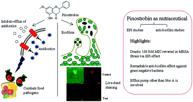
Nutraceutical pinostrobin displayed synergy with ciprofloxacin against diverse bacteria causing drastic 128 fold reduction in ciprofloxacin MIC in MRSA and significant antibiofilm effect against E. coli and P. aeruginosa at subinhibitory concentrations. Studies with NorA mutants of S.aureus revealed efflux inhibition of pinostrobin is mediated by efflux pumps other than NorA.


Phenolic compounds form one of the main classes of plant secondary metabolites. Their diverse structural features contribute to the colour, taste and nutrition of plant based foods. Among phenolics, flavonoids form a vital subclass most of which act as excellent nutraceuticals.1 A nutraceutical could be regarded as any nontoxic food or plant extract supplement that has proven health benefits both therapeutically and also as a prophylactic agent.2 Pinostrobin (flavonoid) has been found in honey and in some plants and is used as a natural food supplement. It is also present in many commercial natural foods like Organika® Bee Propolis liquid, Himalaya® Soliga Forest Honey, Amazon Herbs® etc. which are claimed to be safe food supplements with multiple health beneficiary effects such as improving immunity and skin health and in preventing indigestion etc.3. Pinostrobin has also been proved to exhibit anti-cancer, anti-viral, anti-inflammatory effects.4 In addition, pinostrobin containing extract has been shown to exhibit anti-microbial activity.5

Pinostrobin by virtue of its nutraceutical and antibacterial effect can curtail microbial growth on foods, when it occurs as an incidental food additive/when used as a nutraceutical. Of late, food borne pathogens are becoming increasingly drug resistant which could be attributed to evolutionary selection pressures and indiscriminate use of antibiotics in animal farming, aquaculture and poultry.6 A recent study reported that fermented dairy products like yoghurt was frequently (26%) found to be contaminated with shiga toxin producing Escherichia coli (E. coli) that displayed multidrug resistant (MDR) phenotype.7 Modes to combat drug resistance in food borne pathogens include inhibition of drug efflux and plasmid curing.6 With MDR phenotype being frequently associated with increased drug efflux, recently there is a surge in research for screening and identifying potent efflux pump inhibitors which can mitigate drug resistant bacteria. We explored if pinostrobin could also be effective against drug resistant food borne pathogens. Towards this end, we tested synergy, Minimum Inhibitory Concentration (MIC) reversal, efflux inhibitory effect and antibiofilm effect of pinostrobin on select gram positive and gram negative bacterial pathogens. Finally, we performed membrane permeability studies to discern the mechanism of action of pinostrobin. To the best of our knowledge, we are reporting for the first time a novel efflux inhibitory and biofilm prevention properties of nutraceutical pinostrobin.

Our study revealed that pinostrobin, apart from its nutraceutical effect, also possesses good antimicrobial activity against diverse bacteria (Table S1). Earlier study has indicated that among coumarins from pigeon pea (Cajanas cajan (L.) Millsp.) cajunuslactone, cajaninstilbene acid and pinostrobin, cajanuslactone displayed significant antibacterial effect against Staphylococcus aureus (S.aureus).8,9 Although antibacterial effect of pinostrobin has been reported earlier, its potential to tackle drug resistant food borne bacteria in planktonic and biofilm mode of growth has not been explored earlier. In the present study, synergy testing revealed (Table S1) that pinostrobin displayed synergistic interactions, especially with ciprofloxacin against a wide range of both gram positive and gram negative bacteria and hence, we explored the ability of pinostrobin to reverse the MIC of ciprofloxacin in select gram positive and gram negative bacterial pathogens.

In order to evaluate the ability of pinostrobin to potentiate the effect of ciprofloxacin, pinostrobin was used at 0.5× MIC (15 μg ml−1, for gram positive bacteria and 3.5 μg ml−1, for gram negative bacteria) and its ability to reverse MIC in 7 bacterial strains, 5 ciprofloxacin resistant strains and 2 ciprofloxacin sensitive strains of S.aureus [one in which NorA efflux pump is knocked out (SA-K1758) and another wild type strain of S.aureus (SA-1199)], was evaluated (Table 1). MIC reversal experiments revealed that pinostrobin could effectively restore susceptibility to ciprofloxacin in 4 out of 5 ciprofloxacin resistant bacterial strains tested. A maximal fold reduction in MIC, as reflected by a modulation factor (MF) of 128 was observed in methicillin resistant Staphylococcus aureus (MRSA). On the other hand, upon pinostrobin treatment, an MF of 4 was displayed by wild type strain of S. aureus (SA-1199), NorA knock out strain of S. aureus and Enterococcus faecalis (E. faecalis). Whereas NorA overexpressed strain of S. aureus (SA-1199B) and a laboratory derived mutant strain of Pseudomonas aeruginosa (P. aeruginosa) had a MF of 2. But with E. coli, at the concentration tested, pinostrobin could not reduce the MIC of ciprofloxacin and hence the MF remained at 1. Standard efflux pump inhibitors reserpine and verapamil as positive controls were more effective with E. faecalis wherein they caused a significant reduction in MIC of ciprofloxacin as evidenced by a MF of 64. With MRSA strain, the MF for reserpine and verapamil were 16 and 4 respectively. But neither reserpine nor verapamil could decrease MIC of ciprofloxacin in wild type strain of S. aureus and NorA knock out strain, P. aeruginosa and E. coli. Overall, the trend revealed that relative to standard EPIs, pinostrobin was more effective against S. aureus strains especially against MRSA which is a common food borne pathogen that causes Staphylococcal food borne disease.10

Table 1 Pinostrobin reduces MIC of ciprofloxacin in drug resistant bacterial strains
Strain MIC of Cip (μg ml−1) MIC of Cip (μg ml−1) + Modulation factora (fold change)
PIN RES VER PIN RES VER
a Modulation factor = MIC of drug/MIC of drug in combination.b Laboratory derived stable mutant strain of P. aeruginosa; PIN-pinostrobin, RES-reserpine, VER-verapamil; Cip-ciprofloxacin.
E. faecalis >16 4 0.25 0.25 4 64 64
S. aureus (MRSA)-ATCC 43300 16 0.125 1 4 128 16 4
S. aureus-1199B 8 4 4 8 2 2 1
S. aureus-1199 0.25 0.06 0.25 0.25 4 1 1
S. aureus-K1758 0.5 0.125 0.5 0.5 4 1 1
P. aeruginosab 8 4 8 8 2 1 1
E. coli >16 >16 >16 >16 1 1 1


Efflux pumps belonging to Major Facilitator Superfamily (MFS) class serve predominantly as MDR efflux pumps in gram positive bacteria, and Resistance Nodulation Division (RND) class of efflux pumps serve a similar purpose in gram negative bacteria. These two classes differ from each other structurally, mechanistically and in substrate preferences. Since pinostrobin causes effective MIC reversal in gram positive bacteria, it is likely that pinostrobin interacts efficiently with MFS class of efflux pumps relative to RND class.

When ethidium bromide (EtBr) was used as the efflux substrate, pinostrobin caused only a mild efflux inhibition and a MF of 2 was obtained with E. faecalis, MRSA and P. aeruginosa. It could not decrease MIC of EtBr in other strains tested including NorA over-expression strain (data not shown). Although EtBr is a common substrate for many efflux pumps, it is likely that efflux pump inhibited by pinostrobin preferentially uses ciprofloxacin as the substrate relative to EtBr and this pump might be different from NorA, as increased EtBr efflux was earlier shown to be related to NorA overexpression.11

We have used ciprofloxacin (fluoroquinilone antibiotic) as a model efflux substrate to prove the efflux inhibitory effect of pinostrobin. Ciprofloxacin was chosen because its mechanism of action has been clearly established and in addition, the efflux pumps that use ciprofloxacin as the substrate in S. aureus has been well documented.11 But in a food environment, presence of ciprofloxacin is less likely and hence we checked whether pinostrobin could serve as an efflux inhibitor and reverse the MIC of natural antibacterial agents (ferulic acid and gallic acid) that are common constituents of many foods.12–14 Towards this end, we tested MIC of gallic acid and ferulic acid and evaluated pinostrobin's ability to reverse the MIC of these natural antibacterial agents in all the seven strains employed in this study. MIC determination revealed that both gallic acid and ferulic acid displayed a very high MIC of 2–2.5 mg ml−1 for all the strains tested (Table S2), which is in accordance with earlier literature reports.15 When pinostrobin was used in combination with gallic/ferulic acid, the MIC was reduced in all the seven strains (Table S2). As pinostrobin caused MIC reversal for gallic acid and ferulic acid in all the strains tested, it serves as an effective efflux inhibitor in diverse bacteria, when a natural antibacterial agent is used. The MF of pinostrobin for ferulic acid ranged from 1.25 for P. aeruginosa and E. faecalis, to 1.67 for rest of the strains. On the other hand, the MF of pinostrobin for gallic acid ranged from 1.25 for E. faecalis, S. aureus (SA-1199) and S. aureus (SA-1199 B) to 1.67 for MRSA, NorA knock out strain (K-1758) and E. coli. Pinostrobin caused a maximal MIC reversal (for gallic acid) in P. aeruginosa with MF of 2 (Table S2). Although MF caused by pinostrobin with natural antibacterial agents were much lesser when compared to ciprofloxacin, it has to be noted that MIC for these natural antibacterial agents were 250–312 fold higher than ciprofloxacin and hence a MF of 1.6 to 2.0 implies significant efflux inhibition. Moreover, food is likely to contain high concentrations of these natural antibacterial agents and nutraceutical pinostrobin can very well serve as an EPI and mitigate MDR food borne pathogens effectively.

Pinostrobin displayed synergy especially with ciprofloxacin against diverse bacteria and showed a drastic MIC reversal (for ciprofloxacin) in MRSA, which led to the hypothesis that pinostrobin, brings about these effects by inhibiting efflux pump of S. aureus. An earlier study indicates that pinostrobin could function as an inhibitor for voltage gated sodium channel in mammalian brain.16 To validate the efflux pump Inhibitor (EPI) hypothesis, we performed EtBr cartwheel assay to evaluate EPI effect of pinostrobin on various strains of S. aureus (Fig. 1).


image file: c5ra07165h-f1.tif
Fig. 1 Pinostrobin causes efflux inhibition in wild type and drug resistant strains of S. aureus. Wild type and mutant strains of S.aureus were swabbed in a cartwheel pattern onto nutrient agar plates containing EtBr (0.05 μg ml−1). Control plate lacked pinostrobin and treated plate had 0.5× MIC of pinostrobin. Plates were visualized under UV illumination after 24 h of incubation. Bar diagram depicts the average fluorescence intensity comparisons between control and test computed using open source ImageJ software. All the control and test comparisons were statistically significant by students t test (P < 0.01).

Cartwheel assay qualitatively evaluates the ability of bacteria to extrude EtBr which functions as a substrate for multiple efflux pumps.17 Compounds that inhibit efflux pumps cause EtBr accumulation within cells and triggers fluorescence at much reduced EtBr concentrations. Cartwheel assay was performed with wild type strain of S. aureus, MRSA and laboratory derived mutant strains of S. aureus. Our results very well pointed to the finding that relative to the untreated control, pinostrobin treatment caused notable EtBr accumulation in all the strains of S. aureus tested (Fig. 1). Fluorescence intensity of the control and test plates, when analysed using open source ImageJ software18 revealed that pinostrobin treated plate exhibited statistically significant (P < 0.01) higher fluorescence intensity than the control implying that pinostrobin inhibits efflux activity in S. aureus strains. Hence, it is likely that pinostrobin potentiates the effect of ciprofloxacin in S. aureus probably by inhibiting its efflux activity.

To quantitatively estimate the EPI effect of pinostrobin, real time efflux was performed on three strains namely wild type S. aureus (SA-1199), NorA knocked out mutant strain of S. aureus (SA K1758) and NorA overexpressed strain of S. aureus (SA-1199B) using EtBr as the efflux pump substrate.19 The mutant strains were employed to figure out if pinostrobin induced efflux inhibition is mediated through the NorA efflux pump. Reserpine a plant alkaloid and potent EPI agent that predominantly acts through NorA efflux pump of S. aureus and verapamil that functions by inhibiting ATP dependent efflux pump MsrA of S. aureus were used as a positive controls.20,21 Since EtBr is a substrate for multiple classes of efflux pumps, it was unsurprising that reserpine and verapamil caused an increased accumulation of EtBr in wild type strain (Fig. 2A), followed by NorA knock out strain of S.aureus (Fig. 2B) and finally in NorA overexpressed strain of S.aureus, relative to their respective untreated controls (Fig. 2C). On the other hand, although pinostrobin was less effective than reserpine and verapamil, it could nevertheless inhibit efflux in wild type and knock out strain of S. aureus, relative to the untreated control. When compared among bacterial strains, pinostrobin performed poorly against NorA overexpressed strain of S. aureus as the fluorescence intensity remained below 2 throughout the time course. ANOVA revealed that the different treatments caused statistically significant variation in residual fluorescence values between them with P < 0.001. The ability of pinostrobin to inhibit EtBr efflux in NorA knock out strain, and its inability to effectively cause EtBr retention in NorA overexpressed strain, substantiates the hypothesis that pinostrobin triggered efflux inhibition is probably not mediated through NorA efflux pump of S.aureus and it might involve other MFS pumps other than NorA like NorB, NorC, Mde A etc.11. Interestingly, between 30–35 min, pinostrobin (predominantly) and reserpine (to certain extent) caused a sudden increase in EtBr accumulation in NorA overexpressed strain, which declined slightly by 40 min (especially with pinostrobin treatment), and remained stable thereafter. Although, the reasons for these fluctuations are unclear, since both reserpine and pinostrobin treatment induced a very mild increase in fluorescence intensity by 40 min in all the 3 strains tested, it is likely that exhaustion of energy source (glucose) by 40 min might account for the mild decline in efflux activity. The contention of pinostrobin's action through MFS class of pump other than NorA is substantiated by the fact that even in MIC reversal experiment (Table 1) pinostrobin caused a significant 4 fold reduction in ciprofloxacin MIC in NorA knock out strain. Whereas, with NorA overexpressed strain, only a 2 fold reversal was observed. Hence, based on our cartwheel assay, MIC reversal and real time efflux studies we could infer that pinostrobin effectively functioned as an EPI agent against MFS family of efflux pump other than NorA in S. aureus, which could possibly account for the drastic 128 fold reduction in ciprofloxacin MIC observed in MRSA. Whether the EPI effect of pinostrobin is due to dissipation of Proton Motive Force (PMF) or direct interaction of pinostrobin with the efflux pump of S. aureus remains to be explored in further studies.


image file: c5ra07165h-f2.tif
Fig. 2 Pinostrobin inhibits real time efflux of EtBr in S.aureus (1199) and its mutants (1199B, K1758). Residual fluorescence of EtBr in (A) wild type S. aureus (SA1199), (B) NorA knock out strain of S.aureus (K1758) and (C) NorA overexpressed strain of S.aureus (SA1199B) over the time course of 0–50 min after 1 h treatment with pinostrobin, reserpine and verapamil. Data presented is the average from three independent experiments. Error bars represent standard error of the mean. (PIN – pinostrobin and RES – reserpine, VER – verapamil).

Dose dependent accumulation of EtBr revealed that pinostrobin caused a progressive accumulation of EtBr until 20 μg ml−1. An increase in pinostrobin concentration from 10 to 15 μg ml−1 caused a substantial increase in EtBr accumulation. But, when the concentration of pinostrobin was augmented from 20 to 25 μg no concomitant increase in EtBr accumulation was observed (Fig. S1). Hence, an optimal EtBr accumulation was observed at a pinostrobin concentration of 20 μg ml−1. Standard EPI's verapamil and reserpine did not cause significant difference in EtBr accumulation relative to pinostrobin treatment and in fact at 15 and 20 μg concentrations pinostrobin caused discernible increase in EtBr accumulation, proving that it is more effective than the standard EPI's at those concentrations.

Similarly dose dependent efflux (Fig. S2) revealed that at lower concentration of 10 μg, efflux inhibition caused by pinostrobin was much lower relative to standard EPI's reserpine and verapamil. At concentrations ranging from 15–25 μg ml−1, pinostrobin caused dose dependent increase in efflux inhibition, which was comparable to that caused by standard EPI's. A maximal efflux inhibition was observed at 25 μg. Since at concentrations higher than 15 μg, pinostrobin's antibacterial effects predominates, we have used pinostrobin at 15 μg ml−1 against S.aureus strains for our real time efflux and MIC reversal studies.

Since most of the food contact surfaces/food processing equipment serves as an ideal abiotic support for bacteria to form biofilms,22 the antibiofilm effect of pinostrobin as a food additive against select gram positive and gram negative bacteria was explored. Biofilms were formed with and without pinostrobin using both wild type and drug resistant mutant strains of bacteria. Biofilm biomass was estimated by the standard microtiter based crystal violet assay.23 The results on the effect of pinostrobin on biofilms of wild type bacteria (Fig. 3) revealed that E. coli was most susceptible followed by P. aeruginosa and finally S. aureus. Among the strains tested, E. faecalis was not a good biofilm former since, biofilm biomass in untreated controls was substantially lower and almost 100% inhibition was observed with pinostrobin concentration as low as 0.5 μg ml−1. Significant (>50%) inhibition of E. coli biofilm biomass was observed at concentrations as low as 0.5 μg ml−1, whereas with P. aeruginosa, a dose dependent decline in biofilm biomass was observed with 50% inhibition seen at 1 μg ml−1. At 8 μg ml−1, a notable increase in biofilm biomass of P. aeruginosa was observed the reasons for which remained unclear.


image file: c5ra07165h-f3.tif
Fig. 3 Pinostrobin causes enhanced inhibition of biofilms formed by gram negative bacteria. Biofilm biomass of wild type bacteria, was evaluated by washing to remove unbound cells, staining with 1% crystal violet followed by de staining with acetic acid and determining crystal violet's absorbance at 595 nm and the data was expressed as % inhibition relative to the untreated control. Data presented is a representative data set from three independent experiments. SA – S.aureus, PA – P. aeruginosa, EC – E. coli, EF – E. faecalis, PIN – pinostrobin.

Biofilms formed by gram positive bacterium S.aureus responded slowly to pinostrobin, following an initial rapid decline in biofilm biomass by 45% at 0.5 μg ml−1, further substantial decline in biofilm biomass required high doses of up to 1× MIC (32 μg ml−1) wherein, 73% inhibition was observed and only at 2× MIC (64 μg ml−1) complete biofilm inhibition was noted.

Biofilm bacteria display significantly enhanced resistance to antimicrobial agents relative to its free living counterparts which could be attributed to various factors like presence of Extracellular Polymeric Substances (EPS), slow growth, decreased penetration, heterogeneity in biofilms, altered gene expression etc.24,25. The situation gets exacerbated if biofilms are formed by drug resistant bacteria which are likely to occur in a food contact surface by food borne drug resistant pathogens. Hence, we explored antibiofilm effect of pinostrobin against drug resistant mutant bacteria (Fig. 4). Interestingly, the biofilm formed by drug resistant mutant P. aeruginosa and MRSA exhibited ∼90% inhibition with just 0.5 μg ml−1 of pinostrobin (Fig. 4). Student's t test revealed that there was no significant difference (P = 0.27 at 95% CI) between biofilm formed by mutant P. aeruginosa and wild type P. aeruginosa under untreated conditions (Fig. 3 and 4). Thus pinostrobin at sub inhibitory concentrations was effective in curtailing P. aeruginosa biofilms formed both by wild type and drug resistant mutant. In fact both the clinical isolate of E. coli and MRSA formed only lower biofilm biomass as evidenced by the untreated controls that showed a OD595 value of ∼0.3 and 0.4 respectively. Since the graph is depicted as % inhibition relative to control, it apparently appeared that, lower concentrations of pinostrobin failed to exert antibiofilm effect against the clinical isolate of E. coli (1669). Concentrations from 2 μg to 16 μg caused ∼60% inhibition of biofilm biomass relative to control and further inhibitions of up to 80% was observed only at higher concentrations (32 μg ml−1) of pinostrobin. The apparent resistance displayed by the clinical isolate of E. coli and susceptibility shown by MRSA strain of S. aureus might indicate strain to strain variations in biofilm susceptibility, as reported earlier.26 The overall trend especially with the wild type strain showed that pinostrobin was more effective against biofilms of gram negative bacteria relative to gram positive bacteria, which is contrary to its EPI effect, wherein the efflux pumps of gram positive bacteria were inhibited efficiently relative to gram negative efflux pumps.


image file: c5ra07165h-f4.tif
Fig. 4 Pinostrobin inhibited biofilms formed by mutant (drug resistant) gram negative bacterium. Biofilm biomass of mutant bacteria was evaluated by washing to remove unbound cells, staining with 1% crystal violet followed by de staining with acetic acid and determining crystal violet's absorbance at 595 nm and the data was expressed as % inhibition relative to the untreated control. Data presented is a representative data set from three independent experiments. MRSA-methicillin resistant S.aureus, PCIP2-lab derived stable mutant of P. aeruginosa, 1669-clinical isolate of E. coli, PIN – pinostrobin.

A recent study has shown that EPI agents inhibit bacterial biofilms in Salmonella typhimurium by down regulating expression of curli protein.27 Because we observed an inverse trend in EPI and antibiofilm effect of pinostrobin against gram positive and gram negative bacteria, it is likely that pinostrobin exerts its anti-biofilm effect by a different mechanism that is unrelated to its EPI effect and may not probably involve down regulation of curli genes. Moreover, the concentration at which pinostrobin inhibits 50% of gram negative biofilms (0.5 to 1.0 μg ml−1) is neither bacteriostatic nor bactericidal, hence it could possibly work by interfering bacterial quorum sensing mechanism, which remains to be explored later.

In order to visualize antibiofilm effect of pinostrobin, wild type bacteria (E. faecalis, S. aureus, E. coli and P. aeruginosa) were allowed to form biofilms on the surface of glass slide in the presence and absence of pinostrobin. After 24 h of treatment, the slides were washed twice with sterile PBS to remove unbound cells and were stained with acridine orange and propidium iodide (for determining live dead status of biofilm cells) and were imaged using confocal microscopy. The results (Fig. S3) revealed that under the influence of pinostrobin, E. faecalis (Fig. S3C), E. coli (Fig. S3A) and S. aureus (Fig. S3D) failed to colonize well, with the least colonization ability displayed by E. faecalis, followed by E. coli and S. aureus. Inability of E. faecalis to colonize in the presence of pinostrobin mirrors the results from biofilm assay wherein, E. faecalis failed to form good biofilms (Fig. 3), in case of P. aeruginosa (Fig. S3B), pinostrobin treatment relative to the untreated control caused substantial proportion of dead cells, which probably accounts for its antibiofilm effect. Thus confocal imaging reflects the results of biofilm assay to a greater extent.

Membrane permeabilizers can sensitize food borne pathogens to natural antimicrobial agents (in food) or other agents and would prevent food spoilage. Hence, we explored if pinostrobin as a nutraceutical can exert membrane permeabilizing effect against select gram positive and gram negative bacteria. In addition, membrane permeability studies would shed light on mechanism of EPI/antibiofilm effect of pinostrobin.

Membrane permeability studies were performed using propidium iodide (PI) for gram positive bacteria as reported earlier.28 PI is a nonspecific DNA binding fluorescent probe that cannot cross intact membranes because of its large size and negative charge but membrane perturbing agents like CTAB leads to increased accumulation of PI within cells. Ratio of accumulation of PI in CTAB treated cells to that in CTAB untreated cells is expressed as permeability index and is expressed in percent.29 Our results (Fig. 5) showed that by 1 h, untreated cells of S. aureus showed ∼5% permeability to PI, which declined to negligible values by 2 h. With pinostrobin treatment, 40% increase in membrane permeability in S.aureus, relative to untreated cells was observed. On the other hand E. faecalis unexpectedly displayed high membrane permeability and even CTAB untreated cells exhibited 30% permeability to PI by 1 h, which dropped down to 15% by 2 h. Despite this, pinostrobin caused 35% increase in membrane permeability in E. faecalis, while the detergent CTAB was ineffective in further enhancing permeability of PI in E. faecalis. Thus pinostrobin caused 35–40% increase in membrane permeability of gram positive cells, which could possibly account for EPI effect exerted by pinostrobin against gram positive bacteria.


image file: c5ra07165h-f5.tif
Fig. 5 Pinostrobin causes increased membrane permeability in gram positive bacteria. Cell membrane permeability discerned using propidium iodide uptake studies in S. aureus and E. faecalis. The error bars represents standard deviation of the mean from three independent experiments. SA – S. aureus, EF – E. faecalis, PIN – pinostrobin.

In gram negative bacteria, 1-N-phenyl napthylamine (NPN) was used to assess membrane permeability. NPN is a hydrophobic probe, which displays enhanced fluorescence in lipid environment and exhibits weak fluorescence in aqueous environment. NPN uptake factor is computed by dividing NPN fluorescence in the presence of pinostrobin by NPN fluorescence in buffer. An enhanced value for NPN uptake factor indicates significant changes in outer membrane (OM) permeability in gram negative bacteria.30 Our results (Table 2 and 3) showed that pinostrobin treatment at concentrations as low as 1 μg ml−1, caused altered OM permeability and at 4 μg ml−1 of pinostrobin, near maximal OM permeability in both P. aeruginosa and E. coli was observed. Further increases in pinostrobin concentration caused only marginal increase in fluorescence. Pinostrobin caused an increase in NPN uptake by a factor of ∼4 in E. coli relative to P. aeruginosa (12.51 in E. coli vs. 8.93 in P. aeruginosa) implying that the OM of E. coli was permeabilized more relative to OM of P. aeruginosa, which correlates well with enhanced antibiofilm effect exhibited by pinostrobin against wild type E. coli biofilms relative to wild type P. aeruginosa biofilms (Fig. 3).

Table 2 Change in membrane permeability of P. aeruginosa in the presence of pinostrobin
Sample NPN Fluorescence Net fluorescence NPN factor
Buffer 0.00    
Buffer + 7.36 ± 0.4 7.36 1
+cells 2.07 ± 0.7    
+cells + 35.76 ± 0.7 33.69 4.58
+cells + Pinostrobin (1 μg ml−1) + 40.84 ± 5.7 38.77 5.27
+cells + Pinostrobin (2 μg ml−1) + 59.22 ± 9.1 57.15 7.36
+cells + Pinostrobin (4 μg ml−1) + 65.17 ± 6.2 63.1 8.57
+cells + Pinostrobin (8 μg ml−1) + 67.76 ± 7.1 65.69 8.93


Table 3 Change in membrane permeability of E. coli in the presence of pinostrobin
Sample NPN Fluorescence Net fluorescence NPN factor
Buffer 0.00    
Buffer + 7.36 ± 0.4 7.36 1
+cells 3.03 ± 1    
+cells + 67.57 ± 7.3 64.54 8.77
+cells + Pinostrobin (1 μg ml−1) + 84.2 ± 12.1 81.17 11.03
+cells + Pinostrobin (2 μg ml−1) + 90.86 ± 10.7 87.83 11.93
+cells + Pinostrobin (4 μg ml−1) + 92.95 ± 9 89.92 12.22
+cells + Pinostrobin (8 μg ml−1) + 95.13 ± 7.5 92.1 12.51


Thus pinostrobin caused enhanced membrane permeability in both gram positive and gram negative bacteria, which possibly augments its EPI effect in gram positive bacteria and antibiofilm effect in gram negative bacteria and in turn accounts for the effectivity of pinostrobin in curtailing drug resistant food borne pathogens.

Conclusions

To the best of our knowledge, we are reporting for the first time novel roles for the nutraceutical pinostrobin viz., as an efflux pump inhibitory agent against S. aureus and as an anti-biofilm agent against E. coli and P. aeruginosa. Enhanced membrane permeability caused by pinostrobin in both gram positive and gram negative bacteria correlates well with its EPI and antibiofilm effect. By virtue of its EPI and antibiofilm effect, pinostrobin as a food additive can effectively mitigate drug resistant food borne pathogens like S. aureus, and E. coli. Future approaches would involve elucidating mechanism of action of pinostrobin as an EPI agent and antibiofilm agent.

Acknowledgements

The authors sincerely thank TRR funding provided by management, SASTRA University to NS and AS to carry out this work. DST-FIST funding (grant no SR/FST/ETI-331/2013) provided by DST, Govt of India to SCBT, SASTRA University is gratefully acknowledged. The DST-SERB fast track funding (SR/FT/CS-10/2011) to SA is earnestly acknowledged. We acknowledge the support from DST-FIST programme for the Confocal Microscope facility through SR/FST/LSI-327/2007 grant. Infrastructure facility (CRF) established through R&M funds (grant no R&M/0021/SCBT-007/2012-13) of SASTRA to carry out this work is thankfully acknowledged. Research fellowship provided by UGC to L.R.C. Maulana Azad National Fellowship, UGC, New Delhi, India (MANF-2012-13-CHR-WES-13462) is gratefully acknowledged. R&M grant provided to Department of Microbiology, by SASTRA University is gratefully acknowledged. NorA genetic mutants of S.aureus were kind gift from Dr Glen W Kaatz, Wayne State University, USA and Dr Inshad Ali Khan, IIM (CSIR), India.

Notes and references

  1. A. Tapas, D. Sakarkar and R. Kakde, Trop. J. Pharm. Res., 2008, 7, 1089–1099 CrossRef.
  2. C. J. Dillard and J. B. German, J. Sci. Food Agric., 2000, 80, 1744–1756 CrossRef CAS.
  3. C. Sayre and N. Davies, J. Pharm. Pharm. Sci., 2013, 16, 272–278 CAS.
  4. S. R. Feroz, S. B. Mohamad, Z. S. D. Bakri, S. N. A. Malek and S. Tayyab, PLoS One, 2013, 8, e76067 CAS.
  5. J. P. Dzoyem, A. H. NKuete, V. Kuete, M. F. Tala, H. K. Wabo, S. K. Guru, V. S. Rajput, A. Sharma, P. Tane and I. A. Khan, Planta Med., 2012, 78, 787–792 CrossRef CAS PubMed.
  6. C. Walsh and S. Fanning, Curr. Drug Targets, 2008, 9, 808–815 CrossRef CAS PubMed.
  7. F. S. Dehkordi, F. Yazdani, J. Mozafari and Y. Valizadeh, BMC Res. Notes, 2014, 7, 217 CrossRef PubMed.
  8. Y. Kong, Y.-J. Fu, Y.-G. Zu, F.-R. Chang, Y.-H. Chen, X.-L. Liu, J. Stelten and H.-M. Schiebel, Food Chem., 2010, 121, 1150–1155 CrossRef CAS.
  9. D. Pal, P. Mishra, N. Sachan and A. K. Ghosh, J. Adv. Pharm. Technol. Res., 2011, 2, 207–214 CrossRef PubMed.
  10. J. Kadariya, T. C. Smith and D. Thapaliya, BioMed Res. Int., 2014, 2014, 9 Search PubMed.
  11. I. Couto, S. S. Costa, M. Viveiros, M. Martins and L. Amaral, J. Antimicrob. Chemother., 2008, 62, 504–513 CrossRef CAS PubMed.
  12. M. Srinivasan, A. R. Sudheer and V. P. Menon, J. Clin. Biochem. Nutr., 2007, 40, 92–100 CrossRef CAS PubMed.
  13. N. Kumar and V. Pruthi, Biotechnol. Rep., 2014, 4, 86–93 CrossRef.
  14. M. Brewer, Compr. Rev. Food Sci. Food Saf., 2011, 10, 221–247 CrossRef CAS.
  15. A. Borges, C. Ferreira, M. J. Saavedra and M. Simoes, Microb. Drug Resist., 2013, 19, 256–265 CrossRef CAS PubMed.
  16. R. A. Nicholson, L. S. David, R. L. Pan and X. M. Liu, Fitoterapia, 2010, 81, 826–829 CrossRef CAS PubMed.
  17. M. Martins, M. P. McCusker, M. Viveiros, I. Couto, S. Fanning, J. M. Pages and L. Amaral, Open Microbiol. J., 2013, 7, 72–82 CrossRef CAS PubMed.
  18. C. A. Schneider, W. S. Rasband and K. W. Eliceiri, Nat. Methods, 2012, 9, 671–675 CrossRef CAS PubMed.
  19. N. P. Kalia, P. Mahajan, R. Mehra, A. Nargotra, J. P. Sharma, S. Koul and I. A. Khan, J. Antimicrob. Chemother., 2012, 67, 2401–2408 CrossRef CAS PubMed.
  20. E. Reynolds, J. I. Ross and J. H. Cove, Int. J. Antimicrob. Agents, 2003, 22, 228–236 CrossRef CAS PubMed.
  21. M. Mohtar, S. Johari, A. Li, M. Isa, S. Mustafa, A. Ali and D. Basri, Curr. Microbiol., 2009, 59, 181–186 CrossRef CAS PubMed.
  22. M. Abdallah, C. Benoliel, D. Drider, P. Dhulster and N. E. Chihib, Arch. Microbiol., 2014, 196, 453–472 CrossRef CAS PubMed.
  23. G. A. O'Toole, J. Visualized Exp., 2011, 2437 Search PubMed.
  24. T.-F. C. Mah and G. A. O'Toole, Trends Microbiol., 2001, 9, 34–39 CrossRef CAS PubMed.
  25. P. S. Stewart and J. William Costerton, Lancet, 2001, 358, 135–138 CrossRef CAS.
  26. S. S. Atshan, M. Nor Shamsudin, L. T. Lung, Z. Sekawi, C. Pei Pei, A. Karunanidhi, J. Jeevajothi Nathan, A. Mateg Ali, E. Ghaznavi-Rad, S. A. Abduljaleel and R. Awang Hamat, BioMed Res. Int., 2013, 2013, 515712 Search PubMed.
  27. S. Baugh, A. S. Ekanayaka, L. J. Piddock and M. A. Webber, J. Antimicrob. Chemother., 2012, 67, 2409–2417 CrossRef CAS PubMed.
  28. G. W. Niven and F. Mulholland, J. Appl. Microbiol., 1998, 84, 90–96 CrossRef CAS PubMed.
  29. L. R. Christena, M. Vimalanathan, P. Prabhakaran, K. B. Ayaz Ahmed, S. I. Bastin, V. Mohan, A. Veerappan and S. Nagarajan, RSC Adv., 2015, 5, 12899–12909 RSC.
  30. I. M. Helander and T. Mattila-Sandholm, J. Appl. Microbiol., 2000, 88, 213–219 CrossRef CAS PubMed.

Footnotes

Electronic supplementary information (ESI) available. See DOI: 10.1039/c5ra07165h
Both the authors have contributed equally for the work.
§ Jointly communicated.

This journal is © The Royal Society of Chemistry 2015