DOI:
10.1039/C5RA06481C
(Paper)
RSC Adv., 2015,
5, 59723-59737
Regeneration of dentin–pulp-like tissue using an injectable tissue engineering technique
Received
11th April 2015
, Accepted 23rd June 2015
First published on 23rd June 2015
Abstract
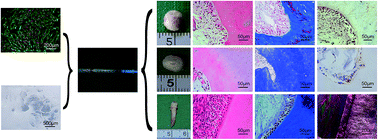
An injectable tissue engineering method was developed for dentin–pulp complex regeneration using an injectable scaffold, crosslinked hyaluronic acid gel (HAG). A cell–scaffold composite composed of HAG, tooth bud-derived dental mesenchymal cells (DMCs), and transforming growth factor-β1 (TGF-β1) was prepared and injected subcutaneously into nude mice. Moreover, β-tricalcium phosphate (β-TCP) and polyglycolic acid (PGA) were chosen as control scaffolds for dentin–pulp regeneration. The suitability of injectable HAG for dentin–pulp complex engineering was further demonstrated in empty tooth slices and the pulp chambers of mini pigs. Histological and immunohistochemical staining was carried out to identify the distinctive tubular dentin and pulp structure, which was further confirmed by the detection of several dentinogenesis-related genes, DSPP, DMP-1, MEPE, and BSP. It was found that a recognizable dentin–pulp-like tissue with a typical well-organized dentinal tubular structure, columnar odontoblast-like cells, was successfully engineered using the injectable HAG scaffold within the subcutaneous area of nude mice, according to histological staining. High expression of the genes DSPP, DMP-1, MEPE and BSP in the above neo-tissue as well as positive immunohistochemical staining for dentin sialoprotein (DSP) confirmed the dentinal characteristics. No typical dentin- or pulp-like tissue was formed when PGA or β-TCP were used as scaffolds. The efficacy of this method was further demonstrated in empty tooth slices and pulp chambers of mini pigs that had been pretreated by the removal of total pulp and partial dentin. Through the successful delivery of DMCs and TGF-β1 by the injectable HAG scaffold, the destroyed dentin was vividly repaired along with the formation of pulp-like tissue. Hence the current strategy to engineer dentin–pulp complex can overcome the difficulty of specific anatomical arrangement of pulp and dentin with minimal invasion, finally leading to regained vitality, which is difficult to realize by the current clinical treatments.
Introduction
Injuries, such as trauma, deep cavity preparation or severe caries, can possibly lead to irreversible pulpitis or necrosis of the dental pulp. Generally, a conventional clinical treatment protocol known as a root canal or endodontic therapy is indicated in such situations,1,2 in which the entire pulp is removed by pulpectomy and the pulp space is then disinfected and replaced with a rubber-like material, “gutta percha”.2 However, in spite of reported clinical success, endodontically treated teeth become devitalized, brittle and susceptible to postoperative fracture or other complications. The loss of pulp vitality in young permanent teeth terminates dentin formation and subsequent tooth maturation.3 Furthermore, pulpless teeth have lost defensive ability and have no sensation to irritation, resulting in caries progression unnoticed by patients and the subsequent extraction of the teeth. Long-term studies have revealed that tooth loss is higher for endodontically treated teeth than non-treated teeth because of secondary caries and complex restoration-associated problems.2,4 Therefore, it is of great importance to regenerate functional dentin–pulp complex to replace the lost pulp and dentin, in which situation the decayed tooth may possibly escape the fate of extraction.
Thanks to the boom and wide usage of tissue engineering technology, it seems that this could provide an attractive strategy for dentin and pulp regeneration.5–10 However, it is thought to be a tough challenge to regenerate the entire pulp and dentin in situ due to the anatomical arrangement of the pulp chamber, which is encased within the dentin and mainly relies on one apical foramen to allow angiogenesis for the engineered tissue.1,2,11 Therefore, in order to translate pulp regeneration to the clinical setting a rigid scaffold is not a good choice, constrained by the inherent character that these scaffolds cannot penetrate and adapt to the dentin walls throughout the entire root canal. Moreover, pulp is a type of soft tissue, while the surrounding dentin is relatively hard. Dentin–pulp complex is a structure containing both hard and soft tissues. Generally, scaffolds often used for soft tissue regeneration, such as polyglycolic acid (PGA) and collagen, are difficult to use for hard tissue engineering, whereas scaffolds such as β-tricalcium phosphate (β-TCP) and bioceramics, are mainly used for hard tissue regeneration, e.g. bone.9,12–17 Therefore, it is of great necessity to develop a suitable and injectable scaffold that can penetrate the entire root canal space1,4,5,8,18–22 and is also suitable for clinical application. Hyaluronic acid (HA) glycosaminoglycans present in the extracellular matrix of the dental pulp are important in maintaining morphologic organization by preserving extracellular spaces. They also contribute to the initial development of dental pulp and dentin.4,23,24 As is known, HA is biocompatible, biodegradable, bioactive, non-immunogenic and non-thrombogenic.25,26 Additionally, HA and its derivatives have been clinically used as medical products for over three decades.27 HA-based gels are usually prepared by crosslinking via different kinds of crosslinkers. 1,4-Butanediol diglycidyl ether (BDDE) is one of the commonly used crosslinkers. It has a significantly lower toxicity than other ether-bond crosslinking chemistry-based agents. Moreover, it is used in the majority of the market-leading HA fillers. The long safety record of BDDE-crosslinked HA gels had spanned more than 15 years, which makes them an industry standard, ahead of other crosslinkers such as divinyl sulfone and 2,7,8-diepoxyoctane.28 Hence, BDDE-crosslinked HA gels (HAGs) in the form of microparticles are proposed to be used as an injectable scaffold to regenerate dentin–pulp complex in this work. Tooth bud-derived dental mesenchymal cells (DMCs) were used as seed cells to be combined with HAG, since DMCs can differentiate into dentin-secreting odontoblasts and have previously been used as seed cells for dentin–pulp complex regeneration based on tooth developmental theory.2,29–36
In this work, HAG, utilized as an injectable scaffold, was combined with DMCs to form a cell–scaffold composite that was then transplanted subcutaneously into nude mice to regenerate dentin–pulp-like tissue. Within the above cell–scaffold composite, transforming growth factor-β1 (TGF-β1) was supplemented as it stimulates matrix secretion, initiates odontoblast cytodifferentiation in vitro and in vivo, and is also essential for the homeostasis of the dentin–pulp complex.37,38 The duration of in vivo construction was also varied to investigate the influence of time on the regenerated tissue. Moreover, β-TCP and PGA were chosen as control scaffolds as they are typical scaffolds for hard and soft tissue regeneration, respectively. The neo-regenerated tissues constructed from the above three scaffolds were compared macroscopically and microscopically. The suitability of the injectable HAG for dentin–pulp complex engineering was further demonstrated in empty tooth slices and in the pulp chambers of mini pigs.
Materials and methods
Culture of porcine DMCs
The surgical procedures were permitted by the Animal Care and Experimental Committee of Shanghai Jiao Tong University, School of Medicine. Tooth buds were isolated from the jaws of newborn pigs and washed with 0.25% chloramphenicol solution 3 times. The tooth bud tissue was then minced into <2 mm3 pieces, and enzymatically treated with 0.15% collagenase (NB4 Standard Grade, SERVA) and 0.5 U mL−1 dispase II (Neutral protease, Roche) in DMEM–F12 (1
:
1) (HyClone) with 15% fetal bovine serum (FBS) (HyClone) for 1.5 h at 37 °C. After that, the digested tooth bud tissues were strained with nylon filter (100 μm pores) and the harvested primary individual dental bud cells were cultured with DMEM–F12 (1
:
1) with 15% FBS and antibiotic/antimycotic solution (300/mL penicillin G, 300 μg mL−1 streptomycin, 0.75 μg mL−1 amphotericin B) (HyClone) in an incubator at 37 °C with 5% CO2.39 These cells were cultured for 5–7 days before reaching confluence and they formed a mixed population of epithelial- and fibroblast-like cells. The cells were then digested with 0.25% trypsin for 1–2 min, during which, dental mesenchymal cells (DMCs) were digested and most of the epithelial cells were still adherent to the plastic surface of the cell culture dish. The digested cells were further cultured in the above medium without any antibiotic/antimycotics. Due to the difference in the sensitivity to trypsin and medium requirement, the residual epithelial cells were gradually lost from the culture, leaving only the mesenchymal cells.40 DMCs of passage 3 were used for further experiments. For the following in vivo studies (nude mice), the cells of passage 3 were cultured with DMEM–F12 (1
:
1) medium supplemented with 10% FBS, 10 ng mL−1 TGF-β1 and 10 mM glycerol 2-phosphate disodium salt hydrate (β-GP) (Sigma).
Identification of cultured DMCs
DMCs of third passage were seeded onto cover slips and cultured to almost 80–90% confluence, and then the cells were fixed in 4% paraformaldehyde for 10 min. The subsequent steps for cell staining were performed according to the manufacturer’s instructions. Briefly, the primary antibody against vimentin, a marker for mesenchymes,41,42 is monoclonal anti-vimentin antibody produced in mice (Sigma), and the second antibody used was Alexa Fluor® 488 Goat Anti-Mouse IgG(H + L) (Invitrogen). Propidium iodide (PI) was used for counterstaining. The sample was then examined using a confocal laser scanning microscope (Nikon, A1).
Preparation of injectable HAG scaffold
HA was first dissolved in 1% NaOH at a concentration of 10 wt%, after which BDDE was added to the HA solution with a final concentration of 0.4 vol%. The solution was then allowed to crosslink at 40 °C for 5 h followed by being dried at room temperature for 3 days. Phosphate buffered saline (PBS
:
NaCl, 9 mg mL−1; KH2PO4, 0.03 mg mL−1; Na2HPO4·2H2O, 0.14 mg mL−1; pH = 7) of 500 mL was then added to the crosslinked HA to make it swell, after which the crosslinked HA was put into a dialysis bag and dialyzed sequentially with excessive deionized water and PBS to remove the residual BDDE. The resulting gel was adjusted with PBS to a concentration of 20 mg mL−1 and then smashed with a homogenizer to prepare gel particles of 0–400 μm. The yielded HAG was then sealed and stored at 4 °C for the standby application. Before being used in cell or animal experiments, HAG was sterilized in a high-pressure steam sterilizer set at 120 °C for 20 min.
Regeneration of dentin–pulp complex subcutaneously in nude mice using injectable HAG scaffold
The above cultured third passage DMCs were mixed with HAG scaffold (pre-incubated at 37 °C for 3 h) at a ratio of 5 × 10⁷ cells per mL scaffold, further supplemented with 1 μg mL−1 TGF-β1. The resulting cell–scaffold composite was immediately implanted subcutaneously into the dorsum of ∼5 week old male nude mice (experimental) for 7 or 10 weeks. At the same time, cell–scaffold complex without TGF-β1 (Control 1) or HAG scaffold only (Control 2) similarly injected into nude mice were used as controls. Each of the above three groups had ten 7 week samples and ten 10 week samples.
Moreover, in the experimental group, the cell concentration was varied (2.5 × 10⁷ cells per mL, 1 × 10⁷ cells per mL and 5 × 10⁶ cells per mL) to investigate its effect on subsequent neo-tissue regeneration. Each condition of these three concentrations had 4 repeated samples.
Preparation of β-TCP scaffold to engineer dentin–pulp complex subcutaneously in nude mice
The sterile β-TCP (Bio-lu, Shanghai, China) was cut to around 5 × 5 × 5 mm3 in size and then coated with pre-prepared 0.1% collagen I (containing 1 μg mL−1 TGF-β1) at 4 °C overnight, after which the coated scaffold was washed with DMEM–F12 (1
:
1) medium once and dried on a clean bench before use. The collagen solution for coating was prepared as follows: powder of collagen I from rat tail (C7661, Sigma) was dissolved in 0.1 M acetic acid and sterilized with chloroform at 4 °C overnight. The sterilized collagen I in the supernate was then gently removed to a sterile centrifugal tube and adjusted to a concentration of 1 mg mL−1 at pH 7.2. TGF-β1 was then added to this collagen solution at a concentration of 1 μg mL−1. The prepared collagen I solution was then stored at 4 °C for subsequent coating.
The above cultured third passage DMCs were then seeded onto this coated β-TCP scaffold with a cell density of 5 × 10⁷ cells per cm3. The composite was then placed in a 6-well plate. 1 mL of the TGF-β1-supplemented DMEM–F12 (1
:
1) medium was added into the well after being incubated for 4 h to allow cell attachment. The β-TCP composite was then implanted subcutaneously into the dorsum of ∼5 week old male nude mice for 7 or 10 weeks, respectively. At each time point, 4 samples were repeated.
Preparation of the PGA scaffold to engineer dentin–pulp complex subcutaneously in nude mice
As described previously,43 fifteen milligrams of non-woven PGA fibers (Shanghai Jurui Biomaterials Company Inc., Shanghai, China) were pressed into meshes approximately 9 mm in diameter and 1 mm in height. To improve its stability, PLA at a concentration of 0.5% in chloroform was added to the PGA mesh dropwise at a ratio of 10% of the total weight. The solvent was allowed to evaporate, and the resulting scaffolds were sterilized using 75% ethanol for 1 h and rinsed 3 times with PBS. Before cell seeding, the scaffolds were immersed in DMEM–F12 (1
:
1) medium overnight before use.43
DMCs of third passage were then seeded onto this stabilized PGA scaffold with a cell density of 5 × 10⁷ cells per cm3. The composite was then placed in a 6-well plate. 1 mL of the TGF-β1-supplemented DMEM–F12 (1
:
1) medium was added into the well after being incubated for 4 h to allow cell attachment. The PGA composite was then implanted subcutaneously into the dorsum of ∼5 week old male nude mice for 7 or 10 weeks. At each time point, 4 samples were repeated.
Regeneration of dentin–pulp complex within pre-treated tooth slices
Preparation of tooth slices. This study was approved by the ethic committee of Shanghai Ninth People's Hospital Affiliated Shanghai Jiao Tong University School of Medicine and informed consent from all of the patients was obtained. Noncarious human second premolars were collected from patients of the Oral Surgery Clinic in Shanghai Ninth People’s Hospital Affiliated Shanghai Jiao Tong University School of Medicine. The residual gingiva and periodontal tissues were removed with a scalpel, and the teeth were then cut at the cervical region with a high-speed turbine to obtain slices approximately 1 mm in thickness. The pulp was thoroughly removed with dental barbed broaches from the above slices. The resulting tooth slices were soaked in 75% ethanol for 1 h and washed with sterile Dulbecco’s phosphate buffered saline (D-PBS) (NaCl: 8.0 g L−1, KCl: 0.2 g L−1, KH2PO4: 0.2 g L−1, Na2HPO4(anhydrous): 1.15 g L−1, pH = 7.2) (Sigma-Aldrich) several times. After that, the tooth slices were preincubated with DMEM–F12 (1
:
1) medium with 15% FBS in an incubator set at 37 °C and 5% CO2 for several hours before subsequent use.DMCs of third passage were mixed with HAG scaffold (pre-incubated at 37 °C for 3 h) at a ratio of 5 × 10⁷ cells per mL scaffold, further supplemented with 1 μg mL−1 TGF-β1. This cell–scaffold composite was first injected into the empty cavity of the prepared tooth slice, and the resulting slice was then implanted subcutaneously into nude mice for 10 weeks. For this tooth slice model, five samples were repeated.
Regeneration of dentin–pulp complex in porcine dental pulp cavity
For dentin–pulp regeneration in mini pigs, the unerupted permanent molar tooth buds of 3 month old mini pigs were surgically removed from the mandibles, and then digested and cultured according to the method mentioned above. For the following in vivo studies, the obtained DMCs of third passage were cultured with DMEM–F12 (1
:
1) medium supplemented with 10% FBS, 10 ng mL−1 TGF-β1 and 10 mM β-GP.
Treatment of porcine dental pulp cavity: after removal of unerupted permanent tooth buds, the same 3 month old pigs were anesthetized with 0.25% pentobarbital sodium. Dental pulp of the second and third premolars in the mandible was removed with dental barbed broaches. The tooth canal space was enlarged and the cavity was temporarily sealed with hydraulic temporary restorative (GC, Japan) according to routine clinical procedures.
One week after the above dental pulp cavity treatment, the third passage DMCs were mixed with HAG scaffold (pre-incubated at 37 °C for 3 h) at a ratio of 5 × 10⁷ cells per mL scaffold, further supplemented with 1 μg mL−1 TGF-β1. For the experimental group in this animal study, the cell–scaffold composite was then injected into the empty porcine dental pulp cavity. Immediately before the injection, the temporary sealing was removed and the pulp cavity was thoroughly washed with 5.25% NaClO, 3% H2O2 and sterile 0.9% NaCl in sequence. The prepared cell–scaffold composite was then injected into the pulp cavity and the hole was sealed with mineral trioxide aggregate (MTA) and glass ionomer cement (GC, Japan) sequentially. Moreover, clinical and blank controls were also conducted. The empty dental pulp cavity was similarly treated except it was filled with gutta percha points, which served as a control for clinical treatment (Clinical control). The empty dental pulp cavity was also similarly treated, except that the cavity was left empty as a blank control. All the pigs were normally raised after the above surgeries. After one month, the above implants were harvested for the following histological analysis. For this mini pig model, each of the three groups (EXP, Clinical and Blank) had 5 samples repeated.
Histology and immunohistochemical analysis
When it was time to harvest the samples, the animals were treated with euthanasia. The samples were then removed and washed with PBS, followed by being fixed in 4% paraformaldehyde at 4 °C for 24 h, and demineralized with 10% EDTA at room temperature for several months. The samples were then processed into paraffin sections, and the sections (5 μm thick) were treated with H&E, Masson (MAIXIN.BIO) and immunohistochemical staining. The primary antibody used for the immunohistochemical analysis was rabbit anti-human dentin sialoprotein (DSP) polyclonal antibody (H-300) (1
:
100 dilution) (Santa Cruz). The second antibody used was Envision + system-HRP-labeled polymer anti-rabbit (Dako), and liquid DAB + substrate chromogen system (Dako) was used as well.
Quantitative real-time reverse transcription polymerase chain reaction (RT-PCR)
To detect the expression of dentinogenesis-related genes DSPP (dentin sialophosphoprotein), DMP-1 (dentin matrix protein 1), MEPE (matrix extracellular phosphoglycoprotein), and BSP (bone sialoprotein), samples of the experimental group (DMC + HAG + TGF-β1) and Control group 1 (DMC + HAG) that were subcutaneously constructed in nude mice for 10 weeks were harvested. The respective total RNA was extracted using TRIzol® Reagent (Invitrogen, USA) from the samples, reverse-transcribed and amplified using a RevertAid First Strand cDNA Synthesis Kit (Thermo) in a 20 μL reaction mixture containing 1 μL RNAase inhibitor, 1 μL reverse transcriptase, 1 μL oligo(dT)15, 2 μL 10 mM dNTPs, buffer and RNAase-free water. The PCR primers used here were purchased form Invitrogen Biotechnology Co., LTD. The detailed primer sequences are shown in Table 1. The expression of β-actin mRNA was used in all real-time PCR reactions as an internal control. The RT-PCR assay was performed using a 7300 Real Time PCR System (Applied Biosystems) with the following profile: 95 °C for 10 min, then cycling for 40 cycles at 95 °C for 15 s and 60 °C for 60 s, and then 60 °C for 5 min. The relative expression of each mRNA was calculated by the ΔCt method. ΔCt is the value obtained by subtracting the Ct value of β-actin mRNA from the Ct value of the target mRNA.44 The experiments were repeated three times and the values shown are the mean of 3 individual samples (n = 3). Data are presented as mean ± SD.
Table 1 Primer sequences used in quantitative real-time RT-PCR
Gene |
Primers |
Length |
DSPP (NM–213777.1) |
Forward: 5′-ATAGAGGACACCCAGAAACCCA-3′ |
295 |
Reverse: 5′-GTCCAGGCTTATTTCCGGGT-3′ |
DMP-1 (NM–001129953.1) |
Forward: 5′-TGAGCAGGACAGCCCATCTG-3′ |
212 |
Reverse: 5′-AGTAGCCGTCCTGGCAGTCATT-3′ |
MEPE (XM–003129339) |
Forward: 5′-ACAGAGTTTTCCAGCCCAAGT-3′ |
115 |
Reverse: 5′-CCCTGGTTCCAATGGTATCTC-3′ |
BSP (XM–003129337.1) |
Forward: 5′-ACAAGCACGCCTACTTCTACCC-3′ |
274 |
Reverse: 5′-TGGAGGGCAGCGAGACCTAT-3′ |
ACTB (XM–003124280) |
Forward: 5′-TTCGAGACCTTCAACACCC-3′ |
201 |
Reverse: 5′-CATGAGGTAGTCGGTCAGGT-3′ |
Morphology of DMCs and injectable HAG scaffold
The cultured DMCs exhibited a typical spindle-shaped appearance, as shown in Fig. 1A. Moreover, these cells were positive for vimentin, which is a characteristic protein of mesenchymal cells (Fig. 1B). The sizes of the prepared HAG particles were in the range of 0–400 μm (Fig. 1C), and these gels in the form of irregular microparticles could be easily injected via a syringe (30G), as shown in Fig. 1D.
 |
| Fig. 1 Microscopic observation of DMCs and injectable HAG scaffold. Optical microscopic observation of DMCs (A) and immunofluorescent staining of DMCs for vimentin (B). Optical microscopic observation of HAG microparticles (C) that could be injected (D) via a syringe (30G). Scale bars: 500 μm in A and C, 200 μm in B. | |
Regeneration of dentin–pulp complex subcutaneously in nude mice
Gross views of samples of the experimental group that had been constructed subcutaneously in nude mice for 7 and 10 weeks are shown in Fig. 2A and D, respectively. Grossly, the engineered tissues of the experimental group were highly mineralized both at 7 and 10 weeks. Therefore, before they were further subjected to histological staining, demineralization was necessary for obtaining paraffin slices. The implanted samples of Control group 1 (briefed as DMC + HAG) in nude mice for 7 and 10 weeks are shown in Fig. 2B and E. From the gross views (Fig. 2B and E), these samples were also mineralized and the mineralization degree was enhanced with time from 7 weeks to 10 weeks. The samples obtained from pristine HAG scaffold (Control 2, briefed as HAG) after 7 and 10 weeks subcutaneous implantation in nude mice are shown in Fig. 2C and F. The harvested samples of this Control group 2 were soft and transparent. A high water content could be seen in the samples for Control group 2.
 |
| Fig. 2 Gross views of regenerated constructs using HAG as a scaffold. Gross view of regenerated tissue constructed subcutaneously with DMC (5 × 10⁷ cells per mL), HAG and TGF-β1 (1 μg mL−1) (experimental group, indicated as EXP) in nude mice for 7 (A) or 10 weeks (D). Gross view of the subcutaneously implanted constructs using DMC (5 × 10⁷ cells per mL) and HAG (Control group 1, indicated as DMC + HAG) in nude mice for 7 (B) or 10 weeks (E). Gross view of the pristine HAG scaffold (Control group 2, indicated as HAG) subcutaneously implanted in nude mice for 7 (C) or 10 weeks (F). | |
Overall views of H&E and Masson staining of the regenerated tissue of the experimental group of 7 weeks are shown in Fig. 3A and D, respectively. The whole sample exhibited island-like features. Within localized areas at a higher magnification (Fig. 3B, C, E and F), there were well-organized dentinal tubules (white arrow heads), columnar odontoblast-like cells (yellow arrow heads) with polarized basal nuclei and blood vessels (black arrow heads). Those cells with polarized basal nuclei were aligned against the regenerated dentin-like tissue, while the dentinal tubules (white arrow heads) were arranged radially from the pulp-like tissue.
 |
| Fig. 3 Histological analysis of the regenerated dentin–pulp complex-like tissue (EXP) constructed subcutaneously in nude mice for 7 weeks. Overall view of H&E (A) and Masson (D) staining of the regenerated tissue of the EXP group. Island-like features can be seen in A and D. B, C, E, and F are higher-magnification observations of the localized square areas within A and D, respectively. It can be seen in B, C, E, and F that in the regenerated construct there are well-organized dentinal tubules (white arrow heads), columnar odontoblast-like cells (yellow arrow heads) with polarized basal nuclei and blood vessels (black arrow heads). Scale bars: 1000 μm for A, D; 50 μm for B, C, E, F. | |
No typical dentin- or pulp-like tissue can be seen in either Control group 1 (Fig. 4A–C) or 2 (Fig. 4D–F). For both groups, HAG (green arrow heads) still occupies most areas, but most of the HAG fragments would be missed during the H&E processing. A few bone-like hard tissues could be located within the DMC + HAG sample (Fig. 4B), whereas only fibrous soft tissue could be identified encapsulating the sample of the HAG group (Fig. 4E and F).
 |
| Fig. 4 Histological analysis of the DMC + HAG (A–C) and HAG (D–F) subcutaneously implanted in nude mice for 7 weeks. No typical dentin- or pulp-like tissue can be seen in either control group. For both groups, HAG (green arrow heads) still occupies most areas and most of the HAG fragments are missed during the H&E processing. A few bone-like hard tissues can be seen within the DMC + HAG sample, whereas only fibrous soft tissue can be identified surrounding the sample of the HAG group. Scale bars: 1000 μm in A, D; 50 μm in B, C, D, E. | |
After 10 weeks, the regenerated tissue of the experimental group exhibited a more mature dentin–pulp-like morphology, as shown in Fig. 5, compared to that of 7 weeks (Fig. 3). From the overall views of H&E (Fig. 5A) and Masson (Fig. 5D) staining of the regenerated tissue, it can be seen that the formed typical island-like tissues are quite homogeneous throughout the whole sample. Moreover, as shown in Fig. 5B, C, E, and F (higher-magnification observations of the localized square areas within A and D), in the regenerated construct there are well-organized dentinal tubules (white arrow heads) arranged radially along the pulp-like tissue and columnar odontoblast-like cells with polarized basal nuclei (yellow arrow heads) juxtaposed along the dentinal wall. Blood vessels could be observed with good distribution within the pulp-like soft tissue (black arrow heads).
 |
| Fig. 5 Histological analysis of the regenerated dentin–pulp complex-like tissue (EXP) constructed subcutaneously in nude mice for 10 weeks. Overall view of H&E (A) and Masson (D) staining of the regenerated tissue of the EXP group. The tissues had typical island-like features in A and D. B, C, E, and F are higher-magnification observations of the localized square areas within A and D. It is shown in B, C, E, and F that in the regenerated construct there are well-organized dentinal tubules (white arrow heads) arranged radially along the pulp-like tissue and columnar odontoblast-like cells with polarized basal nuclei (yellow arrow heads) lining up along the dentinal wall. Blood vessels are distributed throughout the pulp-like soft tissue (black arrow heads). Scale bars: 1000 μm for A, D; 50 μm for B, C, E, F. | |
As for the control groups after 10 weeks (Fig. 6A and D), similar conditions were observed as those after 7 weeks. A relatively high density of cells could still be observed in the samples of Control group 1 (Fig. 6B and C), whereas only residues of HAG scaffolds could be located with fibrous encapsulation at the edge of the implants (Fig. 6E and F).
 |
| Fig. 6 Histological analysis of the DMC + HAG (A–C) and HAG (D–F) subcutaneously implanted in nude mice for 10 weeks. No typical dentin- or pulp-like tissue can be seen in either control group. Quite a few bone-like hard tissues can be seen within the DMC + HAG sample, whereas only fibrous soft tissue can be identified surrounding the sample of the HAG group. HAG fragments (green arrow heads) still occupy most areas of the HAG group. Scale bars: 1000 μm in A, D; 50 μm in B, C, D, E. | |
Based on the above results, immunohistochemical staining for DSP was carried out on the constructs of the experimental group that were implanted for 10 weeks. The results are shown in Fig. 7. Positive staining for DSP throughout the whole structure, especially within the area of soft tissue (Fig. 7A) can be ascertained. From the higher-magnification views (Fig. 7A1 and A2), positive DSP expression by odontoblast-like cells (yellow arrow heads) and dentinal tubules (white arrow heads) can be confirmed. Negative immunohistochemical staining for Fig. 7A, A1 and A2 is shown in Fig. 7B, B1 and B2, respectively, obtained by similar processing except without the use of antibodies, further confirming the specific staining. Moreover, positive control staining was carried out on slices obtained from a normal tooth of a 6 month old pig, as shown in Fig. 7C (Fig. 7D is the respective negative staining for C), exhibiting positive staining for DSP in the pulp and dentinal tubules. The normal mature dentin–pulp structure and the regenerated tissue (experimental group in this study) bear great similarity in their microstructures and tissue distribution.
 |
| Fig. 7 Immunohistochemical analysis of the regenerated dentin–pulp complex-like tissue constructed subcutaneously in nude mice for 10 weeks. (A) Overview of the immunohistochemical staining for dentin sialoprotein (DSP) in the tissue and DSP positive staining can be observed throughout the whole structure, especially within the area of soft tissue. (A1 and A2) are higher-magnification observations of the localized square areas within (A) and (A1), respectively, further confirming the positive DSP expression by odontoblast-like cells (yellow arrow heads) and dentinal tubules (white arrow heads). (B, B1 and B2) are negative controls of A, A1, and A2 respectively, obtained by similar processing except without the use of antibodies. (C) is a slice from a normal tooth of a 6 month old pig, exhibiting positive staining of DSP in the pulp and dentinal tubules as a positive immunostaining control. (D) is negative staining for C. Scale bars: 1000 μm for A and B; 100 μm for A1 and B1; 50 μm for A2, B2, C, and D. | |
A few odonto-osteogenic and dentinogenesis-related markers (DSPP, DMP-1, MEPE and BSP) were examined by RT-PCR, and the results are shown in Fig. 8. The expression of DSPP, DMP-1, MEPE and BSP in the experimental group (EXP) was highly elevated compared to those of Control group 1 (p < 0.0001 for each gene), confirming the odontoblastic differentiation of DMCs and dentin formation within the regenerated tissue of the EXP group.
 |
| Fig. 8 Relative mRNA expression of DSPP, DMP-1, MEPE and BSP by RT-PCR in the experimental group (DMC + HAG + TGF-β1) and Control group 1 (DMC + HAG). EXP represents the experimental group, the samples for which were constructed subcutaneously with DMC (5 × 10⁷ cells per mL), HAG and TGF-β1 (1 μg mL−1) in nude mice for 10 weeks. CTR represents Control group 1, which was constructed subcutaneously with DMC (5 × 10⁷ cells per mL) and HAG in nude mice for 10 weeks. The experiments were repeated three times and the values are the mean of 3 individual samples (n = 3). Data are presented as mean ± SD. | |
The cell concentration was varied from the original 5 × 10⁷ cells per mL to 2.5 × 10⁷ cells per mL, 1 × 10⁷ cells per mL and 5 × 10⁶ cells per mL so as to investigate the effect of cell density within the HAG scaffold on the morphology of the neo-formed tissue of the experimental group. Fig. 9 shows the gross view, histological and immunohistochemical analysis of regenerated tissue constructed subcutaneously in nude mice for 10 weeks with the minimum concentration used (5 × 10⁶ cells per mL). Compared with those constructed from 5 × 10⁷ cells per mL (Fig. 2D and 5A, D), only sporadically-distributed tissue was regenerated within the whole sample (Fig. 9A, B and D). In the higher-magnification observations (Fig. 9C and E) of the localized square areas within Fig. 9B and D, dentinal tubules (white arrow heads) and columnar odontoblast-like cells (yellow arrow heads) could also be identified along the island-like structure, despite their relatively lower density. Moreover, the presence of blood vessels could be ascertained (black arrow heads) as well. According to the immunohistochemical analysis (Fig. 9F), positive DSP expression by the polarized odontoblast-like cells (yellow arrow heads) and dentinal tubules (white arrow heads) in the regenerated tissue could be observed. From the rest of the samples constructed from 2.5 × 10⁷ cells per mL and 1 × 10⁷ cells per mL similar conditions were observed, in that the microstructures of the neo-tissues had similar features to those constructed from 5 × 10⁷ cells per mL. However, the quantity of expected tissue decreased with the reduction in cell density (data not shown).
 |
| Fig. 9 Gross view, histological and immunohistochemical analysis of regenerated tissue constructed subcutaneously with DMC (5 × 10⁶ cells per mL), HAG and TGF-β1 (1 μg mL−1) in nude mice for 10 weeks. (A) Gross view of the constructed sample, and overall view of H&E (B) and Masson (D) staining of the sample, showing the sporadic tissue regeneration morphology within the whole construct. (C and E) are higher-magnification observations of the localized square areas within B and D. A high cell concentration, dentinal tubules (white arrow heads) and columnar odontoblast-like cells (yellow arrow heads) could also be identified along the island-like structures. Moreover, the presence of blood vessels could be ascertained (black arrow heads). According to the immunohistochemical analysis (F), positive but relatively weak DSP expression by the polarized odontoblast-like cells (yellow arrow heads) and dentinal tubules (white arrow heads) in the regenerated tissue could be observed. Scale bars: 1000 μm in B and D; 50 μm for C, E, and F. | |
To further verify the suitability of HAG as a scaffold for dentin–pulp regeneration, two commonly used scaffolds, β-TCP and PGA were applied as scaffold controls. The gross view, histological and immunohistochemical analysis of regenerated tissue constructed with DMC (5 × 10⁷ cell per mL), TGF-β1 (1 μg mL−1) and β-TCP as a scaffold in a mouse subcutaneous model are shown in Fig. 10. Grossly, most of the constructs at either 7 or 10 weeks were still β-TCP scaffolds. The gross shape of the constructs remained the same as that of the original scaffold (Fig. 10A and B). Microscopically, it is shown that no observable pulp- or dentin-like tissue could be identified within this construct from H&E (Fig. 10A1) and Masson staining (Fig. 10A2) except that weak staining for DSP around the cells within the construct was shown from immunohistochemical staining (Fig. 10A3). Even with the increase in in vivo time, the tissue still did not undergo obvious remodeling into the dentin–pulp-like morphology (Fig. 10B1–B3), as observed from the HAG constructed samples (Fig. 5A–F).
 |
| Fig. 10 Gross view, histological and immunohistochemical analysis of regenerated tissue constructed with DMC (5 × 10⁷ cells per mL), TGF-β1 (1 μg mL−1) and β-TCP as a scaffold in a mouse subcutaneous model. Gross view of the regenerated tissue after being implanted in nude mice for 7 (A) and 10 (B) weeks. The insets in A and B are respective overall H&E staining of A and B. H&E staining of the formed tissue using β-TCP as a scaffold after being constructed for 7 (A1) and 10 (B1) weeks. Masson staining of the formed tissue after being constructed for 7 (A2) and 10 (B2) weeks. Immunohistochemical staining for DSP of the formed tissue after being constructed for 7 (A3) and 10 (B3) weeks. No observable pulp- or dentin-like tissue could be identified within this construct, and weak staining for DSP around the cells within the construct is shown. Scale bars: 50 μm for A1–A3 and B1–B3. | |
The gross view, histological and immunohistochemical analysis of regenerated tissue constructed with DMC (5 × 10⁷ cell per mL), TGF-β1 (1 μg mL−1) and PGA as a scaffold in a mouse subcutaneous model are shown in Fig. 11. Grossly, PGA fibers could still be observed within the construct at 7 weeks (Fig. 11A). When it came to 10 weeks, it seemed that most of PGA fibers were degraded, but the tissue was quite soft (Fig. 11B). Microscopically, similar to those observed from Fig. 10 using β-TCP as a scaffold, no typical pulp- or dentin-like tissue could be observed within this construct except weak staining for DSP both at 7 (Fig. 11A1–A3) and 10 weeks (Fig. 11B1–B3). Interestingly, the tissue microstructure varied with time. At 7 weeks, compact and hard bone-like tissue was formed and surrounded by fibrous soft tissue entrapped with a high density of cells (Fig. 11A1–A3), whereas at 10 weeks the previous hard tissue became relatively soft with a cartilage-like appearance in both tissue features and cell distribution.
 |
| Fig. 11 Gross view, histological and immunohistochemical analysis of regenerated tissue constructed with DMC (5 × 10⁷ cells per mL), TGF-β1 (1 μg mL−1) and PGA as a scaffold in a mouse subcutaneous model. Gross view of the regenerated tissue after being implanted in nude mice for 7 (A) and 10 (B) weeks. The insets in A and B are respective overall H&E staining of A and B. H&E staining of the formed tissue using PGA as a scaffold after being constructed for 7 (A1) and 10 (B1) weeks. Masson staining of the formed tissue after being constructed for 7 (A2) and 10 (B2) weeks. Immunohistochemical staining for DSP of the formed tissue after being constructed for 7 (A3) and 10 (B3) weeks. No typical pulp- or dentin-like tissue could be observed within this construct except weak staining for DSP. Scale bars: 50 μm for A1–A3 and B1–B3. | |
Regeneration of dentin–pulp complex subcutaneously in tooth slice model
To further investigate the effect of microenvironment on tissue regeneration, the cell scaffold composite (DMC + HAG + TGF-β1) was injected into a pre-prepared tooth slice and then implanted into the subcutaneous area of a nude mouse, as shown in Fig. 12A–C. After being implanted for 10 weeks, the tooth slice was harvested (Fig. 12D). Grossly, the empty pulp chamber of the tooth slice was filled with regenerated tissue, which was further confirmed by H&E (Fig. 12E) and Masson (Fig. 12F) staining. The regenerated tissue had dentinal tubules (white arrow heads) arranged radially along the pulp chamber (Fig. 13A1 and B1). The neo-dentin-like tissue was consistent with the original dentin. Columnar odontoblast-like cells (yellow arrow heads) with polarized basal nuclei were aligned in a parallel manner along the neo-dentin-like tissue, with blood vessels distributed throughout the construct (black arrow heads) (Fig. 13A1, A2, B1 and B2). Positive expression of DSP by polarized odontoblast-like cells (yellow arrow heads) and dentinal tubules (white arrow heads) could be observed (Fig. 13C1 and C2).
 |
| Fig. 12 The process of dentin–pulp complex-like tissue regeneration in a tooth slice model. (A) The prepared tooth slice with an empty pulp chamber. (B) The pulp chamber of the prepared tooth slice was filled with the cell–scaffold composite (5 × 10⁷ cells per mL DMC, HAG and 1 μg mL−1 TGF-β1). (C) Implantation of the tooth slice subcutaneously into nude mice. (D) The harvested tooth slice after being implanted for 10 weeks. Overall views of H&E (E) and Masson (F) staining of the harvested tooth slice. Scale bars: 1000 μm. | |
 |
| Fig. 13 Histological and immunohistochemical analysis of the tissue regenerated in the pulp chamber of the tooth slice model. H&E (A1 and A2) and Masson (B1 and B2) staining of the regenerated tissue in the pulp chamber of the empty tooth slice. The original empty chamber was filled with regenerated dentin pulp-like tissue where dentinal tubules (white arrow heads) were arranged radially along the pulp chamber. The neo-dentin-like tissue was consistent with the original dentin. Columnar odontoblast-like cells (yellow arrow heads) with polarized basal nuclei were aligned in a parallel manner along the neo-dentin-like tissue, with blood vessels distributed throughout the construct (black arrow heads). (C1 and C2) Immunohistochemical staining for DSP of the regenerated dentin–pulp-like tissue. Positive expression of DSP by polarized odontoblast-like cells (yellow arrow heads) and regenerated dentinal tubules (white arrow heads) could be observed. There were also HAG fragments remaining within the pulp tissue (green arrow heads). Scale bars: 50 μm. | |
Regeneration of dentin–pulp complex in porcine dental pulp cavity
In order to investigate whether the current injectable scaffold met the clinical requirements, the cell scaffold composite (DMC + HAG + TGF-β1) was finally injected into the empty tooth pulp chamber of a mini pig. After one month, the tooth with the injected composite was retrieved, as shown in Fig. 14A and A1. From the higher-magnification H&E (A2, A3) and Masson (A4) observations of the regenerated tissue of this EXP group, multiple layers of columnar odontoblast-like cells (yellow arrow heads) with polarized basal nuclei can be seen well aligned against the regenerated dentin-like tissue. Vivid regeneration of the neo-dentin structure (white arrows) can also be seen, and the neo-tissue is consistent with the original dentin. The tubule morphology in the neo-dentinal structure can be observed clearly (white arrow heads, Fig. 14A4). The distribution of blood vessels (black arrow heads) in the pulp tissue can be observed (Fig. 14A2–A4). As a clinical control, gutta percha points were inserted into the similarly pre-prepared empty pulp chambers of mini pigs and also constructed for one month (Fig. 14B). However, the original dentin remained destroyed or was still in the process of degradation according to the H&E (Fig. 14B1–B3) and Masson (Fig. 14B4) staining. No dentin- or pulp-like tissue was regenerated in this chamber. As for the blank control group (Fig. 14C), in which the empty pulp chamber after removal of pulp tissue via routine clinical procedures was left untreated, no tissue regeneration was seen in the chamber from the overall histological morphology (Fig. 14C1), higher-magnification H&E (Fig. 14C2 and C3) and Masson (Fig. 14C4) observations.
 |
| Fig. 14 Regeneration of dentin–pulp complex in the empty tooth pulp chambers of mini pigs. Gross view (A) and overall histological morphology (A1) of a mini pig tooth implanted with 5 × 10⁷ cells per mL of autologous DMC, HAG and 1 μg mL−1 of TGF-β1 (EXP) for one month, by injection into the pulp chamber. Higher-magnification H&E (A2 and A3) and Masson (A4) observations of the regenerated tissue of the EXP group. Multiple layers of columnar odontoblast-like cells (yellow arrow heads) with polarized basal nuclei are well aligned against the regenerated dentin-like tissue. Vivid regeneration of the neo-dentin structure can be seen and the neo-tissue is consistent with the original dentin (white arrows). The distribution of blood vessels (black arrow heads) in the pulp tissue can be observed. Gross view (B) and overall H&E morphology (B1) of a mini pig tooth implanted with gutta percha points (as a clinical control) for one month. Higher-magnification histological (B2 and B3) and Masson (B4) observations of the tooth of the clinical control group. No dentin- or pulp-like tissue was regenerated in this chamber, and the original dentin remained destroyed or was still in the process of degradation. Gross view (C) and overall H&E morphology (C1) of the tooth with its empty pulp chamber left untreated (blank control). Higher-magnification H&E (C2 and C3) and Masson (C4) observations of the tooth of the blank control group. Similar to those of the clinical control group, nothing was regenerated in the pulp chamber. Scale bars: 1000 μm in A1, B1, C1; 50 μm in A2–A4, B2–B4, C2–C4. | |
The polarized histological morphology of the regenerated dentin–pulp complex in the empty tooth pulp chamber of a mini pig is shown in Fig. 15. Fig. 15A and D are two representative images, while B, C, E, and F are images at higher magnifications. From Fig. 15A and D, we can further confirm the vivid regeneration of the neo-dentin structure. The neo-dentinal tissue integrated well with the original dentin, but has a distinctive polarized morphology. From the higher magnification images (B, C, E, and F), the tubule morphology in the neo-dentinal structure has similar organization and distribution to that in the original dentin.
 |
| Fig. 15 Polarized histological morphology of the regenerated dentin–pulp complex in the empty tooth pulp chamber of a mini pig. The regenerated tissue is of a mini pig tooth implanted with 5 × 10⁷ cells per mL of autologous DMC, HAG and 1 μg mL−1 of TGF-β1 (EXP) for one month, by injection into the pulp chamber. B, C, E, and F are higher-magnification polarized H&E observations of A and D, respectively. Scale bars: 200 μm for A and D; 100 μm for B and E; 50 μm for C and F. | |
Discussion
In this report, an injectable tissue engineering method was developed for dentin–pulp complex structure regeneration via the application of an injectable scaffold, HAG. It was then demonstrated that dentin–pulp-like tissue with typical well-organized dentinal tubular structures and columnar odontoblast-like cells were successfully engineered within the subcutaneous area of nude mice using HAG, DMCs and TGF-β1. It was also found that with time, from 7 to 10 weeks, the neo-tissue was remodeled and further organized. A sufficient number of DMCs is necessary for the formation of the desired tissue, as illustrated by experiments reducing the cell density to 5 × 10⁶ cells per mL scaffold. The efficacy of this injectable tissue engineering method was also demonstrated in empty tooth slices and the pulp chambers of mini pigs that had been pre-treated by thoroughly removing the pulp using dental barbed broaches. Not only vivid growth of neo-dentin and pulp like tissue was realized, but also well integrity with the original dentin was achieved. Hence, the suitability of an injectable scaffold, HAG, for dentin–pulp complex engineering was ascertained, and its superiority for easy penetration and delivery of DMCs and TGF-β1 into the whole pulp canal with minimal invasion over traditional β-TCP and PGA scaffolds with rigid shapes was substantiated. Over the past decades a lot of research effort has been put into dentin–pulp regeneration. However, the dream to engineer the whole pulp has been hampered by the specific anatomical arrangement of pulp and dentin, which can be overcome by the current use of an injectable scaffold, HAG.
In addition to the specific anatomical and clinical requirements that the injectable HAG scaffold meets, the suitability of HAG further lies in its appropriate degradation rate. Based on the histological observation of the experimental group in nude mice, it seems that most of the HAG fragments were already gone, whereas the majority still remained in the control group (HAG only). One indication is that the HAG was completely degraded at a speed keeping pace with the rate of dentin–pulp complex regeneration. The other is that HAG could possibly have the flexibility to adjust its degradation in different environments. That is, cells or extracellular matrix within the HAG scaffold could easily help to adjust its turnover, probably due to the biological nature of HA. What’s more, the degradation product of HAG is HA of different molecular weights. As one type of glycosaminoglycan present in the extracellular matrix, HA was shown to be beneficial to maintain morphologic organization by preserving extracellular space, and was reported to contribute to the initial development of dentin matrix and dental pulp.4,23 Hence, most probably, the degradation products of HA might also facilitate the matrix formation process. As for the two commonly used scaffolds, β-TCP and PGA, no typical dentin- or pulp-like tissue regeneration was observed subcutaneously in nude mice after 7 or 10 weeks. One possible reason for this might be due to their different chemical properties. As is known, when PGA degrades, the acid product might influence the metabolism of the cells in the construct and may thereby hamper the regeneration of dentin–pulp complex under the same circumstances. β-TCP has osteoinductive properties and was shown to be beneficial for bone regeneration,6 but may not be appropriate for dentin–pulp regeneration as the pulp is a type of soft tissue. According to our results, even at 10 weeks subcutaneously, β-TCP still retained its original structure. Such a low degradation rate under such circumstances could be one of the main reasons that hampered the neo-tissue regeneration and reorganization.
DMCs are derived from ectomesenchyme during tooth development, and these cells have the ability to differentiate into dentin-secreting odontoblasts.2,45–47 They can also form dental pulp-like tissue with good vascularization, which is important in the regulation of inflammation, subsequent regeneration of pulp and dentin, and in sustaining the high metabolic demands of odontoblastic cells during active processes of dentin matrix secretion.48,49 Therefore, tooth bud cells have been widely used for tooth development and regeneration research.7,50–53 In this study, DMCs from newborn pigs or from unerupted permanent tooth buds of 3 month old mini pigs were used as seed cells for dentin–pulp regeneration in the subcutaneous area of nude mice or the empty pulp chambers of pigs, respectively. However, no detailed characterization of these cells was carried out to identify their differentiation statuses along with the neo-tissue formation, thereby monitoring their fate and exact roles. This will be one of the next issues to be explored. In order to bridge the gap between research and clinical applications, alternative seed cells should also be developed, such as postnatal dental pulp stem cells (DPSCs), stem cells from human exfoliated deciduous teeth (SHED), stem cells from apical papilla (SCAP), bone marrow-derived mesenchymal stem cells (BMSCs) and adipose-derived stem cells (ASCs).2,54–58
It was shown that the TGF-β family plays an important role in tooth development. Among which, TGF-β1 was shown to promote odontoblast differentiation and the subsequent secretion of extracellular matrix components. It also enhances the formation of reparative dentin and is essential for the homeostasis of the dentin–pulp complex.37,38,59,60 Moreover, as reported, TGF-β1 is crucial in the terminal odontoblast differentiation of pulp cells in vitro.61 Due to its important role in tooth morphogenesis, TGF-β1 was taken as a morphogen in this report to promote dentin–pulp complex regeneration. As confirmed by current results, well-vascularized dentin–pulp complex regeneration in the nude mice model of the experimental group (DMCs + HAG + TGF-β1) was achieved, whereas no dentin or pulp, but only bone-like tissue formation was observed in the control group (DMC + HAG) without any TGF-β1.
The gene expression levels of DSPP, DMP-1, MEPE and BSP, indicative of odontoblast differentiation and dentin–pulp regeneration,5,6,33,62 of the subcutaneously regenerated dentin–pulp-like tissue were assessed. DSPP is one of the key non-collagenous proteins involved in tooth development and is critical for dentin mineralization.63–65 DMP-1, involved in the initial phase of mineralized dentin formation, was shown to induce the undifferentiated pulp cells into matrix-synthesizing cells and to stimulate the formation of mineralized tissues.66,67 MEPE, an extracellular matrix protein that is mainly expressed in mineralized tissues and dental pulp, plays an important role not only in dentinogenesis but also in the physiology of pulp homeostasis.68,69 BSP was reported to stimulate the differentiation of dental pulp cells into odontoblast-like cells and to stimulate regenerative dentin formation.70–72 Quantitatively, their expression was remarkably enhanced in the experimental group. This further helps to confirm the nature of regenerated dentin- and pulp-like tissue, in addition to the observed characteristic tubular dentin morphology, which usually serves as a hallmark of odontoblasts73 as well as the positive immunostaining for DSP.
In this work, three different microenvironments were tested to assess the ability of HAG scaffolds to form dentin–pulp structure and to assess their preliminary clinical suitability. Firstly, the subcutaneous area is usually used to make initial examinations of cell- and tissue-based products in vivo because it offers simple transplantation surgeries compared with others and is easier to access.74 The nude mouse subcutaneous model has also been widely used for partial tooth tissue regeneration, such as dentin, pulp and dentin–pulp complex regeneration.75–77 Hence, in this report, we initially tried to substantiate our hypothesis that HAG could be used as an injectable scaffold to engineer dentin–pulp complex. To make the microenvironment closer to that of the original dentin–pulp, pre-treated tooth slices were analysed. The tooth slice model has been used for several studies, such as for cytotoxicity assessment of dental materials,78 and for the study of dentinogenesis and human dental pulp angiogenesis.79,80 It is a suitable experimental model for mechanistic and translational studies that are focused on the use of stem cells for the understanding of dental pulp biology and dental pulp tissue engineering.81 It is also a well-known model to address the clinical potential of stem cell transplantation, which has provided major insights into the use of scaffolds and stem cells in regenerative endodontics.8,82 Interestingly, growth of neo-dentin along the original dentin was demonstrated, with neo-pulp-like tissues being encased within the regenerated dentin in the current investigation. However, there were few scattered cells entrapped in the regenerated dentin. This phenomenon was reported by previous studies that in the de novo regenerated dentin odontoblast-like cells were sometimes entrapped in the reparative dentin during reparative dentin formation.6,83 Therefore, the newly regenerated dentin-like tissue in this tooth slice model may be similar to tertiary dentin. This second stage evaluation further strengthens our proposal, and finally an in situ regeneration experimental model, an empty pulp chamber after thorough removal of the pulp and part of the dentin, was applied to verify its clinical applicability. Through the successful delivery of DMCs and TGF-β1 by injectable HAG scaffold, the destroyed porcine dentin was visibly repaired along with the formation of pulp-like tissues. It can therefore be concluded that the current injectable engineering method based on injectable HAG scaffolds would make a great contribution to the future clinical regeneration of dentin–pulp complex in regenerative endodontics.
Acknowledgements
This study is financially supported by National “973” Project Foundation (Grant No. 2010CB944804), “the Fundamental Research Funds for the Central Universities”, and Foundations from Shanghai Science and Technology Development Committee (11QB1402200; 12411950700).
References
- F. F. Demarco, M. C. Conde, B. N. Cavalcanti, L. Casagrande, V. T. Sakai and J. E. Nör, Braz. Dent. J., 2011, 22, 3–13 CrossRef PubMed.
- G. T. Huang, Front. Biosci., Elite Ed., 2011, 3, 788–800 CrossRef PubMed.
- H. H. Sun, T. Jin, Q. Yu and F. M. Chen, J. Tissue Eng. Regener. Med., 2011, 5, e1–e16 CrossRef CAS PubMed.
- C. Kitamura, T. Nishihara, M. Terashita, Y. Tabata and A. Washio, Int. J. Dent., 2012, 190561 Search PubMed.
- V. Rosa, Z. Zhang, R. H. Grande and J. E. Nör, J. Dent. Res., 2013, 92, 970–975 CrossRef CAS PubMed.
- G. T. Huang, T. Yamaza, L. D. Shea, F. Djouad, N. Z. Kuhn, R. S. Tuan and S. Shi, Tissue Eng. A, 2010, 16, 605–615 CrossRef CAS PubMed.
- T. F. Kuo, A. T. Huang, H. H. Chang, F. H. Lin, S. T. Chen, R. S. Chen, C. H. Chou, H. C. Lin, H. Chiang and M. H. Chen, J. Biomed. Mater. Res., Part A, 2008, 86, 1062–1068 CrossRef PubMed.
- M. M. Cordeiro, Z. Dong, T. Kaneko, Z. Zhang, M. Miyazawa, S. Shi, A. J. Smith and J. E. Nör, J. Endod., 2008, 34, 962–969 CrossRef PubMed.
- Y. Zheng, X. Y. Wang, Y. M. Wang, X. Y. Liu, C. M. Zhang, B. X. Hou and S. L. Wang, J. Dent. Res., 2012, 91, 676–682 CrossRef CAS PubMed.
- J. Ghoddusi, M. Forghani and I. Parisay, Iran. Endod. J., 2014, 9, 15–22 Search PubMed.
- G. T. Huang, Regener. Med., 2009, 4, 697–707 CrossRef CAS PubMed.
- W. Liu and Y. Cao, Biomaterials, 2007, 28, 5078–5086 CrossRef CAS PubMed.
- C. M. Murphy, F. J. O’Brien and D. G. Little, Eur. Cells Mater., 2013, 26, 120–132 CAS.
- M. X. Peter, Mater. Today, 2004, 7, 30–40 Search PubMed.
- F. Baino and C. Vitale-Brovarone, Acta Biomater., 2014, 10, 3372–3397 CrossRef CAS PubMed.
- C. Liu, Z. Xia and J. T. Czernuszka, Chem. Eng. Res. Des., 2007, 85, 1051–1064 CrossRef CAS.
- T. Garg, O. Singh, S. Arora and R. Murthy, Crit. Rev. Ther. Drug Carrier Syst., 2012, 29, 1–63 CrossRef CAS.
- J. S. Colombo, A. N. Moore, J. D. Hartgerink and R. N. D’Souza, J. Endod., 2014, 40(suppl. 4), S6–S12 CrossRef PubMed.
- E. Piva, A. F. Silva and J. E. Nör, J. Endod., 2014, 40(suppl. 4), S33–S40 CrossRef PubMed.
- S. R. Simon, A. Berdal, P. R. Cooper, P. J. Lumley, P. L. Tomson and A. J. Smith, Adv. Dent. Res., 2011, 23(3), 340–345 CrossRef CAS PubMed.
- K. M. Galler, A. Eidt and G. Schmalz, J. Endod., 2014, 40(suppl. 4), S41–S45 CrossRef PubMed.
- G. Schmalz and A. J. Smith, J. Endod., 2014, 40(suppl. 4), S2–S5 CrossRef PubMed.
- N. Sakamoto, H. Okamoto and K. Okuda, J. Dent. Res., 1979, 58, 646–655 CrossRef CAS PubMed.
- S. Felszeghy, M. Hyttinen, R. Tammi, M. Tammi and L. Módis, Eur. J. Oral Sci., 2000, 108, 320–326 CrossRef CAS.
- J. A. Burdick and G. D. Prestwich, Adv. Mater., 2011, 23, H41–H56 CrossRef CAS PubMed.
- A. La Gatta, C. Schiraldi, A. D. Papa, A. Agostino, M. Cammarota, A. De Rosa and M. De Rosa, Carbohydr. Polym., 2013, 96, 536–544 CrossRef CAS PubMed.
- X. Xu, A. K. Jha, D. A. Harrington, M. C. Farach-Carson and X. Jia, Soft Matter, 2012, 8, 3280–3294 RSC.
- K. De Boulle, R. Glogau, T. Kono, M. Nathan, A. Tezel, J. X. Roca-Martinez, S. Paliwal and D. Stroumpoulis, J. Dent. Res., 1979, 58, 646–655 CrossRef PubMed.
- K. Otsu, R. Kishigami, A. Oikawa-Sasaki, S. Fukumoto, A. Yamada, N. Fujiwara, K. Ishizeki and H. Harada, Stem Cells Dev., 2012, 21, 1156–1164 CrossRef CAS PubMed.
- H. Yamazaki, M. Tsuneto, M. Yoshino, K. Yamamura and S. Hayashi, Stem Cells, 2007, 25(1), 78–87 CrossRef CAS PubMed.
- H. Imai, N. Osumi-Yamashita, Y. Ninomiya and K. Eto, Dev. Biol., 1996, 176, 151–165 CrossRef CAS PubMed.
- M. T. Duailibi, S. E. Duailibi, C. S. Young, J. D. Bartlett, J. P. Vacanti and P. C. Yelick, J. Dent. Res., 2004, 83, 523–528 CrossRef CAS PubMed.
- C. S. Young, S. Terada, J. P. Vacanti, M. Honda, J. D. Bartlett and P. C. Yelick, J. Dent. Res., 2002, 81, 695–700 CrossRef CAS PubMed.
- M. J. Honda, H. Fong, S. Iwatsuki, Y. Sumita and M. Sarikaya, Med. Mol. Morphol., 2008, 41, 183–192 CrossRef PubMed.
- M. J. Honda, T. Ohara, Y. Sumita, T. Ogaeri, H. Kagami and M. Ueda, J. Oral Maxillofacial Surg., 2006, 64, 283–289 CrossRef PubMed.
- I. Miletich and P. T. Sharpe, Birth Defects Res., Part C, 2004, 72, 200–212 CrossRef CAS PubMed.
- W. He, J. Zhang, Z. Niu, Q. Yu, Z. Wang, R. Zhang, L. Su, L. Fu, A. J. Smith and P. R. Cooper, J. Dent. Res., 2014, 93, 496–501 CrossRef CAS PubMed.
- A. J. Sloan and A. J. Smith, Arch. Oral Biol., 1999, 44, 149–156 CrossRef CAS.
- B. R. Snyder, P. H. Cheng, J. Yang, S. H. Yang, A. H. Huang and A. W. Chan, BMC Cell Biol., 2011, 12, 39 CrossRef CAS PubMed.
- S. S. Prime and B. H. Toh, J. Cell Sci., 1978, 33, 329–340 CAS.
- M. J. Honda, Y. Sumita, H. Kagami and M. Ueda, Arch. Histol. Cytol., 2005, 68, 89–101 CrossRef.
- L. Guo, J. Li, X. Qiao, M. Yu, W. Tang, H. Wang, W. Guo and W. Tian, PLoS One, 2013, 8, e62332 CAS.
- G. Zhao, S. Yin, G. Liu, L. Cen, J. Sun, H. Zhou, W. Liu, L. Cui and Y. Cao, Biomaterials, 2009, 30, 3241–3250 CrossRef CAS PubMed.
- Y. Bai, Y. Bai, K. Matsuzaka, S. Hashimoto, E. Kokubu, X. Wang and T. Inoue, Cell Tissue Res., 2010, 342, 221–231 CrossRef PubMed.
- J. Jernvall and I. Thesleff, Mech. Dev., 2000, 92, 19–29 CrossRef CAS.
- H. Peters and R. Balling, Trends Genet., 1999, 15, 59–65 CrossRef CAS.
- H. Yamamoto, E. J. Kim, S. W. Cho and H. S. Jung, J. Electron Microsc. Tech., 2003, 52, 559–566 CrossRef CAS PubMed.
- V. Rosa, T. M. Botero and J. E. Nör, Int. Dent. J., 2011, 61(suppl. 1), 23–28 CrossRef PubMed.
- K. Iohara, M. Murakami, N. Takeuchi, Y. Osako, M. Ito, R. Ishizaka, S. Utunomiya, H. Nakamura, K. Matsushita and M. Nakashima, Stem Cells Transl. Med., 2013, 2, 521–533 CrossRef CAS PubMed.
- K. Nakao, R. Morita, Y. Saji, K. Ishida, Y. Tomita, M. Ogawa, M. Saitoh, Y. Tomooka and T. Tsuji, Nat. Methods, 2007, 4, 227–230 CrossRef CAS PubMed.
- E. Ikeda, R. Morita, K. Nakao, K. Ishida, T. Nakamura, T. Takano-Yamamoto, M. Ogawa, M. Mizuno, S. Kasugai and T. Tsuji, Proc. Natl. Acad. Sci. U. S. A., 2009, 106, 13475–13480 CrossRef CAS PubMed.
- M. Oshima, M. Mizuno, A. Imamura, M. Ogawa, M. Yasukawa, H. Yamazaki, R. Morita, E. Ikeda, K. Nakao, T. Takano-Yamamoto, S. Kasugai, M. Saito and T. Tsuji, PLoS One, 2011, 6, e21531 CAS.
- H. Yamamoto, E. J. Kim, S. W. Cho and H. S. Jung, J. Electron Microsc. Tech., 2003, 52, 559–566 CrossRef CAS PubMed.
- L. Peng, L. Ye and X. D. Zhou, International Journal of Oral Science, 2009, 1(1), 6–12 CrossRef PubMed.
- W. Jing, L. Wu, Y. Lin, L. Liu, W. Tang and W. Tian, Med. Hypotheses, 2008, 70, 540–542 CrossRef CAS PubMed.
- Z. Y. Li, L. Chen, L. Liu, Y. F. Lin, S. W. Li and W. D. Tian, J. Oral Maxillofacial Surg., 2007, 65, 494–500 CrossRef PubMed.
- C. N. Hung, K. Mar, H. C. Chang, Y. L. Chiang, H. Y. Hu, C. C. Lai, R. M. Chu and M. C. Ma, Biomaterials, 2011, 32, 6995–7005 CrossRef CAS PubMed.
- L. Wu, F. Zhu, Y. Wu, Y. Lin, X. Nie, W. Jing, J. Qiao, L. Liu, W. Tang, X. Zheng and W. Tian, Cells Tissues Organs, 2008, 187, 103–112 CrossRef CAS PubMed.
- Y. C. Hwang, I. N. Hwang, W. M. Oh, J. C. Park, D. S. Lee and H. H. Son, J. Mol. Histol., 2008, 39, 153–160 CrossRef CAS PubMed.
- A. Piattelli, C. Rubini, M. Fioroni, D. Tripodi and R. Strocchi, Int. Endod. J., 2004, 37, 114–119 CrossRef CAS PubMed.
- H. Magloire, A. Joffre and F. Bleicher, J. Dent. Res., 1996, 75, 1971–1978 CrossRef CAS PubMed.
- M. Goldberg and A. J. Smith, Crit. Rev. Oral Biol. Med., 2004, 15, 13–27 Search PubMed.
- S. Suzuki, T. Sreenath, N. Haruyama, C. Honeycutt, A. Terse, A. Cho, T. Kohler, R. Müller, M. Goldberg and A. B. Kulkarni, Matrix Biol., 2009, 28, 221–229 CrossRef CAS PubMed.
- S. Y. Lee, S. Y. Kim, S. H. Park, J. J. Kim, J. H. Jang and E. C. Kim, J. Dent. Res., 2012, 91, 407–412 CrossRef CAS PubMed.
- M. Prasad, W. T. Butler and C. Qin, Connect. Tissue Res., 2010, 51, 404–417 CrossRef CAS PubMed.
- D. Martini, A. Trirè, L. Breschi, A. Mazzoni, G. Teti, M. Falconi and A. Ruggeri Jr, Eur. J. Histochem., 2013, 57, e32 CrossRef CAS PubMed.
- R. S. Prescott, R. Alsanea, M. I. Fayad, B. R. Johnson, C. S. Wenckus, J. Hao, A. S. John and A. George, J. Endod., 2008, 34, 421–426 CrossRef PubMed.
- H. Wang, N. Kawashima, T. Iwata, J. Xu, S. Takahashi, T. Sugiyama and H. Suda, Biochem. Biophys. Res. Commun., 2010, 398, 406–412 CrossRef CAS PubMed.
- H. Liu, W. Li, S. Shi, S. Habelitz, C. Gao and P. Denbesten, Arch. Oral Biol., 2005, 50, 923–928 CrossRef CAS PubMed.
- E. Piva, A. F. Silva and J. E. Nör, J. Endod., 2014, 40, S33–S40 CrossRef PubMed.
- F. Decup, N. Six, B. Palmier, D. Buch, J. J. Lasfargues, E. Salih and M. Goldberg, Clin. Oral Invest., 2000, 4, 110–119 CrossRef CAS.
- A. J. Smith and H. Lesot, Crit. Rev. Oral Biol. Med., 2001, 12, 425–437 CAS.
- G. Schmalz and A. J. Smith, J. Endod., 2014, 40, S2–S5 CrossRef PubMed.
- H. Obokata, M. Yamato, S. Tsuneda and T. Okano, Nat. Protoc., 2011, 6, 1053–1059 CrossRef CAS PubMed.
- M. Miura, S. Gronthos, M. Zhao, B. Lu, L. W. Fisher, P. G. Robey and S. Shi, Proc. Natl. Acad. Sci. U. S. A., 2003, 100, 5807–5812 CrossRef CAS PubMed.
- K. M. Galler, A. Eidt and G. Schmalz, J. Endod., 2014, 40, S41–S45 CrossRef PubMed.
- R. S. Prescott, R. Alsanea, M. I. Fayad, B. R. Johnson, C. S. Wenckus, J. Hao, A. S. John and A. George, J. Endod., 2008, 34, 421–426 CrossRef PubMed.
- P. E. Murray, P. J. Lumley, H. F. Ross and A. J. Smith, Biomaterials, 2000, 21, 1711–1721 CrossRef CAS.
- A. J. Sloan, R. M. Shelton, A. C. Hann, B. J. Moxham and A. J. Smith, Arch. Oral Biol., 1998, 43, 421–430 CrossRef CAS.
- S. B. Gonçalves, Z. Don, C. M. Bramant, G. R. Holland, A. J. Smith and J. E. Nör, J. Endod., 2007, 33, 811–814 CrossRef PubMed.
- V. T. Sakai, M. M. Cordeiro, Z. Dong, Z. Zhang, B. D. Zeitlin and J. E. Nör, Adv. Dent. Res., 2011, 23, 325–332 CrossRef CAS PubMed.
- M. T. Albuquerque, M. C. Valera, M. Nakashima, J. E. Nör and M. C. Bottino, J. Dent. Res., 2014, 93, 1222–1231 CrossRef CAS PubMed.
- S. Batouli, M. Miura, J. Brahim, T. W. Tsutsui, L. W. Fisher, S. Gronthos, P. G. Robey and S. Shi, J. Dent. Res., 2003, 82, 976–981 CrossRef CAS PubMed.
|
This journal is © The Royal Society of Chemistry 2015 |
Click here to see how this site uses Cookies. View our privacy policy here.