DOI:
10.1039/C5RA06326D
(Paper)
RSC Adv., 2015,
5, 43508-43514
A novel colorimetric triple-helix molecular switch aptasensor based on peroxidase-like activity of gold nanoparticles for ultrasensitive detection of lead(II)
Received
9th April 2015
, Accepted 8th May 2015
First published on 8th May 2015
Abstract
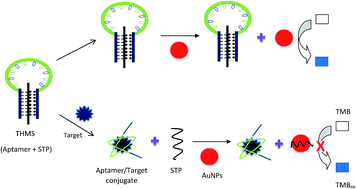
Lead (Pb) is a serious environmental contaminant and one of the most toxic heavy metals. In this study a colorimetric aptasensor was designed for selective, sensitive and rapid detection of Pb2+, based on a triple-helix molecular switch (THMS) and peroxidase-like activity of gold nanoparticles (AuNPs). This sensor inherits the properties of THMS, including high stability and preserving the affinity and selectivity of the original aptamer and properties of peroxidase-like activity of AuNPs, such as fast readout and improvement of the sensitivity. In the absence of Pb2+, THMS is intact, leading to complete peroxidase-like activity of AuNPs and an obvious color change to purplish-blue. In the presence of Pb2+, the aptamer binds to Pb2+, the signal transduction probe (STP) leaves the THMS and adsorbs onto the surface of AuNPs, leading to inhibition of the peroxidase-like activity of AuNPs and no color change is observed. The designed aptasensor showed high selectivity toward Pb2+ with a limit of detection as low as 602 pM for Pb2+. The presented aptasensor was successfully used to detect Pb2+ in water and serum.
1 Introduction
Lead (Pb) is one of the most poisonous heavy metals which could have a serious impact on the environment and human organs.1,2 The accumulation of lead in the human body could induce toxic effects on kidneys, liver, the reproductive system and nervous system.3,4 Based on the International World Health Organization (WHO) and Environmental Protection Agency (EPA), the maximum permitted concentration of Pb2+ in drinking water is 10 μg L−1 and 50 μg L−1, respectively.5,6 The maximum acceptable level of Pb2+ in blood is 10 μg dL−1, according to the Center of Disease Control and Prevention (CDCP).7,8
Atomic absorption spectrometry (AAS) and inductively coupled plasma mass spectrometry (ICP-MS) are the conventional analytical techniques for lead ions detection. These methods are time-consuming and need specialized operator and sophisticated equipment.3,4,9 So selective, sensitive, low-cost and simple methods for determination of Pb2+ are in great demand.
Aptamers are short single-stranded DNA (ssDNA) or RNA oligonucleotides, generated by an in vitro process called SELEX (systematic evolution of ligands by exponential enrichment).10,11 Aptamers could bind to a wide range of targets, from small molecules to proteins and whole cells with high affinity and specificity.12,13 In comparison with antibodies, aptamers have unique advantages, like ease of synthesis and modification, excellent thermal stability, low cost and lack of immunogenicity and toxicity.11,14–16 Because of these unique characteristics, aptamers have received substantial attention as recognition probes in biosensors.
Recently gold nanoparticles (AuNPs) have attracted considerable attention for biomolecular detection, due to their unique properties such as good biocompatibility, high extinction coefficient, large surface area, ease of synthesis and peroxidase-like activity.17–20
Colorimetric assays are common sensing techniques for analytical applications, because the target recognition could be easily detected by the naked eye.21
In this work a colorimetric triple-helix molecular switch (THMS) system was developed for the first time for detection of Pb2+, based on aptamer and peroxidase-like activity of AuNPs. Compared to molecular beacon-based signaling aptamers and known double-helix DNA molecular switches, THMS shows distinct advantages, such as high stability and sensitivity and preserving the affinity and specificity of the original aptamer.22 THMS system generally is composed of a dual-labeled oligonucleotide as a signal transduction probe (STP) and a label-free target specific aptamer sequence with two arm segments,22,23 but in this work a label-free STP was applied which is more cost effective and does not require any special equipment. In this study, a ssDNA aptamer, which selectively binds to Pb2+,24,25 was applied as targeting agent.
2 Materials and methods
2.1 Materials
The sequence of signal transduction probe (STP) and lead-binding aptamer flanked at the 5′- and 3′-ends with two arm segments (Apt) were purchased from Bioneer (south Korea) (Table 1). Plasma from rat, ZnSO4, CuSO4, Ni(OAC)2, Pb(OAC)2, Co(OAC)2, Sn(OAC)2, sodium tetrachloroaurate(III) (HAuCl4), sodium citrate and 3,3′,5,5′-tetramethylbenzidine liquid substrate (TMB) were obtained from Sigma-Aldrich (USA).
Table 1 Oligonucleotide sequences used in this study. The boldface type is the aptamer and the underlined sequences indicate the bases that form a triplex
Entry |
Sequence |
STP |
5′-GAGAGAGAGAGAGA-3′ |
Lead-binding aptamer |
5′-![[C with combining low line]](https://www.rsc.org/images/entities/char_0043_0332.gif) ![[T with combining low line]](https://www.rsc.org/images/entities/char_0054_0332.gif) ![[C with combining low line]](https://www.rsc.org/images/entities/char_0043_0332.gif) ![[T with combining low line]](https://www.rsc.org/images/entities/char_0054_0332.gif) ![[C with combining low line]](https://www.rsc.org/images/entities/char_0043_0332.gif) GGGTGGGTGGGTGGGT![[T with combining low line]](https://www.rsc.org/images/entities/char_0054_0332.gif) ![[C with combining low line]](https://www.rsc.org/images/entities/char_0043_0332.gif) ![[T with combining low line]](https://www.rsc.org/images/entities/char_0054_0332.gif) ![[C with combining low line]](https://www.rsc.org/images/entities/char_0043_0332.gif) ![[T with combining low line]](https://www.rsc.org/images/entities/char_0054_0332.gif) -3′ |
2.2 Synthesis of water resuspended gold nanoparticles
AuNPs were synthesized by the classical citrate reduction of HAuCl4, based on the previously published protocol.26 The prepared AuNPs solution was centrifuged at 15
000 g for 20 min at 4 °C. The supernatant was removed and AuNPs were resuspended in ultrapure water. AuNPs concentrations were measured based on extinction coefficient of 2.7 × 10 8 M−1 cm−1 at λ = 520 nm for 15 nm AuNPs.
The size, zeta potential and morphology of AuNPs were analyzed using a particle size analyzer (Malvern, UK) and transmission electron microscopy (TEM) (CM120, Philips, Holland).
2.3 Preparation of THMS
The THMS was made by incubation of the STP (20 μM final concentration) and the Apt (20 μM final concentration) in binding buffer (20 mM Tris–HCl, pH 6.5) for 60 min at 37 °C.
2.4 Optimizing the concentration of STP
Increasing concentrations of STP (0–4 μM final concentration) were added to each well containing 5 nM AuNPs. After 20 min incubation at 37 °C, 10 μl TMB was added to each well and incubated for 5 min at 37 °C. A651 was measured using a Synergy H4 microplate reader (BioTeK, USA).
2.5 Effect of pH on the formation of THMS
The THMS was made by mixing the STP (20 μM final concentration) and the Apt (20 μM final concentration) in the binding buffer with different pH values (5.5–8.5). After incubation for 60 min at 37 °C, 5 nM AuNPs were added to each well containing THMS (2 μM final concentration, final volume 100 μl) and incubated for 20 min at 37 °C. 10 μl TMB was added to each well. After 5 min incubation at 37 °C, A651 was measured.
2.6 Effect of temperature on the peroxidase-like activity of AuNPs
10 μl TMB was added to each well containing 5 nM AuNPs and incubated for 5 min at different temperatures (22, 37 and 50 °C) and then A651 was recorded.
2.7 Function study of THMS
The interaction of Pb2+ and THMS was assessed by measuring the peroxidase-like activity of AuNPs. 4 μl Pb2+ (30 nM final concentration) was added to the assay solution containing 5 nM AuNPs and 2 μM THMS. After 20 min incubation at 37 °C, 10 μl TMB was added. After incubation for 5 min at 37 °C, A651 was measured.
2.8 Detection of Pb2+ based on colorimetric assay
A range of Pb2+ concentrations, 0–300 nM final concentrations, were added to the THMS (2 μM final concentration) and 5 nM AuNPs (final volume 100 μl) and incubated for 20 min at 37 °C. 10 μl TMB was added to each well. After incubation for 5 min at 37 °C, A651 was recorded.
2.9 Selectivity
The selectivity was investigated in the presence of 100 nM Co2+, Pb2+, Sn2+, Zn2+, Ni2+ and Cu2+, separately. Also the selectivity of the designed aptasensor was performed in the mixture of Co2+, Pb2+, Sn2+, Zn2+, Ni2+ and Cu2+ (the concentration of each ion was 100 nM).
2.10 Pb2+ detection in water and serum
To assess the application of the fabricated aptasensor in serum and water, increasing concentrations of Pb2+ (0–600 nM final concentrations) were spiked to water and rat serum. Proteins of serum were discarded using acetonitrile. 125 μl of cold acetonitrile was gently mixed with 50 μl of serum. After incubation for 60 min at 4 °C, the samples were centrifuged at 9500 g for 10 min at 4 °C. The supernatant was collected and Pb2+ concentrations were recorded.
3 Results and discussion
3.1 Sensing scheme
The designed colorimetric aptasensor is based on the release of STP from Apt in the presence of target (Pb2+), peroxidase-like activity of AuNPs, strong binding of STP (as a ssDNA) to water resuspended AuNPs, and no or very less binding of THMS (as a triple-helix structure) to AuNPs. In the synthesis of AuNPs, removal of sodium citrate and use of water resuspended AuNPs could improve the sensitivity of colorimetric aptasensors.20 Thus, in this study, water resuspended AuNPs were applied.
The THMS is formed by the interaction of the two arm segments of Apt with the sequence of STP through Watson–Crick and Hoogsteen base pairings.
AuNPs could oxidase the peroxidase substrates in the presence of H2O2 to make colored reaction products.27 AuNPs-based colorimetric aptasensors usually use salt-induced aggregation for color change. Complete removal of aptamers from the surface of AuNPs is required for an efficient salt-induced aggregation of AuNPs. So, high amount of target and long-time incubation are needed to completely detach aptamers from the surface of AuNPs.27,28 In this study, a colorimetric THMS system was designed based on peroxidase-like activity of AuNPs, leading to elimination of salt-induced aggregation, faster readout and improvement of the sensitivity of the sensor.
As shown in Scheme 1, in the absence of Pb2+, THMS is intact. THMS could not be adsorbed on the surface of AuNPs. dsDNA has no or little interaction with AuNPs, due to its rigid structure,21,29,30 while in this work THMS was used, a triple-helix structure, which has a more rigid structure than dsDNA. So, AuNPs could oxidize colorless TMB into a purplish-blue product. Addition of Pb2+ causes a conformational change and formation of Apt/target conjugate. So that, THMS is disassembled and the released STP is adsorbed on the surface of AuNPs through electrostatic interaction between the negatively charged AuNPs and positively charged bases of STP.20,31 In this situation, AuNPs lose their peroxidase-like activity, because the surface of AuNPs have been blocked by the STP and AuNPs could not interact with the peroxidase substrate. So that, no color change is observed.
 |
| Scheme 1 Schematic description of Pb2+ detection based on colorimetric THMS and peroxidase-like activity of AuNPs. In the absence of Pb2+, THMS (aptamer + STP) is intact, resulting in the maximum peroxidase-like activity of AuNPs and an obvious color change to purplish-blue. In the presence of target, aptamer binds to its target, STP leaves the THMS and adsorbs on the surface of AuNPs. So the AuNPs lose their peroxidase-like activity and no color change is observed. | |
3.2 Characterization of AuNPs
Particle size and zeta potential of AuNPs were 14.9 ± 0.5 nm and −32.8 ± 1.4 mV, respectively. The prepared AuNPs were characterized using TEM, too. The result showed well-dispersed AuNPs (Fig. 1).
 |
| Fig. 1 TEM image of AuNPs. | |
3.3 Factors involved in the efficacy of designed aptasensor
To determine the optimum concentration of STP for complete reaction with AuNPs and inhibition of peroxidase-like activity of AuNPs, increasing concentrations of STP were added to a fix concentration of AuNPs. The result showed the final concentration of 2 μM STP could almost inhibit the peroxidase-like activity of AuNPs (Fig. 2a).
 |
| Fig. 2 Factors involved in the efficacy of designed aptasensor. (a) Relative peroxidase-like activity of AuNPs in the presence of various concentrations of STP (0–4 μM). (b) Relative peroxidase-like activity of AuNPs in the presence of THMS in the binding buffer with different pH values (5.5–8.5). (c) Relative peroxidase-like activity of AuNPs at different temperatures. | |
PH could affect the stability of the Hoogsteen base pairing. So that, the effect of pH on the formation of THMS was analyzed. The result indicated that in acidic pHs (5.5–6.5), THMS was more stable, leading to maximum peroxidase-like activity of AuNPs (Fig. 2b). Under acidic conditions, two arm segments of the Apt efficiently interact with the STP, because protonation of cytosine residues induces formation of Hoogsteen base pairing between C–G·C+ triplets.22,32 Whereas in higher pHs, the imino groups of cytosines could not realize their protonations. So, the Hoogsteen base pairing becomes unstable, resulting in the formation of triple stem weakly.
Weak acidic condition has only a little effect on the affinity of aptamers, while strong acidic condition could dramatically increase dissociation constant of aptamers.22 So, the other experiments were done in pH 6.5.
To obtain the optimum temperature for peroxidase-like activity of AuNPs, the peroxidase-like activity of AuNPs were investigated at three different temperatures (22, 37 and 50 °C). The result indicated the AuNPs had the maximum peroxidase-like activity at 37 °C (Fig. 2c).
3.4 Monitoring of THMS formation and function
The THMS fabrication and formation were confirmed by absorbance measurement of peroxidase-like activity of AuNPs (Fig. 3). Unlike STP, the THMS could not inhibit the peroxidase-like activity of AuNPs. This result indicated the triple-helix structure (THMS), which has a rigid structure, has formed. Upon addition of target, Pb2+, peroxidase-like activity of AuNPs was dramatically decreased, which confirmed the formation of Apt/target conjugate, release of STP from Apt, adsorption of STP on the surface of AuNPs and good function of THMS system.
 |
| Fig. 3 Relative peroxidase-like activity of AuNPs + STP, AuNPs + THMS and AuNPs + THMS + lead in the binding buffer, pH 6.5. | |
3.5 Pb2+ analysis
In this method, an obvious color change to purplish-blue could be observed as Pb2+ concentration decreased (Fig. 4a).
 |
| Fig. 4 (a) Visual color change upon treatment of AuNPs and THMS with different concentrations of Pb2+ (0, 0.2, 4, 100 nM, and colorless TMB, from left to right) (b) relative peroxidase-like activity of AuNPs as a function of Pb2+ concentration. (c) Pb2+ standard curve. P0 and P are the peroxidase activity (%) before and after addition of various concentrations of Pb2+, respectively. (d) Efficiency of relative peroxidase-like activity of AuNPs in the presence of various metals. | |
Relative peroxidase-like activity of AuNPs at different concentrations of Pb2+ were shown in Fig. 4b. The relative peroxidase-like activity increased and reached to plateau at concentration of 30 nM Pb2+. The developed aptasensor showed a well linear range (0.2–30 nM) toward Pb2+ (Fig. 4c). The limit of detection was determined to be 602 pM, as three times the standard deviation of blank/slope.
Reported detection limits of lead in different experiments were as following: 916 pM for strip immunosensor,9 100 pM for gold nanorods,33 7.67 nM for inductively coupled plasma atomic emission spectroscopy (ICP-AES),34 100 nM for glassy carbon electrode,35 240 pM for DNAzyme-induced catalytic reaction,36 and 1.35 nM for ICP-Mass Spectrometry (ICP-MS).37 Compared to the designed aptasensor most of these approaches are expensive, time-consuming, and have higher LODs.
Selectivity is one of the most important properties of a sensor. To assess the selectivity of the designed aptasensor, other substances, including Co2+, Ni2+, Cu2+ and Zn2+ were analyzed by this approach. No significant inhibition of peroxidase-like activity was observed in the presence of these metal ions. The peroxidase-like activity for Pb2+ was significantly lower than other metal ions (Fig. 4d). Also the designed aptasensor could detect Pb2+ in the mixture of different ions very well. The result confirmed excellent selectivity of the designed aptasensor toward Pb2+.
3.6 Measurement of Pb2+ in tap water and rat serum
The designed aptasensor was used to measure Pb2+ concentration in tap water and rat serum. Different concentrations of Pb2+ were spiked into water and serum, and LODs were calculated to be 0.708 and 2.07 nM, respectively (Fig. 5). The calculated LODs were much lower than the maximum permitted level of Pb2+ in blood and water as regulated by CDCP, WHO and EPA. The results indicated the designed aptasensor could successfully be used for detection of Pb2+ in serum and water.
 |
| Fig. 5 (a) Relative peroxidase-like activity of AuNPs upon the addition of various concentrations of Pb2+ in water. (b) Standard curve of Pb2+ in water. (c) Relative peroxidase-like activity of AuNPs upon the addition of various concentrations of Pb2+ in serum (d) standard curve of Pb2+ in serum. P0 and P are the peroxidase activity (%) before and after addition of various concentrations of Pb2+, respectively. | |
4 Conclusion
In summary, we introduced a selective, sensitive and rapid colorimetric aptasensor based on THMS and peroxidase-like activity of AuNPs for detection of Pb2+. The limit of detection for Pb2+ was determined as low as 602 pM. Furthermore, this aptasensor could well detect Pb2+ in water and serum. It is expected this approach could be extended for detection of other biomolecules and drugs in clinical practice, because of its high affinity and simplicity.
Conflict of interest
There is no conflict of interest about this article.
Acknowledgements
Financial support of this study was provided by Mashhad University of Medical Sciences.
References
- G. Kaur, H. P. Singh, D. R. Batish and R. K. Kohli, Protoplasma, 2013, 250, 53–62 CrossRef CAS PubMed.
- H. Sun, L. Yu, H. Chen, J. Xiang, X. Zhang, Y. Shi, Q. Yang, A. Guan, Q. Li and Y. Tang, Talanta, 2015, 136, 210–214 CrossRef CAS PubMed.
- N. Promphet, P. Rattanarat, R. Rangkupan, O. Chailapakul and N. Rodthongkum, Sens. Actuators, B, 2014, 207, 526–534 CrossRef PubMed.
- Z. S. Qian, X. Y. Shan, L. J. Chai, J. R. Chen and H. Feng, Biosens. Bioelectron., 2015, 68, 225–231 CrossRef CAS PubMed.
- V. K. Gupta and A. Rastogi, J. Hazard. Mater., 2008, 152, 407–414 CrossRef CAS PubMed.
- W. Zhang, B. Xiong, L. Chen, K. Lin, X. Cui, H. Bi, M. Guo and W. Wang, Environ. Toxicol. Pharmacol., 2013, 36, 51–57 CrossRef CAS PubMed.
- S. G. Gilbert and B. Weiss, Neurotoxicology, 2006, 27, 693–701 CrossRef CAS PubMed.
- Center of Disease Control and Prevention (CDCP), 1991.
- H. Kuang, C. Xing, C. Hao, L. Liu, L. Wang and C. Xu, Sensors, 2013, 13, 4214–4224 CrossRef CAS PubMed.
- X. Tang, Y. S. Wang, J. H. Xue, B. Zhou, J. X. Cao, S. H. Chen, M. H. Li, X. F. Wang, Y. F. Zhu and Y. Q. Huang, J. Pharm. Biomed. Anal., 2015, 107, 258–264 CrossRef CAS PubMed.
- B. Zhao, P. Wu, H. Zhang and C. Cai, Biosens. Bioelectron., 2015, 68, 763–770 CrossRef CAS PubMed.
- S. Wu, H. Zhang, Z. Shi, N. Duan, C. Fang, S. Dai and Z. Wang, Food Control, 2015, 50, 597–604 CrossRef CAS PubMed.
- F. Zhao, Q. Xie, M. Xu, S. Wang, J. Zhou and F. Liu, Biosens. Bioelectron., 2015, 66, 238–243 CrossRef CAS PubMed.
- Y. Lian, F. He, H. Wang and F. Tong, Biosens. Bioelectron., 2015, 65, 314–319 CrossRef CAS PubMed.
- P. Luo, Y. Liu, Y. Xia, H. Xu and G. Xie, Biosens. Bioelectron., 2014, 54, 217–221 CrossRef CAS PubMed.
- S. Eissa, M. Siaj and M. Zourob, Biosens. Bioelectron., 2015, 69, 148–154 CrossRef CAS PubMed.
- S. Wang, W. Chen, A. L. Liu, L. Hong, H. H. Deng and X. H. Lin, ChemPhysChem, 2012, 13, 1199–1204 CrossRef CAS PubMed.
- Y. Luo, J. Xu, Y. Li, H. Gao, J. Guo, F. Shen and C. Sun, Food Control, 2015, 54, 7–15 CrossRef CAS PubMed.
- N. Sattarahmady, G. H. Tondro, M. Gholchin and H. Heli, Biochem. Eng. J., 2015, 97, 1–7 CrossRef CAS PubMed.
- J. Liu, Z. Guan, Z. Lv, X. Jiang, S. Yang and A. Chen, Biosens. Bioelectron., 2014, 52, 265–270 CrossRef CAS PubMed.
- S. C. B. Gopinath, T. Lakshmipriya and K. Awazu, Biosens. Bioelectron., 2014, 51, 115–123 CrossRef CAS PubMed.
- J. Zheng, J. Li, Y. Jiang, J. Jin, K. Wang, R. Yang and W. Tan, Anal. Chem., 2011, 83, 6586–6592 CrossRef CAS PubMed.
- A. Verdian-Doghaei, M. R. Housaindokht and K. Abnous, Anal. Biochem., 2014, 466, 72–75 CrossRef CAS PubMed.
- M. Li, X. Zhou, S. Guo and N. Wu, Biosens. Bioelectron., 2013, 43, 69–74 CrossRef CAS PubMed.
- T. Li, S. Dong and E. Wang, J. Am. Chem. Soc., 2010, 132, 13156–13157 CrossRef CAS PubMed.
- J. J. Storhoff, R. Elghanian, R. C. Mucic, C. A. Mirkin and R. L. Letsinger, J. Am. Chem. Soc., 1998, 120, 1959–1964 CrossRef CAS.
- T. K. Sharma, R. Ramanathan, P. Weerathunge, M. Mohammadtaheri, H. K. Daima, R. Shukla and V. Bansal, Chem. Commun., 2014, 50, 15856–15859 RSC.
- P. Weerathunge, R. Ramanathan, R. Shukla, T. K. Sharma and V. Bansal, Anal. Chem., 2014, 86, 11937–11941 CrossRef CAS PubMed.
- J. E. Smith, D. K. Griffin, J. K. Leny, J. A. Hagen, J. L. Chávez and N. Kelley-Loughnane, Talanta, 2014, 121, 247–255 CrossRef CAS PubMed.
- H. Wei, B. Li, J. Li, E. Wang and S. Dong, Chem. Commun., 2007, 3735–3737 RSC.
- C. C. Chang, S. C. Wei, T. H. Wu, C. H. Lee and C. W. Lin, Biosens. Bioelectron., 2013, 42, 119–123 CrossRef CAS PubMed.
- K. M. Vasquez and P. M. Glazer, Q. Rev. Biophys., 2002, 35, 89–107 CrossRef CAS.
- H. H. Cai, D. Lin, J. Wang, P. H. Yang and J. Cai, Sens. Actuators, B, 2014, 196, 252–259 CrossRef CAS PubMed.
- Q. He, X. Chang, X. Huang and Z. Hu, Microchim. Acta, 2008, 160, 147–152 CrossRef CAS.
- Z. Wang, E. Liu and X. Zhao, Thin Solid Films, 2011, 519, 5285–5289 CrossRef CAS PubMed.
- L. Xu, H. Yin, W. Ma, L. Wang, H. Kuang and C. Xu, J. Phys. Chem. B, 2013, 117, 14367–14371 CrossRef CAS PubMed.
- F. A. Aydin and M. Soylak, J. Hazard. Mater., 2010, 173, 669–674 CrossRef CAS PubMed.
|
This journal is © The Royal Society of Chemistry 2015 |
Click here to see how this site uses Cookies. View our privacy policy here.