Formation of highly porous structure in the electrospun polylactide fibers by swelling-crystallization in poor solvents

Renping Tian, Peng Zhang, Ruihua Lv, Bing Na*, Qingxian Liu and Yunhui Ju
Fundamental Science on Radioactive Geology and Exploration Technology Laboratory, School of Chemistry, Biology and Materials Science, East China Institute of Technology, Nanchang, 330013, People's Republic of China. E-mail: bna@ecit.edu.cn; bingnash@163.com; Fax: +86 791 83897982

Received 1st April 2015 , Accepted 20th April 2015

First published on 20th April 2015


Abstract

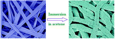
The electrospun fibers made of polylactide (PLA) have been attracting much attention in many applications because of biodegradability and biocompatibility. Introducing porous structure into electrospun PLA fibers is helpful to enhance surface area and thus to extend their applications. Here we report a novel and facile route to produce highly porous structure in the electrospun PLA fibers by simple immersion in poor solvents. It arises from swelling and subsequent solvent-induced crystallization in the electrospun PLA fibers, depending on solvent polarity, immersion temperature and PLA molecular weight. The highly porous PLA fibers have a very large surface area of 137.7 m2 g−1, which is almost unapproachable by other routes.


1 Introduction

Electrospinning is a powerful technique to produce ultrafine fibers or nanofibers from a variety of polymers.1–3 Tailoring of morphology such as porous structure in the electrospun fibers is ongoing pursuits to extend their applications. Owing to large surface area porous polymer fibers, for instance, can be used as the scaffolds of active compounds or catalysts.4

With extensive efforts two main approaches have been developed to fabricate porous polymer fibers. The first is phase separation of polymer solutions induced by evaporation, nonsolvents or thermal gradient during electrospinning.5–10 Namely, it is called as thermally-induced phase separation or vapor-induced phase separation. The solvent-rich regions are transformed into pores in the electrospun fibers that are formed by solvent-poor regions. In most cases, pores are generated on the surface of electrospun fibers by the phase separation approach. The second is post-treatment of electrospun fibers based on selective dissolution.11,12 To this purpose, electrospun fibers consist of two components such as polymer blends, and one component can be selectively removed by solvent extraction to generate pores. However, this approach requires the proper selection of components and co-solvents for electrospinning; and it could be not possible to completely remove the unwanted component from blend fibers.

Polylactide (PLA), derived from renewable sources, is very intriguing in many applications because of superior physicochemical properties. The electrospun PLA fibers have great potentials in drug delivery and tissue engineering.13,14 To date, porous structure has been created in the electrospun PLA fibers by modifying electrospinning conditions or post-treatment such as salt leaching.15–17 Unfortunately, the reported PLA fibers are less porous. Herein, highly porous structure has been achieved by simple immersion of electrospun PLA fibers in poor solvents. It arises from swelling and subsequent solvent-induced crystallization in the electrospun PLA fibers, depending on solvent polarity, immersion temperature and PLA molecular weight. This approach, totally different from previous ones, provides a novel and facile route to highly porous structure in the electrospun PLA fibers. Moreover, residual solvents can be easily removed from highly porous PLA fibers by evaporation, superior to the selective dissolution of the unwanted component from blend fibers.

2 Experimental details

2.1 Fabrication procedures

Four kinds of PLA pellets with various molecular weights were purchased from Changchun Sinobiomaterials Co., Ltd, China. For convenience sample codes of PLAx were used, and the subscript “x” represented the viscosity-average molecular weight of PLA (Table 1). All solvents were analytical reagents and used without further purification. PLA pellets were dissolved at room temperature in the mixed solvents of chloroform and ethanol (5[thin space (1/6-em)]:[thin space (1/6-em)]2, v/v) to generate a transparent solution. Note that adding ethanol with high polarity was to promote the formation of bead-free fibers during electrospinning because of enhanced solution conductivity. The typical PLA concentration in the solutions for electrospinning was given in Table 1, if not specified. The solution was loaded in a 10 ml syringe with a needle and then pumped continuously at a rate of 25 μL min−1. An applied voltage of 11 kV and a distance of 15 cm from the needle to collector (aluminum foils) were adopted. Of note, with trial and error this distance could ensure sufficient evaporation of solvents and thus effectively solidified the electrospun fibers at the adopted conditions. The electrospun fibers were immersed in methanol and acetone at desired temperatures for a certain period, respectively, followed by vacuum drying at room temperature to remove the residual solvents.
Table 1 Molecular weight of PLAs and their typical concentration in the solutions for electrospinning
Sample Viscosity-average molecular weight (g mol−1) PLA concentration in the solution (w/v)
PLA37k 37[thin space (1/6-em)]000 18%
PLA82k 82[thin space (1/6-em)]000 10%
PLA193k 193[thin space (1/6-em)]000 6%
PLA503k 503[thin space (1/6-em)]000 4%


2.2 Characterizations

The morphology was observed by a Nova NanoSEM 450 scanning electron microscope (SEM). The cross-section of fibers was obtained by cryo-fractured in liquid nitrogen. A thin gold layer was sputtered prior to SEM measurements. The crystalline phase was determined by a Bruker D8 ADVANCE X-ray diffractometer (XRD) at room temperature; and the wavelength of the X-ray was 0.154 nm. Thermal behaviors were recorded by a TA Q2000 differential scanning calorimetry (DSC) instrument at a heating rate of 10 °C min−1 in a flowing nitrogen atmosphere. The Brunauer–Emmett–Teller (BET) surface area was measured by using a surface area analyzer (JW-BK132F, Beijing JWGB Sci. & Tech., China). N2 was used and the reported surface area was averaged over three measurements.

3 Results and discussion

Electrospinning of PLA193k solutions produces ultrafine fibers with oblate cross-sections and almost smooth surfaces (Fig. 1a and a′). Immersion of as-spun PLA193k fibers in methanol at 4 °C for 20 h only results in a little change in the surface morphology, and the oblate cross-sections still exist (Fig. 1b and b′). Interestingly, after immersion in acetone at 4 °C for 20 h highly porous structure is created throughout the PLA193k fibers, accompanied by the appearance of nearly round cross-sections (Fig. 1c and c′). As a result, surface area is largely enhanced by formation of highly porous structure in the electrospun PLA193k fibers. Fig. 2 compares surface area of PLA193k fibers before and after immersion in methanol and acetone at 4 °C for 20 h. The as-spun PLA193k fibers have a low surface area of 18.4 m2 g−1 due to their almost smooth surfaces. After immersion in methanol surface area is only enhanced to some extent because of a little change in the surface morphology. In contrast, a very large surface area of 137.7 m2 g−1 is exhibited by the PLA193k fibers after immersion in acetone, consistent with the highly porous structure.
image file: c5ra05738h-f1.tif
Fig. 1 Effect of solvent types on the morphology of PLA193k fibers after immersion at 4 °C for 20 h: (a and a′) as-spun (control), (b and b′) methanol, (c and c′) acetone; (a–c) surface, (a′–c′) cross-section. Scale bars, upper: 10 μm (inset: 2 μm), lower: 1 μm.

image file: c5ra05738h-f2.tif
Fig. 2 Surface area of PLA193k fibers before and after immersion in methanol and acetone at 4 °C for 20 h.

Both methanol and acetone are poor solvents and cannot dissolve the as-spun PLA193k fibers at 4 °C. The distinct morphology induced by methanol and acetone should be correlated with their solubility parameters apart from that of the as-spun PLA193k fibers. Note that the solubility parameter is 14.5 and 9.8 cal1/2 cm−3/2 for methanol and acetone, respectively, whereas that of the as-spun PLA193k fibers is estimated to be about 9.3 cal1/2 cm−3/2 (based on that the as-spun PLA193k fibers can be readily dissolved by chloroform that has a solubility parameter of 9.3 cal1/2 cm−3/2). Therefore, during immersion the as-spun PLA193k fibers are most likely penetrated and swollen by acetone due to their slight difference in solubility parameters, although complete dissolution is impossible. It is the origin for generation of highly porous structure and nearly round cross-sections. On the contrary, large difference in solubility parameters makes methanol have very limited interactions with the as-spun PLA193k fibers and thus the morphology keeps nearly intact.

The strong interactions between the as-spun PLA193k fibers and acetone are further confirmed by rapid formation of highly porous structure only after immersion at 4 °C for 1 h. Fig. 3 gives the corresponding SEM micrographs where numerous pores are observed for the PLA193k fibers. It is same to that obtained by immersion in acetone for 20 h. Even so, immersion period of 20 h is preferentially adopted to ensure enough treatment of as-spun fibers under other conditions. On the other hand, diameter of as-spun PLA193k fibers seems to have some influence on the formation of highly porous structure, as shown by SEM micrographs in Fig. 4. In particular, as for the PLA193k fibers with large diameter generation of porous structure becomes less significant after immersion in acetone at 4 °C for 20 h (Fig. 4b′). It could arise from the suppressed penetration and swelling of PLA193k fibers with large diameter by acetone at the adopted immersion conditions. Of note, PLA193k fibers with small and large diameter were electrospun from the solutions with a PLA concentration of 3% and 8%, respectively.


image file: c5ra05738h-f3.tif
Fig. 3 SEM micrographs of PLA193k fibers after immersion in acetone at 4 °C for 1 h under low and high magnifications, respectively. Scale bar, (a) 10 μm, (b) 2 μm.

image file: c5ra05738h-f4.tif
Fig. 4 SEM micrographs revealing morphology of PLA193k fibers with (a and a′) small and (b and b′) large diameter (a and b) before and (a′ and b′) after immersion in acetone at 4 °C for 20 h. Scale bars, 10 μm (inset: 2 μm).

To illustrate structural changes upon immersion of as-spun PLA193k fibers in methanol and acetone at 4 °C for 20 h, corresponding XRD profiles and DSC heating traces are given in Fig. 5. The as-spun PLA193k fibers are almost amorphous because of rapid evaporation of solvents during electrospinning. It is demonstrated by the nearly absence of diffraction peaks in the XRD profiles and the appearance of a remarkable cold crystallization peak in the DSC heating traces.18 Immersion in methanol only results in some extent of crystallization in the PLA193k fibers, consistent with the limited interactions caused by large difference in solubility parameters. Rather, significant crystallization is induced in the PLA193k fibers by acetone. It corresponds to intense diffraction peaks from PLA crystals in the XRD profiles. For this reason, little cold crystallization occurs in the DSC heating traces, and only melting of PLA crystals generated during immersion is exhibited.


image file: c5ra05738h-f5.tif
Fig. 5 Crystallization induced by solvents in the PLA193k fibers upon immersion at 4 °C for 20 h: (a) XRD profiles, (b) DSC heating traces.

In general, cold crystallization of PLA from amorphous state can only be induced above glass transition temperature where enough molecular mobility is gained.18,19 In the absence of solvents cold crystallization of as-spun PLA193k fibers begins at temperatures above 60 °C (Fig. 5b). Thus, remarkable crystallization in acetone at 4 °C suggests that as-spun PLA193k fibers must be penetrated and swollen by acetone because of similar solubility parameters. It in turn enhances molecular mobility and thus promotes subsequent crystallization of as-spun PLA193k fibers at 4 °C (termed as solvent-induced crystallization).20 At the same time, crystallization of the swollen PLA193k fibers can result in exclusion of acetone to form solvent-rich regions that penetrate throughout the PLA193k fibers, i.e. crystallization-induced phase separation.21,22 After evaporation of acetone, highly porous structure is thus obtained for the PLA193k fibers.

To further solidify the above concept, the as-spun PLA193k fibers were isothermally annealed at 80 °C (above Tg) for 2 h to achieve complete crystallization. It is demonstrated by the intensity diffraction peaks in the XRD profile (Fig. 6a) and the absence of cold crystallization in the DSC heating trace (Fig. 6b). Afterwards, the annealed PLA193k fibers were immersed in acetone at 4 °C for 20 h, same treatment as the as-spun counterparts in Fig. 1. As shown by SEM micrographs (Fig. 6c and d), immersion of annealed PLA193k fibers in acetone only result in the formation of a few small pores, totally different from that observed in the as-spun counterparts with little crystallization. It arises from limited swelling by acetone due to the constraints from already existed PLA crystals with densely molecular packing in the unit cells. Thus, sufficient swelling is a prerequisite for generation of highly porous structure by subsequent crystallization-induced phase separation.


image file: c5ra05738h-f6.tif
Fig. 6 (a) XRD profile and (b) DSC heating trace of annealed PLA193k fibers, and the corresponding SEM micrographs (c) before and (d) after immersion in acetone at 4 °C for 20 h. Scale bars, 10 μm (inset: 2 μm).

In such a sense, formation of porous structure in the electrospun fibers should become insignificant at lower immersion temperature or in the PLA fibers with higher molecular weight, as a result of suppressed swelling of as-spun fibers by acetone. In fact, generation of porous structure is prevented in the PLA193k fibers at −18 °C to some extent (Fig. 7b), and especially in the PLA503k fibers at 4 °C (Fig. 8c). It suggests that swelling is significantly inhibited in the PLA503k fibers with higher molecular weight. On the other hand, swelling becomes severe at higher immersion temperature or in the PLA fibers with lower molecular weight. It results in the disintegration of fibrous morphology, failing to produce highly porous PLA fibers. This is the fact encountered in the PLA193k fibers immersed at 25 °C (Fig. 7a) and particularly in the PLA37k and PLA82k fibers after immersion at 4 °C (Fig. 8a and b).


image file: c5ra05738h-f7.tif
Fig. 7 Effect of immersion temperatures on the morphology of PLA193k fibers in acetone for 20 h: (a) 25 °C, (b) −18 °C. Scale bars, 10 μm (inset: 2 μm).

image file: c5ra05738h-f8.tif
Fig. 8 Effect of PLA molecular weight on the morphology of fibers after immersion in acetone at 4 °C for 20 h: (a) PLA37k, (b) PLA82k, (c) PLA503k; (a′)–(c′), corresponding as-spun fibers (control). Scale bars, 10 μm (inset: 2 μm).

4 Conclusion

Highly porous structure is successfully produced in the electrospun PLA fibers via simple immersion in poor solvents, by properly choosing solvent types, immersion temperatures and PLA molecular weight. It is correlated with swelling of electrospun PLA fibers by poor solvents and subsequent solvent-induced crystallization. The highly porous PLA fibers have a very large surface area of 137.7 m2 g−1. The swelling and crystallization route, facile and advantageous, could be extended to fabricate other porous fibers with large surface area.

Acknowledgements

This work is financially supported by the Major Program of Natural Science Foundation of Jiangxi, China (no. 20133ACB21006), the National Natural Science Foundation of China (no. 21364001) and the Program for Young Scientists of Jiangxi Province (no. 20112BCB23023).

References

  1. D. H. Reneker and I. Chun, Nanotechnology, 1996, 7, 216–223 CrossRef CAS.
  2. H. Li, Y. Xu, H. Xu and J. Chang, J. Mater. Chem. B, 2014, 2, 5492–5510 RSC.
  3. P. Ke, X. N. Jiao, X. H. Ge, W. M. Xiao and B. Yu, RSC Adv., 2014, 4, 39704–39724 RSC.
  4. R. S. Barhate, S. Koeppl and S. Ramakrishna, Chem. Eng. Res. Des., 2011, 89, 621–630 CrossRef CAS PubMed.
  5. L. Li, Z. Jiang, M. Li, R. Li and T. Fang, RSC Adv., 2014, 4, 52973–52985 RSC.
  6. J. T. McCann, M. Marquez and Y. Xia, J. Am. Chem. Soc., 2006, 128, 1436–1437 CrossRef CAS PubMed.
  7. J. Lin, B. Ding, J. Yu and Y. Hsieh, ACS Appl. Mater. Interfaces, 2010, 2, 521–528 CAS.
  8. P. Dayal, J. Liu, S. Kumar and T. Kyu, Macromolecules, 2007, 40, 7689–7694 CrossRef CAS.
  9. X. Y. Ye, F. W. Lin, X. J. Huang, H. Q. Liang and Z. K. Xu, RSC Adv., 2013, 3, 13851–13858 RSC.
  10. S. Megelski, J. S. Stephens, D. B. Chase and J. F. Rabolt, Macromolecules, 2002, 35, 8456–8466 CrossRef CAS.
  11. A. Gupta, C. D. Saquing, M. Afshari, A. E. Tonelli, S. A. Khan and R. Kotek, Macromolecules, 2009, 42, 709–715 CrossRef CAS.
  12. L. Zhang and Y. L. Hsieh, Nanotechnology, 2006, 17, 4416–4423 CrossRef CAS.
  13. D. Ishii, T. H. Ying, A. Mahara, S. Murakami, T. Yamaoka, W. Lee and T. Iwata, Biomacromolecules, 2009, 10, 237–242 CrossRef CAS PubMed.
  14. X. Hu, S. Liu, G. Zhou, Y. Huang, Z. Xie and X. Jing, J. Controlled Release, 2014, 185, 12–21 CrossRef CAS PubMed.
  15. C. H. Kim, Y. H. Jung, H. Y. Kim, D. R. Lee, N. Dharmaraj and K. E. Choi, Macromol. Res., 2006, 14, 59–65 CrossRef CAS.
  16. Z. Qi, H. Yu, Y. Chen and M. Zhu, Mater. Lett., 2009, 63, 415–418 CrossRef CAS PubMed.
  17. Y. H. Lee, J. H. Lee, I. G. An, C. Kim, D. S. Lee, Y. K. Lee and J. D. Nam, Biomaterials, 2005, 26, 3165–3172 CrossRef CAS PubMed.
  18. B. Na, S. Zou, R. Lv, M. Luo, H. Pan and Q. Yin, J. Phys. Chem. B, 2011, 115, 10844–10848 CrossRef CAS PubMed.
  19. P. Pan, W. Kai, B. Zhu, T. Dong and Y. Inoue, Macromolecules, 2007, 40, 6898–6905 CrossRef CAS.
  20. H. Marubayashi, S. Asai and M. Sumita, Macromolecules, 2012, 45, 1384–1397 CrossRef CAS.
  21. J. Yoon, A. J. Lesser and T. J. McCarthy, Macromolecules, 2009, 42, 8827–8834 CrossRef CAS.
  22. C. Schugens, V. Maquet, C. Grandfils, R. Jerome and P. Teyssie, Polymer, 1996, 37, 1027–1038 CrossRef CAS.

This journal is © The Royal Society of Chemistry 2015
Click here to see how this site uses Cookies. View our privacy policy here.