Unusual hierarchical structures of micro-injection molded isotactic polypropylene in presence of an in situ microfibrillar network and a β-nucleating agent
Zhongguo Zhaoa,
Qi Yang*a,
Miqiu Kongb,
Dahang Tanga,
Qianying Chena,
Ying Liua,
Fangli Loua,
Yajiang Huanga and
Xia Liaoa
aCollege of Polymer Science and Engineering, The State Key Laboratory for Polymer Materials Engineering, Sichuan University, Chengdu 610065, PR China. E-mail: yangqi@scu.edu.cn; Fax: +86 28 85405402; Tel: +86 28 85401841
bSchool of Aeronautics and Astronautics, Sichuan University, Chengdu 610065, PR China
First published on 5th May 2015
Abstract
The microstructural and mechanical properties of isotactic polypropylene (iPP), in situ PET microfibrils, and β-nucleating agent blends obtained from micro-injection molding were investigated via polarized light microscopy, differential scanning calorimetry, scanning electron microscopy, and two-dimensional wide-angle X-ray diffraction. The results indicate that addition of PET microfibrils markedly increases crystallization temperatures, and increases the thickness of the final oriented layer. Introduction of PET microfibrils to β-nucleation agent-nucleated iPP samples leads to formation of oriented β-crystals epiphytic on the surface of PET fibers in the inner region; this feature improves adhesion between the fiber and the matrix and simultaneously improves the strength and toughness of the final PP/0.5/15 microparts (e.g., the tensile strength increased by 12 MPa and the elongation at break increased by 1.2%) compared with those of iPP microparts. Taken together, the results of this study introduce an alternative approach to optimize the properties of MIM parts.
1. Introduction
Micro-injection molding (MIM) is a very promising technology for miniaturizing parts because the technique allows integration of functions into smaller spaces.1–4 MIM has been extensively applied in innovative solutions in the fields of microelectronics, micromechanics, telecommunications, and daily life.5 Besides imparting weights in the milligram scale to microparts and achieving dimensions of only several micrometers, the MIM process also offers extremely high injection speeds (1200 mm s−1) and shear rates (>105 s−1), short filling times, and substantially larger temperature gradients. Shear thinning effects will theoretically decrease polymer viscosity, which, in turn, increases the filling length of the microcavity. However, the rheological behavior of flow in micro-cavities can differ from that described by conventional rheology laws. Wall-slip effects reportedly occur in channels with sizes of several micrometers or less and can become more significant in very small microchannels.4,6,7 In the case of semi-crystalline polymers, various processing conditions can affect the crystallization steps, especially those involving nucleation and growth of oriented crystalline morphologies such as shish-kebab structures or lamellae row structures.8 Hence, the characteristics of flow-induced morphological crystallization during MIM may significantly differ from those in conventional molding (CIM), leading to marked variations in the final mechanical performance of the MIM products.3,6,9,10MIM is a good tool for investigating the effects of a complex flow field on the morphology of semi-crystalline polymers during practical polymer processing. However, to the best of our knowledge, MIM investigations mainly focus on mold technologies, special machines, production processes, filling performance analyses, and numerical simulations, among others.11,12 Investigations on the morphological and mechanical properties of microparts obtained through MIM are relatively limited, and most of the available research discusses polypropylene; other polymeric materials (e.g., single components, blends, hybrids, or composites) are rarely studied.6,13,14
Isotactic polypropylene (iPP) is an important engineering thermoplastic commonly used to manufacture microparts, including microgears, micropumps and microfluidic devices. iPP has been extensively studied to improve its mechanical properties by processing with fibers. Such processing yields beneficial properties, including low price and good chemical resistance, among others. However, incorporation of solid fibers into the polymer matrix increases its melt viscosity and causes abrasion on the surface of the processing equipment, thereby resulting in significant reductions in the properties of the resultant composite materials.15 To address these problems, in situ generation of short fibers in an immiscible blend has been proposed.16–20 Despite the advantages presented by this approach, however, addition of in situ microfibrils suppresses β-crystallization of β-iPP.21,22
iPP exhibits four modifications, namely, α, β, γ, and smectic.23–26 Compared with other modification approaches, β-crystallization has received the most attention because it imparts excellent impact toughness and elongation at break.27–29 The impact toughness of β-iPP originates from β-to-α polymorphous transition and the loose structure of β-crystals (compared with α-crystals), which are particularly capable of absorbing impact energy.30–32 Thermodynamically metastable β-forms can be obtained under certain conditions, such as in the presence of a temperature gradient,33 through flow-induced crystallization34 and by addition of a specific nucleating agent.35,36 Considering that the β-nucleating agent required to achieve this effect is extremely low, the practice does not alter processing conditions and can lead to high performance/cost ratios. Hence, addition of a β-nucleation agent (β-NA) is the most effective and accessible method of obtaining large amounts of β-iPP.
Inspired by natural plants such as bamboo and sugarcane, which exhibit excellent overall mechanical properties that may attributed to their unique hierarchical structure, we attempted to manufacture microparts with a similar skin–core structure and improved toughness and strength. To realize this goal, we adopted the combined techniques of β-nucleating agent addition and advanced MIM to prepare in situ microfibrillar poly(ethylene terephthalate) (PET)/iPP parts. We added PET microfibrils to β-NA nucleated samples under MIM conditions and found that oriented β-crystals epiphytic on the surface of PET fibers were formed in the inner region of the micropart. Our study is the first to observe and investigate these oriented β-crystals in microparts. The results of this study suggest an alternative approach to achieve good mechanical properties in polypropylene.
2. Experimental
2.1 Materials
The isotactic polypropylene (iPP, trademarked as T30S) used in this study was supplied by Lanzhou Petrochemical Company, China and featured the following characteristics: melt flow index (MFI) of 2.3 g/10 min (230 °C, 2.16 kg), weight average molecular weight ( ) and distribution index (
 ) is ca. 5.87 × 10⁵ g mol−1 and 5.3, respectively.
PET was friendly donated by LuoYang Petroleum Chemical Co., LuoYang, China, which is a commercial grade of textile polyester with a number average molecular weight of ca. 2.3 × 10⁴ g mol−1. Its melt flow index (MFI) and melting temperature is ca. 42 g/10 min (300 °C, 2.16 kg) and 255 °C. In order to avoid hydrolysis, PET was dried in a vacuum oven at 120 °C for at least 12 h prior to processing.
β-Nucleating agent (β-NA; model TMB-5) was used as the aryl amide compound, which had a similar chemical structure as some aromatic amine β-phase nucleating agent, such as N,N′-dicyclohexyl-2,6-naphthalenedicarboxamide.32 This agent was kindly provided by the Shanxi Provincial Institute of Chemical Industry, China.
2.2 Sample preparation
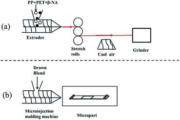
PET granules were dried for 12 h at 120 °C to prevent hydrolytic degradation during extrusion. First, an internal mixer (XSS-300) was used to melt-mix TMB-5 with iPP and form a 5.0 wt% β-nucleating agent master batch at 200 °C. The rotation rate was set to 50 rpm. Then, blends with different compositions were processed in a single-screw extruder with a screw length-to-screw diameter ratio, L/D, of 25. A slit die 43 mm wide and 1.0 mm thick was used. The screw speed was 55 rpm, and the temperature of each section of the extruder was set to 180 °C/250 °C/270 °C/240 °C, from feed to die, respectively. The extrudate was hot-stretched by a take-up device with two pinching rolls to form microfibrils, and the hot stretching ratio was fixed at 8. The experimental setup is shown in Fig. 1.A micro-injection machine (Micropower 5, Battenfeld Co., Austria) was installed on an injection molding machine, and the injection speed, packing pressure, and cooling time were set to 200 mm s−1, 1200 MPa, and 10 s, respectively. The mold temperature was set to 120 °C. Each micropart, bearing dimensions of 8 × 3 × 0.3 mm3, was defined as a part that weighed in the range of several milligrams and with dimensions and tolerances in the micrometer range.
Table 1 gave the detailed designations and compositions of samples of pure iPP and the blends. The preparation of specimens for testing and analysis was illustrated in Fig. 2.
| Code | Composition |
|---|---|
| PP0 | iPP |
| PP/0.5/0 | 99.5 wt% iPP + 0.5 wt% TMB-5 |
| PP/0/15 | 85 wt% iPP + 15 wt% PET |
| PP/0.5/15 | 84.5 wt% iPP + 0.5 wt% TMB-5 + 15 wt% PET |
 | ||
| Fig. 2 Schematic drawing of sampling methods for PLM, SEM, WAXD, and DSC analyses. FD: flow direction, TD: transverse direction, ND: normal direction. | ||
2.3 Measurements
 | (1) |
 | (2) |
The orientations of the crystals of iPP are quantitatively calculated by means of the Herman's orientation function:40
 | (3) |
![[thin space (1/6-em)]](https://www.rsc.org/images/entities/char_2009.gif) φ is an orientation factor defined as:
φ is an orientation factor defined as:
 | (4) |
The 1D-WAXD profiles were obtained from circularly integrated intensities of 2D-WAXD image patterns. Then, by deconvoluting the peaks of the 1D-WAXD profiles, the relative content of the β-crystal, Kβ, can be calculated according to the Turner-Jones' equation:41
 | (5) |
3. Results and Discussion
3.1 Phase morphology of in situ microfibrillar blends
Before exploring the variation of morphological structures and mechanical properties after the addition of β-NA and microfibrillar PET in iPP samples, it is reasonable to investigate morphological development of PET phases during the various stages of the processing. To observe the original state of the microfibrils, we selected hot xylene to partially dissolve iPP. Scanning electron microscopy (SEM) micrographs of iPP/PET blends are presented in Fig. 3. The microstructure of a common blend with a distinct interface is shown in Fig. 3a, which reveals a typical immiscible blend of morphologies comprising the spherical domain in the iPP matrix. After hot-stretching and continuous drawing, elongation of the dispersed phase eventually merges to form long, continuous, and well-orientated PET microfibrils along the flow direction (Fig. 3b). Bare microfibrils, with diameters ranging from 0.5 μm to 1.3 μm at 15 wt% PET, are exposed after iPP is completely etched away. However, the length and aspect ratio of these fibers are unknown, because we cannot observe intact fibers. PET microfibrils in the microparts unexpectedly lose their orientation and become randomly distributed in the iPP matrix. As shown in Fig. 3c, a large number of fibril ends can be seen. Formation of endless PET microfibers occurs when the sample is subjected to high shear stresses during MIM and may lead to fibril breakage.3.2 Thermal properties
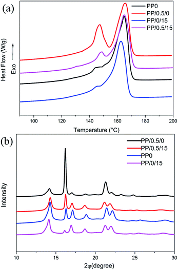
The crystallization exotherms of microfibrillar blends with the same thermal and mechanical history are shown in Fig. 4; in this figure, the influence of β-NA and PET fibers on the crystallization behavior of the samples under mild conditions is clearly reflected. The sole addition of PET fibers markedly improves the onset crystallization temperature (Tonset) from 125.2 °C (PP0) to 130.1 °C (PP/0/15) and the peak crystallization temperature (Tp) from 116.8 °C to 125.3 °C. These findings reveal the fine nucleating ability of the PET fibers. The same phenomenon is observed in microparts with β-NA. Thus, the iPP melt can crystallize at increased temperatures during the cooling process. The PP/0.5/15 micropart presents the highest Tonset and Tp among the MIM specimens studied as a result of the synergetic effects of PET fibers and β-NA.The influence of β-NAs and PET fibers on the melting behavior of iPP is studied using DSC and WAXD. Fig. 5 shows the DSC heating curves, and the WAXD profiles of the samples with the same thermal history. Note that multiple peaks emerge in the DSC heating profiles of all samples. According to previous reports,42 the melting peaks in the temperature range lower than 150 °C are classified as β-phase melting peaks, while the melting peaks that emerge above 165 °C are the α-phase melting peaks. It also can be seen that the positions of the diffraction peaks (2θ = 16.1°) for MIM samples are similar, indicating the existence of β-form crystals in all the microparts, whereas addition of β-NAs can markedly increase the intensity of the β-crystal characteristic peaks.
 | ||
| Fig. 5 (a) DSC heating curves of the microparts, and (b) The 1D-WAXD curves of micro-injection molded samples. | ||
Fig. 6 shows the crystal content of the MIM samples as determined through wide-angle X-ray diffraction (WAXD) and differential scanning calorimetry (DSC). It is obvious that the values of β-crystal content obtained from WAXD analysis are relatively higher than that calculated from DSC analysis. Nevertheless, the values calculated from both DSC and WAXD methods are all available to make a relative comparison. As the amount of β-NA loaded into the micropart increases, the β-crystal content of the micropart also markedly increases, likely because of the strong heterogeneous nucleating ability of β-NA to induce β-crystal formation. However, this content becomes smaller when PET fibers are added to the β-NA nucleated iPP samples. The β-phase content diminishes because some of the PET microfibrils exhibit nucleating capability for the iPP α-phase and shear-induced oriented α-nuclei are preserved by addition of PET fibers. Thus, the content of α-crystals increases and the content of β-crystals decreases.
3.3 Distribution of crystalline morphologies
Discussion of the crystalline morphologies of the matrix formed under the combined effects of shear flow field, β-NA, and PET fibers, which remain poorly understood thus far, is worthwhile. To investigate effects on the crystalline structures of MIM samples, polarized light microscopy experiments were carried out. As shown in Fig. 7, all of the samples possess a typical “skin–core” morphological structure. Addition of β-NA and PET fibers can increase the thickness of the oriented layer (as shown in Fig. 8), thereby inevitably improving the mechanical behavior of the microparts. However, the morphologies in the core layers of the MIM specimens vary considerably. In samples PP0 and PP/0.5/0, the core layers present an isotropic morphology. In other words, the morphology of these samples consists of numerous spherulites. Spherulite sizes in the PP/0.5/0 sample are markedly smaller than those in PP0 because β-NA exerts a strong heterogeneous nucleating effect on the iPP matrix and grain refinement occurs. A pronounced semi-cylindritic layer with high orientation levels is observed between the skin and core layers (indicated by red arrows in Fig. 7a and b). Semi-cylindritic structures that are epiphytic on the surface of the skin layer and grow toward the core layer are composed of β-cylindrulites. Unfortunately, the crystalline structures in microparts with PET fibers are indiscernible in the present case. | ||
| Fig. 7 PLM images of the morphologies of the MIM samples along the flow direction: (a) PP/0/0, (b) PP/0.5/0, (c) PP/0/15, and (d) PP/0.5/15. | ||
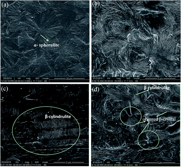
To obtain a clear understanding of the crystalline and oriented structures in the inner region of microparts, Fig. 9 shows images obtained through high-resolution SEM. Considerable divergence in the inner region can be clearly observed in all specimens. The inner region of PP0 and PP/0.5/0 are occupied by α- and β-spherulites, respectively, as shown in Fig. 9a and b. Particularly for PP/0.5/0, the lamellae of β-crystals are nearly randomly oriented. The spherulite size of PP/0.5/0 is evidently smaller than that of PP0 because of the strong nucleating ability of β-NA. According to Fig. 9c, some β-cylindrulites may be observed in the inner region of PP/0/15, a phenomenon that is ascribed to the relaxation time of iPP chains in the inner region becoming longer because of the presence of PET fibers in the blends, which helps maintain the chain orientation and initiates formation of oriented cylindrulite structures.43,44 Fig. 9d presents the inner region morphology of PP/0.5/15; here, the majority of the lamellae is oriented β-crystals, which are packed closely and intersect with one another. Due to the strong ability of β-NA to induce crystallization, the oriented chains that reside adjacent to it can be absorbed to form β-crystals during crystallization. Thus, epitaxial growth of β-crystals on α-nuclei is inhibited and the oriented β-crystals are preserved. Therefore, cylindrulites are scarcely found in the inner region of PP/0.5/15. Unexpectedly, some oriented β-crystals epiphytic on the surface of PET fibers can also be observed. To understand the formation mechanism of oriented structures in blends with PET fibers better, a possible schematic is depicted in Fig. 10. In the beginning, the chains are oriented along the flow direction (Fig. 10a and c). Then, the oriented chains suffer from relaxation because of high temperature. After undergoing the shear flow and adding the PET fibers, the orientation of iPP chains can be maintained, which is beneficial to initiate the formation of cylindrulite structure. As a result, more β-cylindrulites (Fig. 9c and 10b) are formed in the inner region of PP/0/15 and they are surrounded by α spherulites. During the micro-injection molding, the PET microfibrillar network functions effectively as a solid wall and redistributes the flow field.45,46 Thus, iPP molecules undergo confined flow on the surface of the PET fibers, which can induce the formation of oriented β-crystals epiphytic on the surface of PET fibers (Fig. 10b). However, such structures are rarely found in the inner region of PP/0/15 (Fig. 9c). Due to the strong ability of β-NA to induce crystallization, the oriented chains that reside adjacent to it can be absorbed to form β-crystals during crystallization. Hence, the synergetic effects of shear stress and β-NA induce formation of more oriented β-crystals epiphytic on the surface of PET fibers (Fig. 10d), in accordance with the result of Fig. 9d. Such superstructures are seldom directly observed in iPP/PET microparts, and we can reasonably expect that the presence of these structures improves interfacial adhesion and, in consequence, enhances mechanical properties.47
 | ||
| Fig. 9 SEM images reflecting the morphology of the inner region of the MIM samples along the flow direction: (a) PP0, (b) PP/0.5/0, (c) PP/0/15, and (d) PP/0.5/15. | ||
 | ||
| Fig. 10 Structure models of iPP in presence of an in situ microfibrillar network and a β-nucleating agent. “NB” represents the common blends, and “M” represents the flow direction. | ||
3.4 Orientation analyses
The orientation degree of all MIM samples in the thickness direction was characterized by 2D-WAXD, as shown in Fig. 11. The diffraction rings of the MIM samples exhibit a sharp contrast. Varying arc-like diffractions indicate that the c-axes of the iPP lamellae are oriented along the flow direction. The Debye rings consist of (110)α, (300)β, (040)α, (130)α, (111)α, (−131)α/(311), (060)α, and (220)α from the inner to outer circle. The diffraction patterns of the (040)α lattice plane in all samples present sharp arcs, which are indicative of the intensive orientation of molecular chains in these samples. Strong reflections of (hk0) planes of iPP on the equator indicate that the molecular chains of iPP are preferentially oriented along the shear direction, for all compositions. Four (110) reflections around the meridian also emerge in the (110) plane of iPP, indicating a lamellar branching through homoepitaxy between α-crystals themselves.48–51 These arise from the iPP component daughter lamellar regions (a-axis parallel to the meridional direction) and related to the iPP parent lamellar regions (c-axis parallel to the meridian). The epitaxial orientation relationship was first established in α-crystal quadrate some years ago and later explained on a molecular basis by Lotz et al.49 In daughter lamella, the molecular chains are oriented perpendicular to flow direction.In this article, the orientation degree calculated mathematically using Picken's method from the (040) reflection of WAXD for iPP40 is presented in Table 2. The orientation degree obviously decreases with individual addition of PET fibers. iPP melt movement is confined to microchannels or pores formed by the microfibrillar network, that is, the PET fiber network performs an important function in redistributing and homogenizing the flow field of the polymer melt, which impedes the movability of iPP molecules as well as the level of molecular orientation.45 By contrast, addition of β-NA exerts a positive effect on preserving molecular orientation resulting from the effect of shear- and β-NA-induced orientation. With addition of β-NA, nucleation is enhanced under flow conditions; here, higher onset and peak crystallization temperatures (Fig. 4) hinder relaxation of oriented iPP chains.
| Samples | (040)α |
|---|---|
| PP0 | 0.914 |
| PP/0.5/0 | 0.943 |
| PP/0/15 | 0.843 |
| PP/0.5/15 | 0.901 |
Fig. 12 shows the azimuthal spreads of (300)β plane of various samples. It can be easily seen that the (300)β plane of samples with β-NAs presents three peaks. According to the literature,52 we consider that the diffraction peak which is marked by black circle represents the parent crystals, while the other two diffraction peaks which are marked by red circle represent the daughter crystals. The intensity of daughter lamella of PP0 and PP/0/15 is quite feeble. However, the intensity of daughter lamella is comparable to that of parent lamella in PP/0.5/0 and PP/0.5/15 samples. Accordingly, it can be speculated that the relative intensity of parent lamella and daughter lamella can be used to describe the orientation degree of β-crystals. Here, an orientation factor, n(300), is introduced, of which the definition is:53
 | (6) |
 | ||
| Fig. 12 The azimuthal spreads of (300)β plane of various samples: (a) PP0, (b) PP/0.5/0, (c) PP/0/15, and (d) PP/0.5/15. | ||
3.5 Mechanical properties
Changes in mechanical properties with addition of in situ PET microfibrils and β-NA are illustrated in Fig. 13. Individual doping of β-NA considerably increases the toughness of the samples and slightly increases tensile strength. Addition of microfibrillar PET exerts the opposite effect because the abundance of the PET microfibrils acting as stress transfer agents contributes to enhancement in tensile property and diminishment the content of β-crystals (Fig. 6 and 9). Interestingly, addition of microfibrillar PET to β-NA nucleated samples simultaneously improves toughness and tensile strength, which demonstrates the synergetic effects of PET fibers and β-NA. The amount of fibers added, adhesion between the fiber and the matrix, the crystalline morphology, and the content of β-crystals can affect the reinforcement of a composite (matrix and fiber) system. Thus, from the “morphology–property” relation perspective, the observed phenomenon can be explained as follows: the synergetic effects of shear stress and β-NA induce formation of more β-crystals, which can considerably improve toughness (Fig. 6). Second, the oriented layer in PP/0.5/15 is the thickest among the MIM samples studied (Fig. 8). Thicker orientated regions are generally accepted to result in higher tensile strength.54 Third, formation of β-crystals around PET fibers improves adhesion between the fibers and the matrix (Fig. 9). Finally, the molecular chains of PP/0.5/15 are oriented along the flow direction, which is beneficial for elongation along the stretch direction. Thus, mechanical properties markedly increase in PP/0.5/15 microparts. | ||
| Fig. 13 Mechanical properties of β-NA nucleated and PET fiber blended MIM samples: (a) elongation at break; (b) tensile strength. | ||
4. Conclusions and outlooks
In this study, well-defined in situ microfibrillar PET was generated via slit die extrusion, hot-stretching, and quenching. MIM technology was applied to fabricate the corresponding bars, and the effects of in situ PET microfibrils and β-NA on the hierarchical structures and tensile properties of iPP/PET microparts were investigated in detail. Results show that addition of β-NA increases the crystallization temperatures as well as the relative content of β-crystals in the MIM specimens. This phenomenon may be ascribed to the strong heterogeneous nucleating effect of β-NA on the iPP matrix. Introduction of PET microfibrils markedly increases the crystallization peak temperature, and increases the thickness of the oriented layer. This study shows that the combined effects of a strong shear flow field, β-NAs and addition of PET fibers facilitate the formation of oriented β-crystals epiphytic on the surface of PET fibers under MIM conditions. The synergetic effects of PET fibers and β-NA markedly improve the tensile property and toughness of the samples simultaneously. It is worth stressing that the exploration herein gives more insights into the hierarchical structures of iPP/PET microparts during intense shear flow, which might open a promising door to optimizing the properties of MIM parts. Moreover, the exploration also can endow the microparts with multi-functions and higher performances, which can be widely used in practical production.Acknowledgements
This paper was financially supported by State Key Laboratory of Polymer Materials Engineering (Grant no. sklpme2014-2-08), the National Science of China (51421061, 51373109), Sichuan Youth Science and Technology Foundation (2015JQ0012). We are also indebted to the Shanghai Synchrotron Radiation Facility (SSRF) in Shanghai, China for WAXD experiments.Notes and references
- Z. Lu and K. Zhang, Int. J. Adv. Manuf. Technol., 2009, 40, 490–496 CrossRef PubMed.
- S. Abbasi, P. J. Carreau and A. Derdouri, Polymer, 2010, 51, 922–935 CrossRef CAS PubMed.
- M. Heckele and W. Schomburg, J. Micromech. Microeng., 2004, 14, R1 CrossRef CAS.
- J. Giboz, T. Copponnex and P. Mélé, J. Micromech. Microeng., 2007, 17, R96 CrossRef CAS.
- S. C. Tseng, Y. C. Chen, C. L. Kuo and B. Y. Shew, Microsyst. Technol., 2005, 12, 116–119 CrossRef CAS PubMed.
- J. Giboz, T. Copponnex and P. Mélé, J. Micromech. Microeng., 2009, 19, 025023 CrossRef.
- R. D. Chien, W. R. Jong and S. C. Chen, J. Micromech. Microeng., 2005, 15, 1389 CrossRef.
- B. S. Hsiao, L. Yang, R. H. Somani, C. A. Avila-Orta and L. Zhu, Phys. Rev. Lett., 2005, 94, 117802 CrossRef.
- R. Su, J. Su, K. Wang, C. Yang, Q. Zhang and Q. Fu, Eur. Polym. J., 2009, 45, 747–756 CrossRef CAS PubMed.
- M. Fujiyama, H. Awaya and S. Kimura, J. Appl. Polym. Sci., 1977, 21, 3291–3309 CrossRef CAS PubMed.
- A. C. Liou and R. H. Chen, Int. J. Adv. Manuf. Technol., 2006, 28, 1097–1103 CrossRef PubMed.
- W. Michaeli and C. Ziegmann, Microsyst. Technol., 2003, 9, 427–430 CrossRef PubMed.
- F. Liu, C. Guo, X. Wu, X. Qian, H. Liu and J. Zhang, Polym. Adv. Technol., 2012, 23, 686–694 CrossRef CAS PubMed.
- Z. Lu and K. Zhang, Polym. Eng. Sci., 2009, 49, 1661–1665 CAS.
- S. Saikrasun and T. Amornsakchai, J. Appl. Polym. Sci., 2006, 101, 1610–1619 CrossRef CAS PubMed.
- D. Dutta, H. Fruitwala, A. Kohli and R. Weiss, Polym. Eng. Sci., 1990, 30, 1005–1018 CAS.
- A. Isayev and M. Modic, Polym. Compos., 1987, 8, 158–175 CrossRef CAS PubMed.
- A. Mehta and A. Isayev, Polym. Eng. Sci., 1991, 31, 963–970 CAS.
- G. Pawlikowski, D. Dutta and R. Weiss, Annu. Rev. Mater. Sci., 1991, 21, 159–183 CrossRef CAS.
- S. Tjong, Mater. Sci. Eng., R, 2003, 41, 1–60 CrossRef.
- Z. M. Li, L. B. Li, K. Z. Shen, M. B. Yang and R. Huang, J. Polym. Sci., Part B: Polym. Phys., 2004, 42, 4095–4106 CrossRef CAS PubMed.
- C. Wang, Z. Zhang and K. Mai, J. Therm. Anal. Calorim., 2011, 106, 895–903 CrossRef CAS PubMed.
- B. Lotz, J. Wittmann and A. Lovinger, Polymer, 1996, 37, 4979–4992 CrossRef CAS.
- S. V. Meille and S. Brückner, Nature, 1989, 340, 455–457 CrossRef PubMed.
- J. Li, C. Zhou and W. Gang, Polym. Test., 2003, 22, 217–223 CrossRef CAS.
- J. Kang, G. Weng, Z. Chen, J. Chen, Y. Cao, F. Yang and M. Xiang, RSC Adv., 2014, 4, 29514–29526 RSC.
- Y. H. Chen, G. J. Zhong, Y. Wang, Z.-M. Li and L. Li, Macromolecules, 2009, 42, 4343–4348 CrossRef CAS.
- H. Bai, F. Luo, T. Zhou, H. Deng, K. Wang and Q. Fu, Polymer, 2011, 52, 2351–2360 CrossRef CAS PubMed.
- J. Varga and A. Menyhárd, Macromolecules, 2007, 40, 2422–2431 CrossRef CAS.
- H. Chen, J. Karger-Kocsis, J. Wu and J. Varga, Polymer, 2002, 43, 6505–6514 CrossRef CAS.
- D. R. Ferro, S. V. Meille and S. Brückner, Macromolecules, 1998, 31, 6926–6934 CrossRef CAS.
- Y. H. Chen, Z. Y. Huang, Z. M. Li, J. H. Tang and B. S. Hsiao, RSC Adv., 2014, 4, 14766–14776 RSC.
- T. Asano, Y. Fujiwara and T. Yoshida, Polym. J., 1979, 11, 383–390 CrossRef CAS.
- X. Sun, H. Li, J. Wang and S. Yan, Macromolecules, 2006, 39, 8720–8726 CrossRef CAS.
- J. Li, W. Cheung and D. Jia, Polymer, 1999, 40, 1219–1222 CrossRef CAS.
- L. Wang and M.-B. Yang, RSC Adv., 2014, 4, 25135–25147 RSC.
- J. Zhang, L. Zhang, H. Liu, F. Liu and C. Guo, Polym.-Plast. Technol. Eng., 2012, 51, 1032–1037 CrossRef CAS PubMed.
- P. Rizzo, V. Venditto, G. Guerra and A. Vecchione, Macromol. Symp., 2002, 185, 53–63 CrossRef CAS.
- P. Rizzo, A. Spatola, A. De Girolamo Del Mauro and G. Guerra, Macromolecules, 2005, 38, 10089–10094 CrossRef CAS.
- S. J. Picken, J. Aerts, R. Visser and M. G. Northolt, Macromolecules, 1990, 23, 3849–3854 CrossRef CAS.
- A. Turner-Jones and A. Cobbold, J. Polym. Sci., Part B: Polym. Phys., 1968, 6, 539–546 CrossRef CAS PubMed.
- M. Fujiyama and K. Azuma, J. Appl. Polym. Sci., 1979, 23, 2807–2811 CrossRef CAS PubMed.
- Y. An, L. Gu, Y. Wang, Y.-M. Li, W. Yang, B.-H. Xie and M.-B. Yang, Mater. Des., 2012, 35, 633–639 CrossRef CAS PubMed.
- Y. An, R.-Y. Bao, Z.-Y. Liu, X.-J. Wu, W. Yang, B.-H. Xie and M.-B. Yang, Eur. Polym. J., 2013, 49, 538–548 CrossRef CAS PubMed.
- G. J. Zhong, L. Li, E. Mendes, D. Byelov, Q. Fu and Z.-M. Li, Macromolecules, 2006, 39, 6771–6775 CrossRef CAS.
- X. Yi, C. Chen, G. J. Zhong, L. Xu, J. H. Tang, X. Ji, B. S. Hsiao and Z.-M. Li, J. Phys. Chem. B, 2011, 115, 7497–7504 CrossRef CAS PubMed.
- M. Zhang, J. Xu, Z. Zhang, H. Zeng and X. Xiong, Polymer, 1996, 37, 5151–5158 CrossRef CAS.
- F. Padden Jr and H. Keith, J. Appl. Phys., 1973, 44, 1217–1223 CrossRef PubMed.
- B. Lotz and J. Wittmann, J. Polym. Sci., Part B: Polym. Phys., 1986, 24, 1559–1575 CrossRef CAS PubMed.
- R. Su, K. Wang, P. Zhao, Q. Zhang, R. Du, Q. Fu, L. Li and L. Li, Polymer, 2007, 48, 4529–4536 CrossRef CAS PubMed.
- B. He, X. Yuan, H. Yang, H. Tan, L. Qian, Q. Zhang and Q. Fu, Polymer, 2006, 47, 2448–2454 CrossRef CAS PubMed.
- Z. Cai, Y. Zhang, J. Li, F. Xue, Y. Shang, X. He, J. Feng, Z. Wu and S. Jiang, Polymer, 2012, 53, 1593–1601 CrossRef CAS PubMed.
- Y. Zhang, L. Zhang, H. Liu, H. Du, J. Zhang, T. Wang and X. Zhang, Polymer, 2013, 54, 6026–6035 CrossRef CAS PubMed.
- S. Liang, K. Wang, H. Yang, Q. Zhang, R. Du and Q. Fu, Polymer, 2006, 47, 7115–7122 CrossRef CAS PubMed.
| This journal is © The Royal Society of Chemistry 2015 |


