Combined experimental and computational insights into the key features of L-alanine L-alaninium picrate monohydrate: growth, structural, electronic and nonlinear optical properties

Shabbir Muhammad ac, Mohd. Shkir *ac, S. AlFaify ac, Ahmad Irfan bc and Abdullah G. Al-Sehemi bc
aDepartment of Physics, College of Science, King Khalid University, P.O. Box 9004, Abha 61413, Saudi Arabia. E-mail: shkirphysics@gmail.com; shkirphysics@kku.edu.sa; Fax: +966 7 241 8319; Fax: +91 9911769335; Tel: +966 530683673
bDepartment of Chemistry, Faculty of Science, King Khalid University, P.O. Box 9004, Abha 61413, Saudi Arabia
cResearch Center for Advanced Materials Science (RCAMS), King Khalid University, P.O. Box 9004, Abha 61413, Saudi Arabia

Received 28th March 2015 , Accepted 1st June 2015

First published on 1st June 2015


Abstract

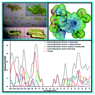
In the current work, we spotlight the novel key features of L-alanine L-alaninium picrate monohydrate (LALAPM) using a dual approach comprised of experimental and computational techniques. Single crystals of LALAPM have been grown indigenously in three different ratios (1[thin space (1/6-em)]:[thin space (1/6-em)]1, 1[thin space (1/6-em)]:[thin space (1/6-em)]5, 2[thin space (1/6-em)]:[thin space (1/6-em)]1) through a slow cooling technique. The formations of different types of crystals were recorded during the growth process and found to vary significantly from each other. The grown crystals were subjected to single crystal powder X-ray diffraction analysis to confirm their respective crystal structures. Additionally, ultraviolet-visible-near infrared, diffuse reflectance measurements and optical parameter analysis were performed. The state-of-art computational techniques were used to get the ground state molecular geometry of LALAPM at the B3LYP/6-31G* level of theory. Different important electro-optical parameters (complementary to the experimental results) including IR, Raman, and UV-visible spectra have been calculated at the same level of theory. The polarizability and first hyperpolarizability (both static and dynamic) were calculated to see the potential applications of LALAPM in nonlinear optics. Furthermore, several novel molecular level insights have been obtained in the form of the total and partial density of states, the HOMO–LUMO gap and electrostatic potential maps etc. The obtained quantum chemical findings were compared with experimental results. The static and frequency dependent dynamic first hyperpolarizability values of the LALAPM molecule are found to be 8.06 × 10⁻³⁰ and 10.24 × 10⁻³⁰ esu that are about 37 times and 59 times larger than those of the prototype urea molecule, respectively, at the same B3LYP/6-31G* level of theory. The obtained results indicate that the titled compound contains good nonlinear optical properties and can be treated as a good contender for optoelectronic device fabrications.


1. Introduction

The field of linear and nonlinear optics has gained significant momentum since the discovery of the photon as the fastest carrier of information. Over the past few decades, several types of materials have been functionalized to match the demand of functional materials with efficient optical and nonlinear optical (NLO) properties.1,2 Organic materials are excellent candidates for hi-tech laser applications due to their fast and giant nonlinear response over an extensive frequency range, inherent synthetic flexibility and large optical damage threshold for laser power and low frequency dispersion. One of the key advantages of these organic materials is that they permit one to modify the chemical structure with large physical structural diversity and properties for the desired NLO properties.3–9 Optical single crystals are being used in devices for high optical data storage, frequency conversion, fusion research etc. So it is very important to grow new as well as existing optical single crystals with a high level of perfection for device fabrication because of their needful applications. Crystals of the amino acid family are of great interest due to their attractive nonlinear optical properties.10–12 When an organic acid is mixed with amino acid, usually its nonlinear optical properties increase due to the zwitterionic nature associated with an enhanced transparency range.13,14 Picric acid is an organic acid which forms molecular charge transfer complexes with aromatic compounds through electrostatic or hydrogen bonding interactions,15,16 and there are many reports containing picric acid with many organic molecules which have interesting properties,17–20 due to the presence of active π and ionic bonds.21

Picric acid and L-alanine are good NLO materials that show excellent NLO efficiencies better than that of KDP.22–25 These compounds also have a good tendency to form new compounds by reacting with each other as well as with other organic and inorganic materials such as diglycine picrate, L-leucine L-leucinium picrate,6,18,26,27 β-alanine β-alaninium picrate,28 2-aminopyridinium picrate,4,29DL-phenylalanine DL-phenylalaninium picrate,30 and DL-methionine DL-methioninium picrate.31 Furthermore investigations of these crystals were also carried out in some previous studies,32,33 including DL-valine DL-valinium picrate.34 Salts of amino acids are an interesting class from different points of view i.e. as species with very short hydrogen bonds, crystals in which phase transitions are possible due to presence of dimeric cations, as well as many of them are nonlinear optical materials. There are more than 20 new salts that have been recently reported with dimeric cations.35

Recently, along the above lines for picrate compounds, Ghazaryan et al.,36 reported the L-alanine L-alaninium picrate. They obtained single crystals of L-alanine L-alaninium picrate monohydrate. They also performed IR and single crystal analyses of the L-alanine + picric acid + H2O system, characterized the final product as an L-alanine L-alaninium picrate system with two more phases and revisited the previous findings on the L-alanine + picric acid + H2O system.37 Very recently, another experimental study was performed to highlight the nucleation kinetics, growth and hardness parameters of LALAPM.38

The above recent reports specify the preliminary experimental findings about LALAPM. Nevertheless, the first use of a dual approach comprising of experimental and computational techniques will spotlight the several novel features of LALAPM including its geometrical parameters, configuration of frontier molecular orbitals (FMOs) as well as its nonlinear optical properties, which are still not known to the scientific community. For instance, the corner stone of the present investigation is to provide molecular level insights to explore the potential of LALAPM for advanced functional materials in the framework of combined computational and experimental investigations. Furthermore, bulk crystal growth using a slow cooling technique, morphology, and optical analysis using UV-vis-NIR and diffuse reflectance (DR) is performed. Thus the aims of present investigation are manifold. The growth of L-alanine L-alaninium picrate monohydrate (LALAPM) single crystals is performed using a slow cooling technique in three different ratios and subsequently the indigenously grown single crystals are subjected to the single crystal and powder X-ray diffraction analyses. During the single crystal growth process, the morphology is also recorded randomly with an inverted microscope. The grown crystals were subjected to UV-vis-NIR spectroscopy measurements. Additionally, the molecular geometry optimization and calculation of electro-optical properties have been performed using different advanced theoretical methods to shed light on the bonding features among L-alanine, L-alaninium, picric acid and water molecules. The IR, Raman and UV-visible spectra, polarizability, and first hyperpolarizability are calculated using DFT, TD-DFT and finite field (FF) methods, respectively. The obtained computational and experimental results are discussed and compared with each other wherever possible.

2. Experimental and computational details

2.1. Synthesis, solubility and crystal growth

For the synthesis of the titled compound, L-alanine and picric acid of high purity were purchased from Loba Chemie Pvt. Ltd and were taken in different ratios such as 1[thin space (1/6-em)]:[thin space (1/6-em)]1, 1.5[thin space (1/6-em)]:[thin space (1/6-em)]1 and 2[thin space (1/6-em)]:[thin space (1/6-em)]1, and mixed in double distilled water. The calculated amounts of both materials were weighed by an electronic balance (METTLER TOLEDO) of high accuracy and taken into three good quality Borosil glass beakers. For complete dissolution, homogenization and proper chemical reaction of all three materials, they were stirred well at a temperature of 38 °C with the help of a Eurotherm temperature controlled magnetic stirrer. Finally, all the prepared solutions were kept at room temperature and allowed to evaporate to yield the yellow crystalline powder salt of LALAPM of three different ratios. The chemical reaction of LALAPM (2[thin space (1/6-em)]:[thin space (1/6-em)]1) formation is given below:
image file: c5ra05533d-t1.tif

Before pursuing the crystal growth of the titled material it is very important to study its solubility. Therefore we determined the solubility curve of LALAPM using deionized water as a solvent in the temperature range of 28–58 °C. A gravimetric method39 was used to calculate the required amount of LALAPM which was added to saturate the aqueous solution at 28 °C and we repeated this process for different temperatures. The solubility curve is represented in Fig. 1(a) and found to be in close agreement with an earlier report.38 LALAPM shows a positive solubility-temperature gradient in an aqueous solution and this is a positive point for growing the bulk crystals. From its solubility curve it is clear that the titled material is highly soluble in water hence it will be easy to grow the bulk single crystals of LALAPM from the solution technique. The bulk growth of L-alanine L-alaninium picrate monohydrate (LALAPM) was carried out using a slow cooling technique. For the growth of single crystals of LALAPM, the saturated solutions of all the synthesized ratios were prepared above room temperature i.e. 32 °C using double distilled water with continuous stirring for more than 2 days to get the homogeneous and transparent solution of yellow color.


image file: c5ra05533d-f1.tif
Fig. 1 The (a) solubility curve and (b) grown single crystals of LALAPM and LA from the same solution (2[thin space (1/6-em)]:[thin space (1/6-em)]1).

The prepared solutions were filtered in three other beakers, covered with a perforated lid and kept in a constant temperature bath (CTB) at the same prepared temperature, with a temperature accuracy of ±0.01 °C, and left for one day. After one day, we started to reduce the temperature of the CTB by 0.5 °C per day for slow cooling and during this time the solutions were observed with an inverted microscope. The nucleation started in all the beakers after four days when the temperature reached 30°. We then reduced the temperature reduction rate to 0.1 °C per day for further growth. Good quality, large size and different shaped single crystals were harvested from the mother solutions after a span of 20 days as shown in Fig. 1(b). They were isolated and found to be large size L-alanine (containing picric acid) as well as comparatively small size LALAPM, as was also mentioned by Ghazaryan et al. in their recently published article on the titled compound. The well-known floatation method was applied to determine the density of the grown crystals using a liquid-mixture of bromoform and carbon tetrachloride and found to be 1.5168 g cm−1 which is in good agreement with the reported value i.e. 1.517 g cm−1 (ref. 36) and warrants that the grown crystals are LALAPM.

2.2. Characterization

The morphology of the growing crystals for all three solutions was randomly recorded with the help of an inverted microscope made by Motic, which interfaced with a computer system. The pictures were captured using a camera (Moticam 2300, 3.0 Mpixel USB 2.0) attached to the microscope. The objective lens of magnification PH 10×/0.25, ∞/1.1WD7.5 was used in our experiments for the present investigation. The random morphology of the growing crystals is shown in Fig. 2 and indicates that there are different types of crystal formations happening including L-alanine (with/without modified morphology due to the addition of picric acid)40,41 and LALAPM in the same solution. These crystals can also be matched with grown crystals as shown in Fig. 1(b).
image file: c5ra05533d-f2.tif
Fig. 2 Randomly recorded morphology of the LALAPM (2[thin space (1/6-em)]:[thin space (1/6-em)]1) solution.

To confirm the crystal system and lattice parameters of grown crystals, their single crystal X-ray diffraction analyses were performed. Single crystals of suitable size (0.17 × 0.20 × 0.45 mm) were cut from a larger specimen. The crystal structure was confirmed by using the intensity data collected using a Bruker Kappa Apex II diffractometer (graphite-monochromated, MoKα = 0.71073 Å) at 296 K. The least-squares refinement of the diffraction angles of 25 reflections was performed to obtain the cell data. For further confirmation of the crystal system and to determine the lattice parameters of both types of grown single crystals, they were crushed into a fine powder of ∼150 μm size and subjected to a PW3710 based Philips Analytical Powder X-ray diffractometer with nickel filtered CuKα radiation operated at 35 kV, and 30 mA, and the scanning was performed with a step size of 0.02° in the angular range of 5–70° for 2-theta.

To calculate the various optical parameters of the LALAPM single crystal and LA crystal (for comparison), their optical absorbance values were recorded in the wavelength range of 200–800 nm at 300 K by using a JASCO V-570 UV-vis-NIR spectrophotometer. A Shimadzu UV-vis-NIR spectrophotometer model (UV-3600) was used to record the diffuse reflectance (DR) in the wavelength range from 230–2300 nm of the powdered samples of the grown crystals with an integrating sphere attachment.

2.3. Computational details

All the calculations were performed using the Gaussian suite of programs.42 The molecular geometry of LALAPM was optimized by density functional theory (DFT) using the B3LYP/6-31G* level of theory. In addition to B3LYP, a highly correlated method MP2 and a recently developed M06 functional were also applied to optimize the initial geometry for comparison of the results with B3LYP and experiment. The equilibrium geometry of LALAPM was successfully achieved corresponding to the true minimum on the potential energy surface (PES) by solving a self-consistent field equation. The optimized structural parameters of LALAPM were used to characterize all the stationary points as minima using IR and Raman frequency calculations. The IR and Raman spectra of LALAPM were calculated by taking the second derivative of the energy that was computed analytically. Similarly, all other electronic properties including the UV-visible spectra, dipole moment, polarizability, and first hyperpolarizability of LALAPM were calculated at the same level of theory. Time dependent density functional theory (TD-DFT) was used to calculate the excitation energies with a relatively larger basis set of 6-31+G*. The static first hyperpolarizability (βtot) and its components for LALAPM were calculated with the finite field (FF) method. The FF method has been broadly applied to investigate the NLO properties of organic materials because this methodology can be used in concert with the electronic structure method to calculate β values. Recently, several βtot amplitudes calculated with this method were found to be in semi-quantitative agreement with the experimental structure property relationship.8,43 In the FF method, a molecule is subjected to a static electric field (F), and the energy (E) of the molecule is expressed by the following equation:
 
image file: c5ra05533d-t2.tif(1)
where E(0) is the energy of the molecule in the absence of an electric field, μ is the components of the dipole moment vector, α is the linear polarizability, β and γ are the first and second hyperpolarizabilities, respectively, while i, j and k label the x, y and z components respectively. It is clear from the above equation that the values of μ, α, β, and γ can be obtained by differentiating E with respect to F. Furthermore the chemical hardness (η) of the LALAPM molecule was calculated using its HOMO and LUMO energy values. These parameters of the titled compound were obtained using Koopman’s theorem44 for closed-shell molecules, the chemical hardness of any molecule can be calculated by the following relation (an approximation for absolute hardness η was developed earlier45–47) as given below:
 
image file: c5ra05533d-t3.tif(2)
where A (= −EHOMO) is the ionization potential and I (= −ELUMO) is the electron affinity of the molecule. The values of A and I of LALAPM were calculated with the B3LYP/6-31G* level of theory.

3. Results and discussion

3.1. Single crystal and powder X-ray diffraction (PXRD) studies

Single crystal and powder X-ray diffraction (PXRD) analysis were performed for the confirmation of the crystal system, space group, and lattice parameters of LALAPM as well as L-Alanine and compared with the reported values.36,48 The recorded PXRD data was used as the input for calculating the lattice parameters using different softwares like ‘CHECKCELL’,49 and ‘POWDERX’,6,49 from the obtained results it was confirmed that the grown crystals of LALAPM belong to a monoclinic crystal system with a space group of P21. The unit cell parameters determined by single crystal XRD as well as refined by the above said software and the reported values are listed in Table 1. The calculated parameters were found to be in good agreement with the earlier reported data.36
Table 1 Lattice parameters of the LALAPM crystals
Lattice parameters Reported work Present work (LALAPM)
L-Alanine51 LALAPM36 Single crystal XRD data Powder XRD refined data by POWDERX Powder XRD refined data by CHECKCELL
a 5.784(1) Å 8.268(2) Å 8.268(5) Å 8.29811 Å 8.2345 Å
b 6.032(1) Å 7.510(2) Å 7.510(6) Å 7.51048 Å 7.5696 Å
c 12.343(1) Å 15.540(3) Å 15.540(4) Å 15.60090 Å 15.5254 Å
V 430.636 Å3 931.0(3) Å3 931.0 Å3 936.749 Å3 937.243 Å3
α 90 90° 90° 90° 90°
β 90 105.23° 105.23° 105.540° 104.42°
γ 90 90° 90° 90° 90°
System Orthorhombic Monoclinic Monoclinic Monoclinic Monoclinic
S.G P212121 P21 P21 P21 P21


3.2. Molecular geometry analysis

The initial molecular geometry of the LALAPM molecule was extracted from single crystal data and subjected to full optimization at three different levels of theory including B3LYP/6-31G*, M06/6-31G* and MP2/6-31G* as presented in Table 2 (labeling is according to Fig. 3). The important bond parameters are presented in Table 2 and the experimentally reported values are also given for comparison. Overall there is reasonable agreement among the bond lengths at different levels of theory as well as with the experimental bond values except hydrogen bonds involving H2O molecules, which perhaps do not have a well-defined position in the experimental structure. In the present study, the optimized unit contains one picrate anion, one zwitterionic L-alanine and one L-alaninium cation. The hydrogen bond between the zwitterionic L-alanine and the L-alaninium cation is about 1.727 Å, near to its experimental value of 1.70 Å. The dimeric distance between O1–O3 is about 2.498 Å, which is also in semi-quantitative agreement with its experimental value of 2.553 Å as seen from Table 3.
Table 2 The bond lengths [Å] and bond angles [°] of the optimized LALAPM molecule at different functionals with a 6-31G* basis set
Bond lengths Exp.36 Cal. Bond angles Exp.36 Cal.
B3LYP M06 MP2 B3LYP M06 MP2
O1–O13 2.553 2.498 2.489 2.518 O1–O3–O2 125.96 125.65 128.167 128.02
C16–O13 1.255 1.257 1.253 1.264 O13–C16–O15 125.65 128.86 128.77 128.97
C16–O15 1.258 1.238 1.251 1.264 O13–C16–C17 112.43 115.15 115.59 115.86
C28–O27 1.255 1.247 1.237 1.262 C16–C17–N23 107.8 105.14 105.36 105.18
C41–N42 1.466 1.447 1.463 1.459 O27–C28–C29 123.47 125.85 125.65 127.68
O15–O45 2.836 2.718 2.723 2.723 C29–C28–C41 112.24 112.351 112.10 111.46
H14⋯O13 1.703 1.727 1.446 1.459 C28–C41–C39 124.85 124.06 124.25 125.79
H11⋯O45 1.948 1.493 1.545 1.544 O1–H14–O13 171.00 169.51 167.45 168.64
H46⋯O15 2.01 1.743 1.763 1.760 O31–N30–O32 120.67 122.19 120.95 121.17
H47⋯O32 2.09 2.04 2.03 2.024 O37–N36–O38 123.97 124.40 124.62 124.41
H25⋯O27 1.950 1.638 1.657 1.634 O44–N42–O43 121.03 124.66 124.91 125.49


image file: c5ra05533d-f3.tif
Fig. 3 The optimized structure of LALAPM monohydrate at the B3LYP/6-31G* level of theory with white, red, gray and blue atoms representing the H, O, C, and N atoms, respectively.
Table 3 The calculated IR and Raman frequencies at the B3LYP/6-31G* level of theory and experimentally reported IR and Raman frequencies with their appropriate assignments for LALAPM
IR frequencies (cm−1) Raman frequencies (cm−1) Assignments
Calculated Experimental36 Calculated Experimental36
3499 3570 3505 3567 OH (H2O) asymmetric stretching
3386, 3308 3416 3393, 3314 3423 OH (H2O) symmetric Stretching
3239 3192 3245 3210 NH (NH3+) asymmetric stretching
3161 3104 3167, 3141 3104 NH (NH3+) symmetric stretching
3054, 3028, 3011 3000 CH (CH) stretching vibration
2954, 2936 2943 2960, 2942 2982, 2971, 2950 CH (CH3) stretching vibration
2730 2730, 2603, 2546 2725 2748, 2606 Sum tones
2175, 2054 1934, 1884 2180, 2080 2258 Sum tones
1760 1837 1763 1839 Sum tones
1708 1703 1685 1702 C[double bond, length as m-dash]O (COOH) stretching vibration
1656 1633 1659 1634 ν(C[double bond, length as m-dash]C) ring; νas(COO)
1630 1614 1628 1611 ν(C[double bond, length as m-dash]C) ring; δas(NH3+)
1578 1580 1558 NH3+ bending
1561, 1526 1566, 1539 1564 1554 ν as(NO2); δas(NH3+)
1483 1501, 1490 1512 1493 δ s(NH3+)
1478, 1458 1460 1467 δ as(CH3)
1422 1429 1420 ν s(COO)
1396, 1362 1384, 1366 1350 1367 δ s(CH3)
1327 1340, 1321 1330, 1312 1334, 1318 ν s(NO2)
1275, 1258 1299, 1260 1260 1301, 1271 ν(C–O) phenolic; ν(C–OH)
1197, 1180, 1154 1194, 1163 1200, 1156 1169, 1147 δ(CH) in plane
1137, 1111 1118 1115 1123 ρ(NH3+)
1065 1083 1065 1085 N(CN)
972 1003 1000, 975 1010, 992 N(CN)
930 946 931 949
905 915 910 920 P(CH3)
851 844 850 846 ν(C–C)
821 801 825 δ(NO2)
799 790, 772 793 δ(CH) out-of-plane
765, 747 746 750 764, 751 ω(NO2)
713 712, 696 706 721, 698 Δ(COO)
695, 678, 652, 626 656, 618 670, 630 676, 660, 622
574 552 575 555
522 529, 514 532 530517 ρ(NO2)
427 472 437 427 ρ(COO)
358 Not mentioned 367 400, 380, 361 Skeletal deformation
332 Not mentioned 324 338
298 Not mentioned 300 302, 276
272 Not mentioned 185 203 Lattice vibrations
220 Not mentioned 160 164
124 Not mentioned 99, 56, 13


The nitro (NO2) groups of the picrate ion do not completely follow the pattern with their torsion angles of 122°, 124° and 125° as in the usual picrate molecule. The H2O molecule is held between zwitterionic L-alanine, one L-alaninium cation and a picrate anion through hydrogen bonds. The water molecule forms one hydrogen bond with the picrate anion while two hydrogen bonds with the L-alanine and L-alaninium molecules. The water molecule can be envisaged as a connecting bridge among L-alanine, the L-alaninium cation and the picrate anion through hydrogen bonding.

3.3. IR and Raman spectroscopic analyses

A vibrational spectroscopic study is a highly effective tool in identifying the functional groups present in any molecule as well as in molecular conformational analysis and in the analysis of reaction kinetics.50 The vibrational spectra (IR and Raman) of the LALAPM molecule were determined using the B3LYP/6-31G* level of theory as shown in Fig. 4(a) and (b), respectively. It is well known that due to the combination of electron correlation effects and basis set deficiencies, calculated harmonic frequencies were found to be higher than the experimentally observed frequencies. Notwithstanding with the level of calculations, it is usual to scale down the calculated harmonic frequencies in order to develop the model with experimental values. The scaled frequencies minimize the root mean square difference between the calculated and experimental frequencies for bands with definite identifications. All the observed wavenumbers and their corresponding assignments along with the experimentally reported assignments are listed in Table 3. From Fig. 4, it is clear that there are characteristic vibrations in the titled molecule due to the presence of zwitterionic L-alanine and L-alaninium moieties as well as the picrate anion and water molecules.36 It is expected that the stretching vibrations of the OH bonds of the water molecules, N–H and C–H bonds of the NH3 and CH3 and CH groups of the alanine and alaninium moieties as well as the C–H bonds in the picrate anions appear in the high-frequency region. The pictorial presentation of some of the main vibrations such as O–H, N–H, C–H, C–C, CH3, NO2 are shown in Fig. 5. Because of strong hydrogen bonding O–H⋯O in the dimeric cation, absorption due to the stretching vibration of O–H bonds in the L-alaninium moieties is not expected in the high frequency region.36
image file: c5ra05533d-f4.tif
Fig. 4 Calculated (a) IR and (b) Raman spectra of the LALAPM molecule at the B3LYP/6-31G* level of theory.

image file: c5ra05533d-f5.tif
Fig. 5 The pictorial representation of the vibrational modes with their displacement vectors (light blue arrows) at the B3LYP/6-31G* level of theory.

The O–H stretching vibrations of the crystallization water are assigned to the absorption bands observed at ∼3499, 3386, and 3308 cm−1 and the respective Raman-bands. While experimentally these bands were observed at 3570, and 3416 cm−1 in the IR and 3567 and 3423 cm−1 in the Raman spectra.36 The above assigned band positions are in accordance with the relatively weak hydrogen bonds formed by water molecules (see Fig. 5). The bands observed in the IR and Raman spectra both just below 3300 cm−1 are mainly caused by the hydrogen bonded symmetric and asymmetric stretching of the NH3 groups, respectively (see Fig. 5). The peaks observed at ∼3054, 3028, 3011, 2960, 2942 cm−1 in the Raman and 2954, 2936 cm−1 in the IR spectra are assigned to the C–H stretching vibrations (see Fig. 5). While experimentally these bands were observed at 2943 cm−1 in the IR and 2982, 2971, and 2950 cm−1 in the Raman spectra.36 The bands observed at ∼2730, 2175, 2054, and 1760 cm−1 in the IR and ∼2725, 2180, 2080, and 1763 cm−1 in the Raman spectra are assigned as sum tones, these bands correlate well with an earlier report.36

Now, we discuss the bands observed below 1800 cm−1. The absorption band at ∼1708 cm−1 in the IR spectrum and the respective Raman-band at 1685 cm−1 are assigned to the stretching vibrations of the C[double bond, length as m-dash]O bond in the COOH group. While these bands were observed experimentally at 1703 and 1702 cm−1 respectively in the IR and Raman spectra.36 In the range of 1650–1400 cm−1 several characteristic bands are expected due to the picrate anion. The peaks at ∼1656, and 1630 cm−1 in the IR and 1659, and 1628 cm−1 in the Raman spectra are assigned to the C[double bond, length as m-dash]C ring stretching (see Fig. 5), asymmetric stretching and deformation of COO and NH3+ vibrations. While these bands were observed experimentally at 1633, 1614 and 1634, 1611 cm−1 respectively in the IR and Raman spectra.36 The bands at 1578 cm−1 in the IR and 1580 cm−1 in the Raman spectra are assigned to the NH3+ bending vibration. Bands at ∼1561, and 1526 cm−1 in the IR and 1564 cm−1 in the Raman spectra are assigned to the asymmetric vibration and deformation of NO2 and NH3+ respectively. While these bands were observed experimentally at 1566, 1539 and 1554 cm−1, respectively in the IR and Raman spectra.36 The bands at 1483 cm−1 and 1512 cm−1 in the IR and Raman spectra, respectively, are assigned to the symmetric deformation of NH3+ vibrations. While these bands were observed at 1501, 1490 and 1554 cm−1, respectively experimentally in the IR and Raman spectra.36 The band at ∼1460 cm−1 in the Raman spectrum is assigned to the asymmetric deformation of CH3 vibration. However, experimentally this band was found in both the IR and Raman spectra at 1478, 1458 and 1467 cm−1 respectively.36 The bands observed at 1422 cm−1 and 1420 cm−1 are assigned to the symmetric vibration of the COO group. While experimentally this band was observed only in the IR spectrum at 1429 cm−1.36 The bands at ∼1396, and 1362 cm−1 in the IR and 1350 cm−1 in the Raman spectra are assigned to the asymmetric deformation of CH3 vibrations (see Fig. 5). While these bands were observed experimentally at 1384, 1366 and 167 cm−1, respectively in the IR and Raman spectra.36 The most intensive peaks in the Raman spectrum at 1327, 1330, 1312, and 801 cm−1 and the respective peaks in the IR spectrum are assigned to the vibrations of the nitro groups. While these bands were observed experimentally at 1340, 13[thin space (1/6-em)]321 and 825 cm−1 in the Raman spectrum with respective peaks in the IR spectrum.36 This group has characteristic vibrations active in both the IR and Raman spectra. The bands at 1275, 1258 cm−1 in the IR and 1260 cm−1 in the Raman spectra are assigned to the C[double bond, length as m-dash]O phenolic and C–OH vibrations, respectively. While these bands were observed at 1299 and 1260, and 1301 and 1271 cm−1, respectively in the IR and Raman spectra experimentally.36 The bands observed at 1197, 1180 and 1154 cm−1, and 1200 and 1156 cm−1 are assigned to the in plane deformation of the CH group. While these bands were observed at 1194 and 1163, and 1169 and 1147 cm−1, respectively in the IR and Raman spectra experimentally.36 The bands at 1137, and 1111 cm−1 in the IR and 1115 cm−1 in the Raman spectra are assigned to ρ(NH3+), while these bands were observed experimentally at 1118 and 1123 cm−1, respectively. Bands at 1065 and 972 cm−1, and 1065, 1000 and 975 cm−1 in the IR and Raman spectra are assigned to the CN group vibration, while these bands were observed experimentally at 1083 and 1003, and 1085, 1010 and 992 cm−1, respectively.

The bands at 905 cm−1 and 910 cm−1 are due to ρ(CH3), 851 and 850 cm−1 are due to ν(C–C), 801 cm−1 is due to δ(NO2), 799 cm−1 is due to δ(CH) out-of-plane, 765, 747 cm−1 and 750 cm−1 are due to ω(NO2) (see Fig. 5), while these bands experimentally were observed at 746, 764 and 751, respectively, 713 and 706 cm−1 are due to δ(COO), 522 and 532 cm−1 are due to ρ(NO2), and 427 and 437 cm−1 are due to ρ(COO) vibrations. The bands observed below 400 cm−1 are assigned to skeletal and lattice vibrations in the molecule. All the vibrations approve the confirmation of LALAPM and agreed well with the experimental values.36

3.4. Optical analysis

3.4.1. UV-vis-NIR spectroscopic study. To recognize the suitability of the grown single crystals of LALAPM for optical applications the optical light absorbance is an essential factor to assess. The optical absorption spectra of the grown single crystals of L-alanine and LALAPM were recorded in the wavelength range of 200–800 nm as shown in Fig. 6(a). The optical transmission was calculated from the absorption data, which shows that the grown crystals are highly transparent in the entire testing range as shown in Fig. 6(b). The lower cut-off wavelength of LA was observed at ∼230 nm while for LALAPM two cut-off wavelengths were observed at 286 and ∼435 nm. The high percentage of transmission in the entire tested region (from 450 to 800 nm) is an essential requirement for optically active materials. The transparency of the titled compound can be compared with other reported picrate, organic, metal-organic as well as L-alanine compounds.4–7,18,51–60
image file: c5ra05533d-f6.tif
Fig. 6 UV-vis-NIR (a) absorbance (b) transmittance and (c) band gap plots of the LA and LALAPM crystals.

TD-DFT is considered to be a reliable quantum chemical technique for studying the UV-visible absorption spectra (they correspond to vertical electronic transitions) of different organic compounds.61–63 For the LALAPM molecule, different TD-DFT methods (TD-B3LYP, TD-PBE0, TD-M06, TD-CAM-B3LYP and TD-B2PLYPD) were applied to study its excitation energies in its ground state geometry, which were optimized at the B3LYP/6-31G* level of theory. The experimental electronic absorption spectrum of the titled crystal shows two bands at 286 and 435 nm (Fig. 6(a)). The UV-visible absorption spectra of LALAPM using five different TD-DFT methods with the 6-31+G* basis set are shown in Fig. 7. It can be seen from Fig. 7 that all the TD-DFT methods (except TD-CAM-B3LYP) reasonably reproduced two absorption bands as observed in the experimental absorption spectrum. TD-CAM-B3LYP is already well-known to reproduce excitation energies with a long-range charge transfer effect which is not the situation in the titled compound. Among all the five TD-DFT methods, the excitation energies calculated with TD-B3LYP are closest to the experimental values. The two predicted electronic absorption wavelengths are 312 and 414 nm with oscillator strengths (f0) of 0.133 and 0.066 at the TD-B3LYP/6-31+G* level of theory, which are in reasonably good agreement with the experimental values of 286 and 435 nm. Calculations of the optimized molecular orbital geometry of LALAPM show that the visible absorption maxima are related to the electronic transition between frontier orbitals such as the transition from the HOMO to the LUMO which will be explained in Section 3.6. The lower energy visible band observed at 435 nm (exp.) and 414 nm (theor.) could be attributed to the redistribution of electronic intramolecular charge involving π–π* transitions.


image file: c5ra05533d-f7.tif
Fig. 7 Calculated UV-vis spectra of the LALAPM molecule with different TD-DFT functionals and the 6-31+G* basis set.
3.4.2. Band gap analysis. Investigation of the optical band gap of any material is a very important parameter to gain a deep perception about their optical properties and application in optoelectronic devices. So, the understanding of optical processes is an important factor in the design and optimization of devices.

The optical band gap of the titled compound was calculated using the optical absorption data of the LA and LALAPM crystals. Initially, the optical absorption coefficient was calculated for both the crystals from the absorbance using the following relation:

 
image file: c5ra05533d-t4.tif(3)

(Eg)crys was calculated from the optical absorption coefficient αcrys near the absorption edge according to following general relation:

 
image file: c5ra05533d-t5.tif(4)
where A is a constant, h is Planck’s constant, and ν is the frequency of the incident photon. The process of optical absorption is explained by the value of an index indicated as s in the above relation. For direct allowed, indirect allowed, indirect forbidden and direct forbidden transitions the values of s correspond to 1/2, 2, 3 and 3/2, respectively.

When s = 1/2, which is assigned to a direct allowed transition then the equation will be:

 
(αhν)2 = A[ − (Eg)crys](5)

The (Eg)crys of the LA and LALAPM single crystals was calculated by extrapolating the straight line to the x-axis () as shown in Fig. 6(c). The band gap of LA was found to be 5.47 eV, while two band gaps, ∼2.86 and 4.68 eV, were found for LALAPM. Because of the large band gap of the grown crystal it can be considered as a suitable applicant for optoelectronic applications.52,58,64–66

3.4.3. Frontier molecular orbital (FMO) analysis. The frontier molecular orbitals (FMOs) play a crucial role in the reactivity of any molecule. Among FMOs, the highest occupied molecular orbital (HOMO) and lowest unoccupied molecular orbital (LUMO) are very important. These FMOs of a molecule determine the way that it interacts with other species. For the LALAPM molecule, its calculated energy gaps between the HOMO and LUMO (HOMO − 1 and LUMO + 1) are 3.517 and 4.943 eV, which are in semi-quantitative agreement with the experimental band gap values of 2.86 and 4.68 eV, respectively. The HOMO and LUMO (H–L) energy gap describes the chemical reactivity, kinetic stability and chemical softness of a molecule. According to the softness–hardness rule, molecules with larger energy gaps are known as hard molecules and they usually possess higher thermal and kinetic stabilities. Based on the H–L energy gap of the LALAPM molecule, its chemical hardness value was found to be 1.758 eV, which indicates that LALAPM has significant kinetic stability (see Table 4). As the H–L energy gap is relatively larger and the transition energy exists in the UV and near visible region, an advantage of significant transparency is obtained.
Table 4 The calculated energy values of the frontier molecular orbitals (FMOs) and their orbital energy gaps at the B3LYP/6-31G* level of theory for LALAPMa
Orbital eV
a 1 a.u. of energy = 27.211396 eV.
E HOMO −6.317
E HOMO−1 −6.964
E LUMO −2.799
E LUMO+1 −2.021
ΔEHOMO−LUMO 3.517
ΔEHOMO−1−LUMO+1 4.943


In present study, the experimental UV-visible spectrum has shown two maximum absorption wavelengths i.e. 435 and 286 nm, which are reasonably well reproduced as 414 and 312 nm at the TD-B3LYP/6-31G* level of theory, respectively. The TD-DFT calculations on the LALAPM molecule show that the lower energy electronic transition corresponds to the transition of an electron from the ground state to the first excited state with HOMO to LUMO major orbital contributions (67% configuration interaction) and the second transition involves HOMO to LUMO + 1 major orbital contributions (43% configuration interaction). The three-dimensional plots of the FMOs including the HOMO, HOMO − 1, LUMO, and LUMO + 1 orbitals are shown in Fig. 8. From Fig. 8, it can be seen that the HOMO and LUMO orbitals are mainly located on the picrate ion, which is also evident from the DOS graph. It can be seen that the electronic density is redistributed on the picrate ion during the transition, which perhaps causes the non-zero amplitude of the first hyperpolarizability. According to the composition of the second transition, a somewhat similar pattern of charge redistribution can also be seen for the HOMO and LUMO + 1 orbitals, which are involved in the second transition with relatively higher energy.


image file: c5ra05533d-f8.tif
Fig. 8 The 3-D plot of the frontier molecular orbitals of the LALAPM molecule with counter values of ±0.02 a.u.
3.4.4. Density of states. The total density of states (TDOS) and partial density of states (PDOS) were investigated for the LALAPM molecule using the AOMix wave function analysis program.67 The TDOS and PDOS were calculated to understand the role of individual molecular fragments in the bonding and electro-optical properties of the LALAPM molecule. We generate the TDOS and PDOS plots by dividing the LALAPM molecule into four fragments including L-alanine, L-alaninium, the picrate ion and water molecule fragments as shown in Fig. 9. The TDOS plot shows the population analysis per orbital and demonstrate a modest view of the makeup of the molecular orbitals in a certain energy range while the PDOS plot shows the percentage contribution of each group to each molecular orbital in the final molecule. A careful analysis of Fig. 9 shows that the picrate anion contributes more to the total number of states per interval of energy compared with the other fragments. Most of the high energy occupied states and lower energy unoccupied states around the band gap are composed of the picrate anion fragment. Similarly the contribution of L-alaninium is significant over the energy range of −6 to −7 eV and there is a further significant contribution at relatively lower energy levels around −10 to −11 eV. Additionally, it can be seen that the contributions of L-alanine and water are the smallest compared to the other fragments of the picrate anion and the L-alaninium cations. Thus, from the DOS diagram, it can be observed that the picrate anion is the main fragment in the LALAPM molecule that has significant influence on its electro-optical and nonlinear optical properties because any change in the picrate anion fragment will significantly influence the frontier orbitals (as evident from DOS graph) which will ultimately lead to a change in its electro-optical properties. Furthermore, the HOMO–LUMO (H–L) orbital energy gap was found to be dependent on the number of states per interval of energy originating from the picrate anion and can be tuned by modifying the picrate anion fragment according to the DOS diagram. Thus, the picrate anion plays a crucial role to tune the optical and nonlinear optical properties of LALAPM.
image file: c5ra05533d-f9.tif
Fig. 9 Total density of state (TDOS) and partial density of state (PDOS) plots for the LALAPM molecule.
3.4.5. Diffuse reflectance study. The diffuse reflectance of the titled crystal was measured, as shown in Fig. 10(a), and its band gap evaluated. The diffuse reflectance is a destructive method for surface measurements using a mirror like reflection from the surface of the sample. Generally the Kubelka–Munk theory is used for diffuse reflectance spectra analysis from weakly absorbing samples. In such a case the Kubelka–Munk equation at any wavelength is given by:
 
image file: c5ra05533d-t6.tif(6)
where R is the absolute reflectance of the sample and F(R) is called the Kubelka–Munk function. For this measurement, we have powdered our grown crystals very finely and loaded them into the sample holder and the recorded DR spectra is shown in Fig. 10(a).

image file: c5ra05533d-f10.tif
Fig. 10 (a) Diffuse reflectance spectrum and (b) optical band gap plot of LALAPM.

Eqn (6) can be written in terms of F(R) as follows:

 
image file: c5ra05533d-t7.tif(7)
where the symbols have their usual meanings and the value of t of the loaded circular sample is 0.5 mm. To determine the optical band gap of the sample using diffuse reflectance data the eqn (7) can be written as:
 
image file: c5ra05533d-t8.tif(8)
where the symbols have their usual meanings. The band gaps were found to be 2.64 and 4.4 eV calculated by the same procedure as mentioned above and the graph of hν vs. [F(R)/t]2 is shown in Fig. 10(b). The optical band gap calculated from the UV-vis-NIR data was found to be similar to the diffuse reflectance.

3.5. Polarizability and first hyperpolarizability

In our present investigation, we have calculated the electronic dipole moment (μ), molecular polarizability and first hyperpolarizability. For a molecule, its μ is defined as follows:
 
μ = (μx2 + μy2 + μz2)1/2(9)

The average polarizability (α0) can be calculated using the following equations:

 
image file: c5ra05533d-t9.tif(10)

For the anisotropy of polarizability (Δα)

 
image file: c5ra05533d-t10.tif(11)

Similarly, the magnitude of the first static hyperpolarizability (β0) can also be calculated using the following equation

 
β0 = (βx + βy + βz)1/2(12)
where
 
image file: c5ra05533d-t11.tif(13)

The second-order polarizability (β) is a third rank tensor that can be described by a 3 × 3 × 3 matrix. According to Kleinman symmetry (βxyy = βyxy = βyyx, βyyz = βyzy = βzyy,… likewise other permutations also take the same value), the 27 components of the 3D matrix can be reduced to 10 components.68 The details of these 10 components are shown in Table 5. It is a well acknowledged fact that the importance of the polarizability and hyperpolarizability of a molecular system is dependent on the electronic communication of two different parts of the molecule. The calculated values of dipole moment (a.u.), average polarizability (α0), anisotropy of polarizability (Δα) and hyperpolarizability (β) are given in Table 5.

Table 5 The calculated values of polarizability (α), hyperpolarizability (β) and dipole moment (μ) along with their individual tensor components for LALAPM
Components a.u. (×10−24) esu Component a.u. (×10−30) esu
a Units of μ in Debye (D) for 1 Debye (D) = 1 × 10⁻¹⁸ esu cm, for α, 1 a.u. = 0.1482 × 10⁻²⁴ esu, for β, 1 a.u. = 0.008629 × 10⁻³⁰ esu. b Exp. (ref. 70).
α xx 139 20.60 β xxx −10 −0.09
α xy 8 1.19 β xyy −24 −0.21
α yy 267 39.56 β xzz −181 −1.56
α xz −14 −2.07 β xxy −952 −8.23
α yz −5 −0.74 β yyy 22 0.19
α zz 262 38.82 β yzz 79 0.68
α 0 222 32.99 β xxz 670 5.79
Δα 384 56.90 β yyz 22 0.19
μ x 1.174 2.98a β zzz 61 0.53
μ y 0.249 0.63D β xxx −622 −5.37
μ z −4.006 −10.18D β tot 933 8.06
μ tot 4.182 10.63D β 0 559 4.84
μ tot (urea) 1.66 4.24D (4.56D)b β 0 (urea) 26 0.22


The total μ value for the LALAPM molecule was observed to be 10.63 D. The highest component of μ is μz having a value of −10.18 D. This clearly indicates that the μ of LALAPM is entirely directed from the picrate ion towards the L-alanine L-alaninium dimer along the negative z-axis as shown in Fig. 3. In a similar way, the average polarizability (α0), anisotropy of polarizability (Δα) and hyperpolarizability (βtot) of the LALAPM molecule have non-zero values of 32.99 × 10⁻²⁴, 56.90 × 10⁻²⁴ and 8.06 × 10⁻³⁰ esu, respectively. The non-zero value of βtot shows that the titled molecule possesses microscopic first static hyperpolarizability. The first hyperpolarizability value of the LALAPM molecule is 37 times larger than that of urea as calculated in the present investigation at the same B3LYP/6-31G* level of theory. Furthermore, a careful analysis of the individual tensor components of the βtot amplitude indicates that the off-diagonal components (βxxz, βxxy) have larger values compared to the diagonal components. These larger off-diagonal components indicate good nonlinear anisotropy, which is a ratio (η) between the off-diagonal and diagonal components.70,71 The NLO chromophores with good nonlinear anisotropy ratios have several advantages (better phase matching and increased stability in the polar order in pole polymers) over traditional linear chromophores with donor–π-conjugated-acceptor configurations.72 Why is the β amplitude of LALAPM relatively larger than that of urea? To trace the origin of the relative β amplitude of LALAPM, we considered the widely used two-level approximation.73–76

 
image file: c5ra05533d-t12.tif(14)
where Δμ is the change in dipole moment from the ground to excited state, f0 is the oscillator strength and ΔE is the transition energy. According to the two-level approximation, the third power of the transition energy is inversely proportional to the β amplitude. The transition energy can be considered as a decisive factor to determine the β amplitude. From Table 6, it can be seen that the transition energy of LALAM is significantly lower than that of urea. Thus according to the two-level approximation, it is reasonable that the β amplitude of LALAPM is several times larger than that of the urea molecule.

Table 6 The transition energy ΔE, oscillator strength (f0) change in dipole moment from the ground to excited state (Δμ) and static hyperpolarizability (β0) calculated at the B3LYP/6-31G* level of theory
Systems ΔE (eV) f 0 Δμ (Debye) β 0 (a.u.)
LALAPM 2.996 0.133 0.813 559
Urea 7.902 0.001 2.383 26


3.5.1. Frequency dependent polarizability and first hyperpolarizability. In addition to the static first hyperpolarizability values, we also calculated the dynamic (frequency dependent) electric field induced SHG (EFISHG) first hyperpolarizabilities (βω and μβ), which are usually approximated as a complement to the experimental first hyperpolarizability values. The frequency dependent coupled-perturbed Kohn–Sham (CPKS) method with the B3LYP functional was applied to calculate the dynamic first hyperpolarizability values. In the case of EFISHG experiments, the measurements provide information on the projection of the vector part of β on the dipole moment vectors as given by the following equation
 
image file: c5ra05533d-t13.tif(15)
where μ is the norm of the dipole moment vector and μζ and βζ are the components of the μ and β vectors. The product of the first hyperpolarizability and dipole moment vectors (μβω) can be finally calculated using the following relationship:
 
image file: c5ra05533d-t14.tif(16)

All the μβω values have been given in electrostatic units (10−48 esu) within the T-convention of reference.77 In the CPKS method, the matrices of the CPKS equation are expanded in a Taylor series of the external dynamic electric field and are solved analytically order by order. According to experimental setup for the EFISHG first hyperpolarizability (μβω) measurement, the frequency dependent calculations were carried out using different optical wavelengths. We calculated the dynamic frequency dependent values of polarizability including isotropic polarizability and anisotropic polarizability as well as the first hyperpolarizability (μβω) for LALAPM. The dynamic frequency dependent values of the first hyperpolarizability are complementary to the experimental values that are determined using the electric field induced second harmonic generation (EFISHG) technique (dynamic frequency dependent first hyperpolarizability). A number of laser frequency values have been used to determine the effect of frequency on the μβω values. In the present investigation, a number of optical wavelengths have been used to determine the effect of incident laser wavelength on polarizability (αiso, and αiso), βω and μβω.

From Table 7, it can be seen that for polarizability both its isotropic and anisotropic values show a gradual increase with the decreasing optical wavelength of the laser. A similar trend can also be seen for all the frequency dependent EFISHG values. The βω values were also found to be larger than the static first hyperpolarizability (β0). The βω and μβω hyperpolarizabilities show a gradual increase in their amplitude with a decrease in the optical wavelength. Five different optical wavelengths were used in the present investigation, ranging from the lowest at 487 nm to the highest at 1064 nm. The frequency dependent βω value for the urea molecule was also calculated at the same level of theory that is often used for a standard NLO molecule. The frequency dependent βω value of the urea molecule was found to be 0.173 × 10⁻³⁰ esu which is a slight underestimation compared to the experimental SHG value of 0.45 × 10⁻³⁰ esu in water at 1064 nm. According to some previous computational studies, this underestimation is due to improper solvent effects.78 A comparison between the LALAPM and urea βω values shows that the LALAPM value is about 59 times larger than that of urea. The different frequency dependent values of βω and μβω were plotted over a range of wavelengths which shows that the frequency dependent hyperpolarizabilities gradually increase towards a lower wavelength or a higher frequency of the incident laser. The graphical representation of the frequency dependent hyperpolarizabilities (βω and μβω) with different wavelengths is shown in Fig. 11.

Table 7 The calculated values of the frequency dependent polarizabilities including isotropic and anisotropic polarizability along with dynamic EFISHG hyperpolarizability (βω, in ×10−30 esu) and the product μβω (μβ = 5/3μβω, in ×10−48 esu) for LALAPM
Frequency (nm) α iso (× 10−24 esu) α aniso (× 10−24 esu) β ω (× 10−30 esu) μβ ω (× 10−48 esu)
a Experimental SHG value of urea as calculated at ω = 1064 nm in water.69
487 44.48 33.88 22.45 490.14
543.7 41.51 29.27 18.59 405.81
632.8 39.46 26.33 14.89 325.25
799.4 37.92 24.23 11.92 260.35
1064 37.03 23.62 10.24 223.56
1064 (urea) 4.05 4.51 0.173 (0.45)a 1.228


image file: c5ra05533d-f11.tif
Fig. 11 The graphical representation of the frequency dependent hyperpolarizabilities (βω and μβω) with different wavelengths.

3.6. Molecular electrostatic potential (MEP)

To have molecular level understanding, we calculated 3-D plots of the molecular electrostatic potential (MEP) of the LALAPM molecule as shown in Fig. 12. The MEP is the measurement of the electrostatic potential on a constant electron density surface. The 3-D plots of MEP surface overlap on the top of the total energy density. The MEP is a helpful property to investigate the reactivity of a molecular species by predicting whether an approaching nucleophile is attracted to a positive region of the molecule. In a MEP plot, the maximum positive region that is the preferred site for nucleophilic attack is indicated with blue color. Similarly, a maximum negative region which is the preferred site for electrophilic attack is indicated as a red surface. The MEP of LALAPM is illustrated in Fig. 12 to obtain simultaneous information about its molecular size and shape along with its positive, negative and neutral electrostatic potential regions in terms of color grading.
image file: c5ra05533d-f12.tif
Fig. 12 Molecular electrostatic potential (MEP) plot of the LALAPM molecule with an iso-value of 0.0400 a.u.

A careful analysis of Fig. 12 provides important information about the LALAPM structure–property relationship. For instance, the maximum positive potential regions are the phenyl ring of the picrate anion, and the H-atoms of the methyl groups of L-alanine and L-alaninium. The positive potential of the phenyl group is perhaps due to the three strongly withdrawing NO2 groups, which have significantly attracted the electron density making the phenyl ring susceptible to external nucleophiles. On the other hand, the important negative potential regions are the H-atoms of the NH3 and COOH groups as well as the three NO2 groups of the phenyl ring. Unlike the H atoms of the NH3 and COOH groups, the negative potential of the NO2 groups are delocalized equally on all three atoms of each NO2 group. The sites with positive and negative potentials on the LALAPM molecule also provide information about the intermolecular and intramolecular hydrogen bonds (between different colors) mainly including O13⋯H14, O27⋯H25 and O15⋯H46etc. Using MEP, the relative polarity can also be understood for the LALAPM molecule. For example, the dimer of L-alanine and L-alaninium shows localized negative potential sites on the left side (red color) while the phenyl ring has delocalized positive potential (blue color) over the whole ring resulting in a larger molecular polarity, which can lead to significant solvatochromism with the external dielectric environment as well as larger polarizability by interacting with external electric fields.

4. Conclusion

We have successfully applied a dual approach comprised of experimental and computational techniques to investigate LALAPM. The single crystals of L-alanine L-alaninium picrate monohydrate (LALAPM) were grown successfully using a slow cooling technique for the first time. The crystal morphology was recorded during its growth, which was found to vary, confirming the different types of crystal formation i.e.L-alanine and LALAPM. The grown crystals were subjected to single crystal, and powder X-ray diffraction analyses to confirm its structure. The obtained lattice parameters from the single crystal XRD analysis as well as the refined parameters from the powder XRD data were found to be in close agreement with the reported values. Additionally, the grown crystals were cut and polished for ultraviolet-visible-near infrared and diffuse reflectance measurements using optical parameter analyses. The optical transmission spectrum showed that the grown crystals are highly transparent in the entire testing range. The lower cut-off wavelength of LA was observed at ∼230 nm while for LALAPM two cut-off wavelengths were observed at ∼286 and ∼435 nm respectively. The diffuse reflectance spectra showed one absorption band at ∼285 nm similar to the transmission spectra. Two optical band gaps were found for LALAPM, i.e., ∼2.86 and ∼4.68 eV, and ∼2.64 and ∼4.4 eV, calculated from the absorbance data and diffuse reflectance data respectively, which are very close to each other. Furthermore, using different computational methods, the ground state molecular geometry of LALAPM was optimized at the B3LYP/6-31G*, M06/6-31G* and MP2/6-31G* levels of theory. The structural, spectroscopic, linear and nonlinear optical properties were calculated and the obtained theoretical findings were compared with reported as well as our experimental results. The calculated values of static (βtot) and dynamic first (βω) hyperpolarizabilities of the titled compound were found to be 37 and 59 times larger than a typical prototype urea, respectively. The state of the art calculations of the molecular electrostatic potential, and total and partial densities of states analyses provided several novel features of the individual molecular components in the LALAPM complex. The obtained results indicate that the titled compound has good nonlinear optical properties and can be treated as a good contender for optoelectronic device fabrication as well.

Acknowledgements

The authors are thankful to the King Khalid University and Research Center for Advanced Materials Science (RCAMS), King Khalid University, Abha 61413, P.O. Box 9004, Saudi Arabia for providing the research infrastructure.

References

  1. B. F. Levine and C. G. Bethea, J. Chem. Phys., 1975, 63, 2666–2682 CrossRef CAS.
  2. S. Muhammad and M. Nakano, Nanoscience and Computational Chemistry: Research Progress, 2013, p. 309 Search PubMed.
  3. N. Zaitseva and L. Carman, Prog. Cryst. Growth Charact. Mater., 2001, 43, 1–118 CrossRef CAS.
  4. M. Shkir, B. Riscob and G. Bhagavannarayana, Solid State Sci., 2012, 14, 773–776 CrossRef CAS.
  5. M. Shkir and H. Abbas, Spectrochim. Acta, Part A, 2014, 118, 172–176 CrossRef CAS PubMed.
  6. G. Bhagavannarayana, B. Riscob and M. Shakir, Mater. Chem. Phys., 2011, 126, 20–23 CrossRef CAS.
  7. J. Badan, R. Hierle, A. Perigaud, J. Zyss and D. Williams, American Chemical Society Symposium Series, American Chemical Society, Washington, DC, 1993 Search PubMed.
  8. S. Muhammad, A. Irfan, M. Shkir, A. R. Chaudhry, A. Kalam, S. AlFaify, A. G. Al-Sehemi, A. Al-Salami, I. Yahia and H. L. Xu, J. Comput. Chem., 2015, 36, 118–128 CrossRef CAS PubMed.
  9. S. Muhammad, J. Mol. Graphics Modell., 2015, 59, 14–20 CrossRef CAS PubMed.
  10. M. Kitazawa, R. Higuchi, M. Takahashi, T. Wada and H. Sasabe, Appl. Phys. Lett., 1994, 64, 2477–2479 CrossRef CAS.
  11. M. Shkir, S. AlFaify, H. Abbas and S. Muhammad, Spectrochim. Acta, Part A, 2015, 147, 84–92 CrossRef CAS PubMed.
  12. M. Shkir, S. Muhammad and S. AlFaify, Spectrochim. Acta, Part A, 2015, 143, 128–135 CrossRef CAS PubMed.
  13. M. N. Bhat and S. M. Dharmaprakash, J. Cryst. Growth, 2002, 236, 376–380 CrossRef CAS.
  14. J. Nicoud and R. Twieg, Nonlinear Opt. Prop. Org. Mol. Cryst., 1987, 1, 227–296 CAS.
  15. Y. In, H. Nagata, T. Ishida and A. Wakahara, Acta Crystallogr., Sect. C: Cryst. Struct. Commun., 1997, 53, 367–369 Search PubMed.
  16. P. Zaderenko, M. S. Gil, P. López, P. Ballesteros, I. Fonseca and A. Albert, Acta Crystallogr., Sect. B: Struct. Sci., 1997, 53, 961–967 Search PubMed.
  17. P. Srinivasan, T. Kanagasekaran, R. Gopalakrishnan, G. Bhagavannarayana and P. Ramasamy, Cryst. Growth Des., 2006, 6, 1663–1670 CAS.
  18. M. Shakir, S. Kushwaha, K. Maurya, M. Arora and G. Bhagavannarayana, J. Cryst. Growth, 2009, 311, 3871–3875 CrossRef CAS.
  19. M. Shakir, B. Singh, B. Kumar and G. Bhagavannarayana, Appl. Phys. Lett., 2009, 95, 252902–252903 CrossRef.
  20. M. Shakir, G. Bhagavannarayana, B. K. Singh and B. Kumar, NPG Asia Mater., 2010 Search PubMed.
  21. S.-i. Yamaguchi, M. Goto, H. Takayanagi and H. Ogura, Bull. Chem. Soc. Jpn., 1988, 61, 1026–1028 CrossRef CAS.
  22. L. Koreneva, V. Zolin and B. Davydov, Molecular crystals in nonlinear optics, Nauka, Moscow, Russian, 1975 Search PubMed.
  23. J. Baran, A. J. Barnes and H. Ratajczak, Spectrochim. Acta, Part A, 1995, 51, 197–214 CrossRef.
  24. L. Misoguti, A. Varela, F. Nunes, V. Bagnato, F. Melo, J. Mendes Filho and S. Zilio, Opt. Mater., 1996, 6, 147–152 CrossRef.
  25. S. Boomadevi and K. Pandiyan, Phys. B, 2014, 432, 67–70 CrossRef CAS.
  26. V. V. Ghazaryan, M. Fleck and A. M. Petrosyan, Spectrochim. Acta, Part A, 2011, 78, 128–132 CrossRef CAS PubMed.
  27. K. Anitha, S. Athimoolam and R. Rajaram, Acta Crystallogr., Sect. E: Struct. Rep. Online, 2005, 61, 1604–1606 Search PubMed.
  28. K. Anitha, B. Sridhar and R. Rajaram, Acta Crystallogr., Sect. E: Struct. Rep. Online, 2004, 60, o1530–o1532 CAS.
  29. M. Sivaramkumar, R. Velmurugan, M. Sekar, P. Ramesh and M. Ponnuswamy, Acta Crystallogr., Sect. E: Struct. Rep. Online, 2010, 66, o1821–o1821 CAS.
  30. K. Anitha and R. K. Rajaram, Acta Crystallogr., Sect. E: Struct. Rep. Online, 2005, 61, o589–o591 CAS.
  31. K. Anitha, S. Athimoolam and R. K. Rajaram, Acta Crystallogr., Sect. E: Struct. Rep. Online, 2006, 62, o8–o10 CAS.
  32. M. Briget Mary, V. Sasirekha and V. Ramakrishnan, Spectrochim. Acta, Part A, 2006, 65, 414–420 CrossRef CAS PubMed.
  33. M. B. Mary, V. Sasirekha and V. Ramakrishnan, Spectrochim. Acta, Part A, 2006, 65, 955–963 CrossRef PubMed.
  34. K. Anitha, S. Annavenus, B. Sridhar and R. K. Rajaram, Acta Crystallogr., Sect. E: Struct. Rep. Online, 2004, 60, o1722–o1724 CAS.
  35. V. V. Ghazaryan, M. Fleck and A. M. Petrosyan, International Conference on Laser Physics, International Society for Optics and Photonics, 2010, p. 79980F Search PubMed.
  36. V. Ghazaryan, M. Fleck and A. Petrosyan, J. Mol. Struct., 2012, 1015, 51–55 CrossRef CAS.
  37. T. Freeda, T. Vela and P. Selvarajan, J. Exp. Sci., 2010, 1 Search PubMed.
  38. D. Shanthi, P. Selvarajan and R. J. Mani, Optik, 2014, 125, 2531–2537 CrossRef CAS.
  39. M. Shkir, S. Alfaify, M. Ajmal Khan, E. Dieguez and J. Perles, J. Cryst. Growth, 2014, 391, 104–110 CrossRef CAS.
  40. D. Lechuga-Ballesteros and N. Rodriguez-Hornedo, Pharm. Res., 1993, 10, 1008–1014 CrossRef CAS.
  41. C. Razzetti, M. Ardoino, L. Zanotti, M. Zha and C. Paorici, Cryst. Res. Technol., 2002, 37, 456–465 CrossRef CAS.
  42. M. J. Frisch, G. W. Trucks, H. B. Schlegel, G. E. Scuseria, M. A. Robb, J. R. Cheeseman, G. Scalmani, V. Barone, B. Mennucci and G. A. Petersson, there is no corresponding record for this reference.
  43. S. Muhammad, H. Xu, M. R. S. A. Janjua, Z. Su and M. Nadeem, PCCP, 2010, 12, 4791–4799 RSC.
  44. T. Koopmans, Physica, 1933, 1, 104–113 CrossRef CAS.
  45. R. G. Parr and P. K. Chattaraj, J. Am. Chem. Soc., 1991, 113, 1854–1855 CrossRef CAS.
  46. R. G. Pearson, J. Am. Chem. Soc., 1986, 108, 6109–6114 CrossRef CAS.
  47. R. G. Parr and R. G. Pearson, J. Am. Chem. Soc., 1983, 105, 7512–7516 CrossRef CAS.
  48. V. Bisker-Leib and M. F. Doherty, Cryst. Growth Des., 2003, 3, 221–237 CAS.
  49. M. Shkir, B. Riscob, V. Ganesh, N. Vijayan, R. Gupta, J. Plaza, E. Dieguez and G. Bhagavannarayana, J. Cryst. Growth, 2013, 380, 228–235 CrossRef CAS.
  50. A. Teimouri, A. N. Chermahini, K. Taban and H. A. Dabbagh, Spectrochim. Acta, Part A, 2009, 72, 369–377 CrossRef PubMed.
  51. B. Riscob, M. Shakir, J. Kalyana Sundar, S. Natarajan, M. Wahab and G. Bhagavannarayana, Spectrochim. Acta, Part A, 2011, 78, 543–548 CrossRef CAS PubMed.
  52. M. Shkir, B. Riscob, M. Hasmuddin, P. Singh, V. Ganesh, M. Wahab, E. Dieguez and G. Bhagavannarayana, Opt. Mater., 2014, 36, 675–681 CrossRef CAS.
  53. M. L. Caroline, M. Prakash, D. Geetha and S. Vasudevan, Spectrochim. Acta, Part A, 2011, 79, 1936–1940 Search PubMed.
  54. M. L. Caroline and S. Vasudevan, Mater. Lett., 2008, 62, 2245–2248 CrossRef CAS.
  55. A. L. Rose, P. Selvarajan and S. Perumal, Mater. Chem. Phys., 2011, 130, 950–955 CrossRef.
  56. D. R. Babu, D. Jayaraman, R. M. Kumar and R. Jayavel, J. Cryst. Growth, 2002, 245, 121–125 CrossRef.
  57. M. Shkir, S. Muhammad, S. AlFaify, A. Irfan and I. Yahia, Spectrochim. Acta, Part A, 2015, 137, 432–441 CrossRef CAS PubMed.
  58. M. Shkir, H. Abbas, S. Kumar, G. Bhagavannarayana and S. AlFaify, J. Phys. Chem. Solids, 2014, 75, 959–965 CrossRef CAS.
  59. M. Shkir, S. Muhammad and S. AlFaify, Spectrochim. Acta, Part A, 2015, 143, 128–135 CrossRef CAS PubMed.
  60. M. Shkir, S. AlFaify, H. Abbas and G. Bhagavannarayana, Mater. Chem. Phys., 2015, 155, 36–46 CrossRef CAS.
  61. D. Jacquemin, J. Preat and E. A. Perpète, Chem. Phys. Lett., 2005, 410, 254–259 CrossRef CAS.
  62. D. Jacquemin, J. Preat, M. Charlot, V. Wathelet, J.-M. André and E. A. Perpète, J. Chem. Phys., 2004, 121, 1736–1743 CrossRef CAS PubMed.
  63. M. Cossi and V. Barone, J. Chem. Phys., 2001, 115, 4708–4717 CrossRef CAS.
  64. M. Shakir, B. Riscob, K. Maurya, V. Ganesh, M. Wahab and G. Bhagavannarayana, J. Cryst. Growth, 2010, 312, 3171–3177 CrossRef CAS.
  65. M. Shakir, S. Kushawaha, K. Maurya, S. Kumar, M. Wahab and G. Bhagavannarayana, J. Appl. Crystallogr., 2010, 43, 491–497 CrossRef CAS.
  66. M. Shakir, S. Kushwaha, K. Maurya, G. Bhagavannarayana and M. Wahab, Solid State Commun., 2009, 149, 2047–2049 CrossRef CAS.
  67. S. I. Gorelsky and A. B. P. Lever, J. Organomet. Chem., 2001, 635, 187–196 CrossRef CAS.
  68. D. Kleinman, Phys. Rev., 1962, 126, 1977 CrossRef CAS.
  69. I. Ledoux and J. Zyss, Chem. Phys., 1982, 73, 203–213 CrossRef CAS.
  70. S. Muhammad, M. R. S. A. Janjua and Z. Su, J. Phys. Chem. C, 2009, 113, 12551–12557 CAS.
  71. S. Muhammad, H. Xu, Z. Su, K. Fukuda, R. Kishi, Y. Shigeta and M. Nakano, Dalton Trans., 2013, 42, 15053–15062 RSC.
  72. M. A. Ramírez, A. M. Cuadro, J. Alvarez-Builla, O. Castaño, J. L. Andrés, F. Mendicuti, K. Clays, I. Asselberghs and J. J. Vaquero, Org. Biomol. Chem., 2012, 10, 1659–1669 Search PubMed.
  73. J. L. Oudar and D. S. Chemla, J. Chem. Phys., 1977, 66, 2664–2668 CrossRef CAS.
  74. S. Muhammad, H. Xu, Y. Liao, Y. Kan and Z. Su, J. Am. Chem. Soc., 2009, 131, 11833–11840 CrossRef CAS PubMed.
  75. S. Muhammad, H. Xu and Z. Su, J. Phys. Chem. A, 2011, 115, 923–931 CrossRef CAS PubMed.
  76. S. Muhammad, T. Minami, H. Fukui, K. Yoneda, R. Kishi, Y. Shigeta and M. Nakano, J. Phys. Chem. A, 2012, 116, 1417–1424 CrossRef CAS PubMed.
  77. A. Willetts, J. E. Rice, D. M. Burland and D. P. Shelton, J. Chem. Phys., 1992, 97, 7590–7599 CrossRef CAS.
  78. A. Alparone and S. Millefiori, Chem. Phys. Lett., 2005, 416, 282–288 CrossRef CAS.

Footnote

Electronic supplementary information (ESI) available. See DOI: 10.1039/c5ra05533d

This journal is © The Royal Society of Chemistry 2015
Click here to see how this site uses Cookies. View our privacy policy here.