Micro/nano-structured polyaniline/silver catalyzed borohydride reduction of 4-nitrophenol

Bing Ma, Man Wang, Di Tian, Yanyan Pei and Liangjie Yuan*
College of Chemistry and Molecular Sciences, Wuhan University, Wuhan, China. E-mail: ljyuan@whu.edu.cn; Fax: +86-27-68752800; Tel: +86-27-68752800

Received 26th March 2015 , Accepted 27th April 2015

First published on 27th April 2015


Abstract

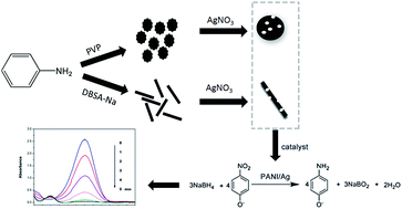
Micro/nano-structured polyaniline/silver (PANI/Ag) composites have been prepared by simply mixing nitric acid doped polyaniline with silver nitrate. A reducing agent was added to accelerate the reaction process and enhance the conversion rate of silver nitrate. As the support material of silver nanoparticles, nitric acid doped polyaniline was synthesized under turbulent flow and surfactants were adopted to control the morphology and uniformity of the particles. Various analysis techniques were adopted to confirm the composition and structure of the composites, including scanning electron microscopy (SEM), X-ray diffraction (XRD), transmission electron microscopy (TEM), Fourier transform infrared spectrometry (FTIR) and energy dispersive X-ray analysis (EDX). The PANI/Ag particles were used as a catalyst in the borohydride reduction reaction of 4-nitrophenol (4-NP), which was online monitored by UV-vis absorption spectroscopy. Results demonstrated that PANI microspheres loaded with 18.30 wt% of silver showed a comparable catalytic performance, and the apparent rate constant is 0.0256 s−1. Moreover, the stability and reusability of the catalyst were also investigated. The present work highlights the incorporation of well-dispersed silver nanoparticles with the conductive polyaniline matrix, and the accelerated electron transfer during the catalytic process owing to the high electrical conductivity of the support material.

Introduction

Composites consisting of conducting polymers and noble metal nanoparticles have aroused considerable attention in recent years because of their potential applications in fields of chemical sensors, catalysis and memory devices.1–5 Among all the noble metal nanoparticles, silver has been frequently employed in the area of catalysis especially as a catalyst in reduction of 4-nitrophenol, which has been listed as priority pollutant by US Environmental Protection Agency (EPA) due to its high solubility and stability in water.6,7 And the catalytic activity of conducting polymer/metal particles was investigated by reduction of 4-nitrophenol (4-NP) into 4-aminophenol (4-AP) in the presence of excessive NaBH4.8 This reaction has been used extensively as a benchmark to quantify the catalytic activity of various metallic nanoparticles, as the reaction dynamics can be easily monitored by UV-vis absorption spectroscopy.9–11 On the other hand, the product of the catalytic reaction, namely 4-NP is of great use in industries, for example, as a potent intermediate for many analgesic and antipyretic drugs.12

For conducting polymers, polyaniline (PANI) remains one of the most important conducting polymers owing to its ease of preparation, high conductivity, good environmental stability under ambient conditions, promising chemical, electrical and optical properties, as well as its unique redox tenability.14,15 What's more, the doping level of PANI can be easily controlled through an acid–base doping–dedoping process.16 Usually PANI is synthesized by the traditional chemical oxidative polymerization approach.17 For a better control of the dispersion and structure of PANI, surfactant-assisted synthesis of PANI was explored, since surfactants can serve as templates for the preparation of PANI. For example, the water-soluble polymer, poly(vinylpyrrolidone) (PVP) has been reported to be effective for dispersion polymerization of aniline in aqueous acidic condition.18 A novel 2D leaf-like PANI hierarchical structures using sodium dodecyl benzene sulfonate (SDBS) was reported by Jian Xu et al.19

Several methods were adopted for the preparation of PANI/metal composites: (1) by synthesis of PANI in the presence of metal nanoparticles,20,21 (2) using PANI as a reducing agent of noble-metal salts or acids,22,23 (3) by the oxidation of aniline with noble-metal compounds.24,25 PANI/Ag with core–shell structure (Ag as the core, and PANI as the shell) has been synthesized in the last few years by the first method to combine the advantage of silver and PANI.26 Nevertheless, it has been shown that the presence of a densely packed capping layer on the metal nanoparticle surface may compromise the catalytic activity since the surface active sites become inaccessible.27 While, the last two methods are lack of efficiency in the reduction of silver compounds since the reaction continues for many hours to accomplish, and the conversion rate of silver compounds is low.28

In the present work, micro/nano-structured polyaniline (PANI) particles with regulated morphology were synthesized by surfactant-assisted chemical oxidative polymerization method under turbulent circulation. The as-synthesized polyaniline microparticles were utilized as the reducing agent of silver nitrate, and additional reducing agent was introduced in the reaction of PANI with AgNO3 to accelerate the reaction process and enhance the conversion rate of Ag. Various analysis techniques were used to confirm the composition and structure of the composites, including scanning electron microscope (SEM), transmission electron microscope (TEM), X-ray diffraction (XRD), Fourier transform infrared spectrometer (FTIR) and energy dispersive X-ray analysis (EDX). The catalytic performance of micro/nano-structured PANI/Ag composites was evaluated by borohydride reduction reaction of 4-nitrophenol (4-NP), and comparable catalytic performance was achieved.

Experimental

Materials

All reagents were analytical grade in purity. Aniline, ammonium persulfate (APS), poly-vinylpyrrolidone (PVP, K30, MW = 40[thin space (1/6-em)]000), sodium dodecyl benzene sulfonate (SDBS), silver nitrate (AgNO3), nitric acid, 4-nitrophenol (4-NP), sodium borohydride (NaBH4) were purchased from Sinopharm Chemical Reagent corporation, Ltd. In all the process, anhydrous ethanol and distilled water were used.

Preparation of PANI microparticles

In a typical process, 9.3 g of aniline, 100 ml of 1 M nitric acid, and a certain amount of surfactant (PVP or SDBS) were mixed in a stainless steel container and stirred for about 10 minutes. 100 ml of 1 M APS was added drop-wise into the container while the mixture was strongly stirred under turbulent flow. The reaction was taken in ice bath to slow down the polymerization process. About 1 hour later, the solution was centrifuged at the speed of 10[thin space (1/6-em)]000 rpm and washed several times with ethanol, acetone and deionized water, respectively.

Preparation of micro/nano-structured PANI/Ag composites

0.45 g of PANI particles were added in aqueous AgNO3 solution at a certain concentration. The mixture was under ultrasound for 10 minutes and mechanically stirred for 1 hour in a 250 ml round-bottom flask. Then 120 ml of 0.025 M NaBH4 was slowly added into the flask for about 1 h. After being stirred for another 1 hour, the solution was filtered using a Buchner funnel under reduced pressure. Finally, the as synthesized particles were dried at 60 °C in a vacuum oven for 24 hours.

Catalysis experiments of borohydride reduction of 4-nitrophenol

1 mg of PANI/Ag was added into a quartz cuvette. Then 2.8 ml of 0.1 mM 4-nitrophenol and 0.2 ml of 0.1 M NaBH4 which was freshly prepared were added into the quartz cuvette in sequence. Rapidly the mixture in the quartz cuvette was put into the UV-vis absorption spectrophotometer to monitor the reaction.

Characterizations

The morphologies of the PANI and PANI/Ag particles were observed by scanning electron microscopy (SEM, FEI QUANTA 200) and transmission electron microscopy (TEM, JEM2010(HT)). The powder X-ray diffraction patterns of the particles were obtained by Bruker D8 advanced powder X-ray diffractometer with Cu Kα1 radiation (λ = 1.54060 Å) at the scanning speed of 6° min−1 (2θ). The scanning scope is 20–80° at tube voltage of 40 kV and tube current of 40 mA. Fourier transform infrared (FT-IR) spectra were recorded on a Nicolet NEXUS670 spectrum analyzer, which were collected from the range of 4000 to 400 cm−1. The energy dispersive X-ray analysis was conducted by the EDX analyzer equipped with SEM in surface mode. The UV-vis absorption spectra were recorded by UV-vis spectrophotometer (UV-vis, SHIMADZU UV-3600) from the scale range of 250–550 nm. The size distribution of the particles were obtained by Zetasizer Nano Series ZEN3600 with a 633 nm laser and 173° backscatter detection optics, MALVEN Instrument. The as-synthesized PANI particles were made into tablets under the pressure of 10 MPa by an infrared tablet press, and the conductivity of them was examined using a RTS-9 four-point probe tester. The Brunauer–Emmett–Teller surface area (BET) was measured by a Gemini2390 system using N2 adsorption and desorption at liquid-nitrogen temperature (77 K).

Results and discussion

Characterizations of PANI and PANI/Ag

Fig. 1 depicts the FTIR spectra of PANI and PANI/Ag particles. The main bands observed for PANI at 1562 and 1483 cm−1 stand for the characteristic C[double bond, length as m-dash]C stretching of the quinoid and benzenoid rings, respectively. The measurements show bands at 1292 and 1236 cm−1 for the characteristic C–N stretching of the benzenoid and quinoid rings. Peak at 1120 cm−1 is the bending vibration of C–H in benzene ring. The relative small peaks at 790 and 505 cm−1 can be attributed to the plane bending vibration of C–N for 1, 4 substitute position of benzene ring and bending vibration of benzene ring, respectively. The spectrum of PANI/Ag shows a slight change in comparison with that of PANI. Vibrations of quinoid and benzenoid rings were shifted from 1562 and 1483 cm−1 to 1600 and 1500 cm−1, respectively. It can be explained that the electrons of silver atoms tend to conjugate quinoid and benzenoid rings which lead a shift of infrared absorption.25,28 The FTIR spectrum of a representative sample of PANI/Ag was chosen, since the FTIR spectra of each composite (PANI/Ag) were almost the same.
image file: c5ra05396j-f1.tif
Fig. 1 FTIR of PANI and PANI/Ag particles.

As illustrated in Fig. 2, the broad reflection coming from PANI is the so called amorphous halo,15 which is ascribed to the periodicity parallel to the polymer chains of PANI.28 A more detailed low angel XRD analysis of PANI was explained in the ESI. While, for the XRD pattern of PANI/Ag composite, there exist four peaks at 2θ angles of 38.1°, 44.3°, 64.4° and 77.4°, respectively corresponding to the reflection of (111), (200), (220) and (311) crystalline planes of Ag NPs (JCPDS no. 04-0783). The results confirm the presence of silver in the composite. PANI/Ag-3 was chosen as the representative sample, since the XRD patterns of each composite (PANI/Ag) were almost the same. The estimated average crystal size was 59.3 nm, according to Scherrer equation. And the detailed derivation process was shown in ESI.


image file: c5ra05396j-f2.tif
Fig. 2 X-ray diffraction patterns of PANI and PANI/Ag particles.

Two kinds of surfactants were taken as templates for the preparation of PANI. As displayed in Fig. 3, the morphologies of the PANI particles change from microspheres to micro-rods when taking PVP or SDBS as the surfactant, respectively. With the amount of PVP ranges from 0.50 g to 1.00 g, the morphology and dispersion of PANI particles gradually have an improvement, which can be seen in Fig. 3(a–c). And when higher concentration of PVP was added in the synthesis of PANI, the change of morphology and dispersion of PANI particles become stable. Likewise, with the addition of SDBS as the surfactant from 1.00 g to 2.00 g, the morphology of PANI particles turns into micro-rods and the dispersion of PANI particles has been improved, which can be shown in Fig. 3(d–f). After a series of experiments, it was concluded that 2.00 g of SDBS was the proper amount to achieve optimum morphology and dispersion. The SEM images suggest that the size of PANI microparticles is of about several microns.


image file: c5ra05396j-f3.tif
Fig. 3 SEM image of PANI prepared with the addition of (a) 0.5 g of PVP, 5000×, (b) 0.75 g of PVP, 5000×, (c) 1.0 g of PVP, 5000×, (d) 1.0 g of SDBS, 5000×, (e) 1.5 g of SDBS, 5000×, (f) 2.0 g of SDBS, 5000×.

Simultaneously, turbulent circulation method was taken instead of conventional mechanical stirring29 for a better control of the reaction. To explore the mechanism of the stirring method, pulse method was adopted to simulate the stirring process. Scheme 1 shows the typical diagrammatic sketch of turbulent circulation. Fluid under the dramatic stirring of turbulent flow can be evenly mixed by controlling the moving pathway and distance of particles in the reactor. PANI particles synthesized under turbulent circulation exhibit better size distribution than results reported in literature.13,20 Particle size distribution of PANI with different morphology is illustrated in the ESI, which suggests that the PANI particles are normally distributed of a few microns, consistent with the SEM images of PANI particles.


image file: c5ra05396j-s1.tif
Scheme 1 Diagrammatic sketch of turbulent circulation.

Fig. 4(a and b) shows the morphologies of PANI/Ag composites (PANI/Ag-1 and PANI/Ag-3, respectively), and it can be seen that Ag nanoparticles are scattered on the surface of PANI particles. To further observe the morphologies of PANI/Ag, transmission electron microscope was utilized, and the images are shown in Fig. 4(c and d). The silver nanoparticles were evenly loaded on the surface of PANI. For a clear observation of silver nanoparticles loaded on PANI, HRTEM images were taken, which can be seen in Fig. 4(e and f). The estimated diameter of silver nanoparticles on microsphere structured PANI is about 50–70 nm, and the size of silver nanoparticles on micro-rod structured PANI is about 60–70 nm. They are all spherical in shape. To certify the content of Ag loaded on PANI, elemental analysis by EDX was conducted. And the results are demonstrated in Table 1.


image file: c5ra05396j-f4.tif
Fig. 4 SEM images of (a) microsphere structured PANI/Ag-3, 7000×, (b) micro-rod structured PANI/Ag-1, 7000×, TEM images of microsphere structured PANI/Ag-3, (c) 30[thin space (1/6-em)]000×, (e) 40[thin space (1/6-em)]000×, TEM images of micro-rod structured PANI/Ag-1, (d) 30[thin space (1/6-em)]000×, (f) 40[thin space (1/6-em)]000×.
Table 1 Preparation of PANI/Ag composites
Sample name AgNO3 (mol l−1) NaBH4 (mol l−1) Morphology of PANI Conductivity of PANI (S cm−1) BET surface area (m2 g−1) Silver content (wt%)
PANI/Ag-1 0.0050 0.025 Micro-rod 18.26 11.7165 10.19
PANI/Ag-2 0.0005 0.025 Microsphere 19.28 13.1032 2.19
PANI/Ag-3 0.0050 0.025 Microsphere 19.28 13.2750 10.81
PANI/Ag-4 0.0500 0.025 Microsphere 19.28 14.1285 18.30
PANI/Ag-5 0.0050 0.025 Microsphere Nonconductive 13.2310 10.64


BET surface area of PANI/Ag has been showed in Table 1. The specific surface area of bare microsphere PANI and micro-rod PANI are 10.7890 m2 g−1 and 10.1796 m2 g−1, respectively. When loaded with silver, the specific surface area of each sample showed an increase, which may ascribe to the effect of large amount of silver nanoparticles.

Catalytic performance of PANI/Ag composites

The UV-vis absorption spectra recorded during the process of 4-NP reduction are shown in Fig. 5. The colour of aqueous 4-nitrophenol solution changed from light yellow to green yellow with the addition of excessive NaBH4. Without adding a catalyst, the colour of the solution remains yellow-green. The maximum absorption band at about 400 nm corresponds to 4-nitrophenolate ions in the presence of NaBH4. While, after PANI/Ag particles were added, the peak at about 400 nm gradually disappeared, and a new peak at about 300 nm was gradually under development, corresponding to the absorption of 4-aminophenol. With the difference of catalytic performance of PANI/Ag composites, the solution turned into colourless at different times. The reaction formula of borohydride reduction of 4-NP can be expressed in the Scheme 2.
image file: c5ra05396j-f5.tif
Fig. 5 Time dependent absorption spectra for catalytic reduction of 4-NP by NaBH4 in presence of (a) PANI/Ag-1, (b) PANI/Ag-2, (c) PANI/Ag-3, (d) PANI/Ag-4, (e) PANI/Ag-5, (f) plot of ln(At) versus time for PANI/Ag particles.

image file: c5ra05396j-s2.tif
Scheme 2 Mechanism of Langmuir–Hinshelwood mechanism for the reduction of 4-NP to 4-AP in presence of NaBH4 by catalyst of PANI/Ag.

For the initial concentration of NaBH4 greatly exceeded that of 4-nitrophenol, so it was assumed that the concentration of NaBH4 remained constant during the reaction. In other words, the reduction of 4-nitrophenol can be considered as pseudo-first-order reaction. And the reaction rate follows first order kinetics,19 which can be illustrated by equation: −dc/dt = kc, where t represents time, k represents the apparent rate constant, c represents the concentration of 4-nitrophenol. There exists a linear correlation between ln(At) and the reaction time. ln(At) represents the natural logarithm of absorption value at a time. The derivation process was presented in the ESI.

The catalytic performance of PANI/Ag with different morphologies, namely Ag loaded on micro-rod structured PANI particles and Ag loaded on microsphere structured PANI particles was tested. They were reacted with 0.0050 mol l−1 of AgNO3, respectively. EDX result shows that the silver loading of them are 10.19 wt% for PANI/Ag-1 and 10.81 wt% for PANI/Ag-3, respectively. From the results of UV-vis absorption spectra shown in Fig. 5(a and c), we can conclude that the catalytic reaction finishes in 30 minutes for PANI/Ag-1 which in contrast PANI/Ag-3 takes only 5 minutes. Furthermore, according to the slopes of the lines shown in the Fig. 5(f), the values of the calculated apparent rate are 0.0024 s−1 for PANI/Ag-1 and 0.0151 s−1 for PANI/Ag-3, respectively. Accordingly, the correlation coefficients (R2) are 0.9374 and 0.9919, respectively, which indicate that the results are credible. Since silver nanoparticles loaded on the surface of PANI microspheres showed a better catalytic performance than on PANI micro-rods, PANI microspheres were employed as the supporting material to explore the influence of silver content on the catalytic performance of PANI/Ag. The content of silver loaded at the surface of PANI was adjusted by altering the concentration of AgNO3 from 0.0005 M to 0.0500 M, and the corresponding silver content is shown in Table 1. As the results illustrated in Fig. 5(b–d), with the increase of AgNO3 concentration, the catalytic performance of PANI/Ag has an enhancement. According to the slopes of the lines showed in Fig. 5(f), the maximum apparent rate constant (0.0256 s−1) is obtained when the concentration of AgNO3 is 0.0500 M. The apparent rate constant of PANI/Ag-4 particles is comparable to the previous findings in literatures, as demonstrated in Table 2. To certify whether the conductivity of PANI makes a difference on the catalytic performance, PANI microspheres were de-doped by mixing with 5% of ammonium hydroxide for 24 hours and then decorated with Ag. Comparing the results in Fig. 5(c and e), obviously it can be concluded that the conductivity of the PANI make a difference on the performance of PANI/Ag microspheres. As shown in Fig. 5(f), the apparent constant of PANI/Ag-3 and PANI/Ag-5 are 0.0151 s−1 and 0.0025 s−1, respectively.

Table 2 Rate constant of other catalysts compared with PANI/Ag
Catalysts k (s−1) Reference
Au–PAMAM 5.9 × 10⁻⁴ Kunio Esumi et al. (2004)3
Ag-DENs 7.0 × 10⁻³ Mulisa Nemanashi et al. (2012)2
Ag–SiO2 3.56 × 10⁻³ Man Wang et al. (2013)10
Ag–TiO2 2.11 × 10⁻³ Agileo et al. (2015)11
Ag–Al2O3 5.0 × 10⁻³ Bhanudas Naik et al. (2012)9
Ag–PANI 2.56 × 10⁻² This paper


Stability and reusability of the PANI/Ag catalyst

The physical and chemical stability of the catalyst are quite important owing to its practical application in industry. The Stability and reusability of PANI/Ag-3 particles were investigated by recycling them in borohydride reduction of 4-nitrophenol under the same catalytic condition. The recycle experiment was taken for 10 times. Each time when the catalytic reaction finished, the particles were collected. After being filtered and washed for several times, the particles were used in another new catalytic reaction. The catalytic performance were measured by UV-vis. In Fig. 6(a), X-axis stands for recycling time, and Y-axis stands for efficiency (we take the efficiency of the first recycle as 1). As shown in Fig. 6(a), the catalytic performance exhibited a slight decrease when reused for 10 times. X-ray diffraction pattern of the recycled particles was shown in Fig. 6(b), which indicated that silver was well crystallized after being recycled for 10 times. What's more, the morphology and silver content of the particles reused for 10 times were investigated. Fig. 6(c) shows the SEM image of the recycled particles and the result shows that silver nanoparticles are still stabilized on the surface of polyaniline. As shown in Fig. 6(d), though recycled for 10 times, silver content of PANI/Ag-3 is 9.91 wt%, which remains nearly steady compared with the result in Table 1. Therefore, we can conclude that the as prepared PANI/Ag composite is a kind of efficient and stable catalyst in the borohydride reduction of 4-nitrophenol.
image file: c5ra05396j-f6.tif
Fig. 6 (a) Cycling experiments results of PANI/Ag-3, (b) XRD patterns of PANI/Ag-3 cycled for 10 times, (c) SEM image of PANI/Ag-3 cycled for 10 times, 5000×, (d) EDS spectra of PANI/Ag-3 cycled for 10 times.

Insight on the catalytic mechanism of 4-NP reduction

The mechanism of the catalytic reaction could be explained by Langmuir–Hinshelwood mechanism.30,31 NaBH4 ionizes in water to offer BH4, which provides surface hydrogen for the reaction. BH4 and 4-NP are both adsorbed on Ag nanoparticles. The surface hydrogen firstly transfers to silver nanoparticles, and then reacts with 4-NP to yield 4-nitrophenol.32–34 In the catalytic process, BH4 is acted as the electron donor, while 4-nitrophenol is acted as the electron acceptor. Ag particles serve as the catalyst to provide active sites to promote the reaction. BH4 is oxidized into BO2, and when the reaction finishes, the resultant of the reaction in the form of BO2 and 4-aminophenol broke away from the surface of PANI/Ag particles, and entered into the solution. The mechanisms can be illustrated in Scheme 2. The advantages of loading silver on the surface of PANI microparticles are that (1) polymers have been proved as excellent hosts for trapping nanoparticles of metals and semiconductors because of their ability to act as stabilizer or the surface capping agent to prevent nanoparticles from aggregation. The dispersity and stability of silver particles are enhanced due to the presence of PANI microparticles, which is of great importance to metal nanoparticles, since the agglomeration of metal nanoparticles will have a bad influence on the catalytic performance; (2) meanwhile, the loose structure and large surface area of PANI tend to form more active sites for the catalytic reaction to occur. The results of BET surface area of PANI/Ag samples were shown in Table 1. The high surface area can enhance the absorption of BH4 and 4-NP on the catalyst, which would help to concentrate reactant molecules for the catalytic reaction. In the present work, the micro/nano-structured PANI/Ag composites displayed relatively high catalytic performance of 4-NP reduction than the previous works in literature; (3) furthermore, the electrical conductivity of PANI as the substrate of silver promotes the electron transfer in the catalytic process, and the catalytic performance of PANI/Ag has a remarkable improvement.

Conclusions

In this paper, micro/nano-structured polyaniline/silver (PANI/Ag) composites have been prepared by simply mixing nitric acid doped polyaniline with silver nitrate. A reducing agent was added to accelerate the reaction process and enhance the conversion rate of silver nitrate. As the support material of silver nanoparticles, nitric acid doped polyaniline was synthesized under turbulent flow and surfactants were adopted to control the morphology and uniformity of the particles. The structure and composition of the composites were characterized by SEM, TEM, XRD, EDX, FTIR methods. The composites were employed as catalysts for the borohydride reduction of 4-nitrophenol and showed excellent catalytic performance with the loading level of 18.3 wt% of silver. The reduction reaction of 4-nitrophenol follows pseudo-first-order kinetics, and the calculated apparent rate constant is 0.0256 s−1 under the condition that the PANI particles is doped with 1 M nitric acid and micro-spherical in shape. What's more, the stability and reusability of the catalyst were also investigated. The PANI/Ag composites still showed comparable catalytic performance though recycled for 10 times. It is believed that PANI microparticles will be a promising candidate as the substrate of loading metal nanoparticles for use in the field of catalysis.

Acknowledgements

This work was supported by grants of the National Nature Science Foundation of China (no. 21071112 and 21471119), and large-scale Instrument and Equipment Sharing Foundation of Wuhan University (no. LF20110063).

Notes and references

  1. P. Xu, X. J. Han, B. Zhang, Y. C. Du and H. L. Wang, Chem. Soc. Rev., 2014, 43, 1349–1360 RSC.
  2. M. Nemanashi and R. Meijboom, J. Colloid Interface Sci., 2013, 389, 260–267 CrossRef CAS PubMed.
  3. K. Esumi, H. Houdatsu and T. Yoshimura, Langmuir, 2004, 20, 2536–2538 CrossRef CAS.
  4. M. Guerra-Balcázar, R. Ortega, F. Castaneda, L. G. Arriaga and J. Ledesma-Garcia, Int. J. Electrochem. Sci., 2011, 6, 4667–4675 Search PubMed.
  5. M. A. Gracia-Pinlla, M. Villanueva, N. A. Ramos Delgado, M. F. Melendrez and J. Menchaca-Arredondo, Dig. J. Nanomater. Bios., 2014, 9, 1389–1397 Search PubMed.
  6. N. V. Blinova, J. Stejskal, M. Trchova and J. Prokes, Polymer, 2008, 57, 50–56 Search PubMed.
  7. G. Nesher, G. Marom and D. Avnir, Chem. Mater., 2008, 20, 4425–4432 CrossRef CAS.
  8. X. J. Liu, L. G. Li, M. X. Ye, Y. Xue and S. W. Chen, Nanoscale, 2014, 6, 5223–5529 RSC.
  9. B. Naik, V. S. Prasad and N. N. Ghosh, Powder Technol., 2012, 232, 1–6 CrossRef CAS PubMed.
  10. M. Wang, D. Tian, P. P. Tian and L. J. Yuan, Appl. Surf. Sci., 2013, 283, 389–395 CrossRef CAS PubMed.
  11. A. Hernández-Gordillo and V. R. González, Chem. Eng. J., 2015, 261, 53–59 CrossRef PubMed.
  12. J. F. Corbett, Dyes Pigm., 1999, 41, 127–136 CrossRef CAS.
  13. Y. Lu, Y. Mei, M. Drechsler and M. Ballauff, Angew. Chem., Int. Ed., 2006, 45, 813–816 CrossRef CAS PubMed.
  14. S. G. Chen, Z. D. Wei, X. Q. Qi, L. C. Dong, Y. G. Guo, L. J. Wan, Z. G. Shao and L. Li, J. Am. Chem. Soc., 2012, 134, 13252–13255 CrossRef CAS PubMed.
  15. X. F. Du, Y. L. Xu, L. L. Xiong, Y. Bai, J. B. Zhu and S. C. Mao, J. Appl. Polym. Sci., 2014, 18, 40827–40834 Search PubMed.
  16. J. Han, L. Li and R. Guo, Macromolecules, 2010, 43, 10636–10644 CrossRef CAS.
  17. M. D. Yilmaz, S. H. Hsu and D. N. Reinhoudt, Angew. Chem., Int. Ed., 2010, 49, 5938–5941 CrossRef CAS PubMed.
  18. R. Murugesan, G. Anitha and E. Subramanian, Mater. Chem. Phys., 2004, 85, 184–194 CrossRef CAS PubMed.
  19. H. Fan, H. Wang, J. Guo, N. Zhao and J. Xu, J. Colloid Interface Sci., 2014, 414, 46–49 CrossRef CAS PubMed.
  20. B. B. Wang, B. Li, B. Zhao and C. Y. Li, J. Am. Chem. Soc., 2008, 130, 11594–11595 CrossRef CAS PubMed.
  21. F. Ruffino, M. G. Grimaldi, F. Giannazzo, F. Roccaforte and V. Raineri, Nanoscale Res. Lett., 2009, 4, 262–268 CrossRef CAS PubMed.
  22. Z. H. Li, Y. B. Li, J. T. Lu, F. Zheng, J. Laven and A. Foyet, J. Appl. Polym. Sci., 2013, 10, 3933–3938 CrossRef PubMed.
  23. X. Zhang and Z. H. Su, Adv. Mater., 2012, 24, 4574–4577 CrossRef CAS PubMed.
  24. M. Schrinner, S. Proch, Y. Mei, R. Kempe, N. Miyajima and M. Ballauff, Adv. Mater., 2008, 20, 1928–1933 CrossRef CAS PubMed.
  25. B. L. Moore, A. Lu, D. A. Longbottomb and R. K. O'Reilly, Polym. Chem., 2013, 4, 2304–2312 RSC.
  26. D. M. Bedre, S. Basavaraja, B. D. Salwe, V. Shivakumar, L. Arunkumar and A. Venkataraman, Polym. Compos., 2009, 30, 1668–1677 CrossRef PubMed.
  27. W. Y. Ko, W. H. Chen, C. Y. Cheng and K. J. Lin, Nanoscale Res. Lett., 2009, 4, 1481–1485 CrossRef CAS PubMed.
  28. Q. Wu, Y. X. Xu, Z. Y. Yao, A. R. Liu and G. Q. Shi, ACS Nano, 2010, 4, 2342–2349 Search PubMed.
  29. C. C. Ai, Y. Xiao, W. Wen and L. J. Yuan, Powder Technol., 2011, 210, 323–327 CrossRef CAS PubMed.
  30. M. E. Leyva, F. G. Garcia, A. A. Queiroz, D. Artur and W. Soares, J. Mater. Sci.: Mater. Electron., 2011, 22, 376–383 CrossRef CAS PubMed.
  31. M. G. Murali, U. Dalimba and K. Sridharan, J. Mater. Sci., 2012, 47, 8022–8034 CrossRef CAS.
  32. S. Y. Bao, M. Zhang, M. L. Du, H. Zhu and P. Wang, Soft Mater., 2014, 12, 179–184 CrossRef CAS PubMed.
  33. D. N. Yang, R. M. Wang, M. S. He, J. Zhang and Z. Liu, J. Phys. Chem. B, 2005, 109, 7654–7658 CrossRef CAS PubMed.
  34. V. K. Shukla, P. Yadav, R. S. Yadav, P. Mishraa and A. C. Pandeya, Nanoscale, 2012, 4, 3886–3893 RSC.

Footnote

Electronic supplementary information (ESI) available. See DOI: 10.1039/c5ra05396j

This journal is © The Royal Society of Chemistry 2015
Click here to see how this site uses Cookies. View our privacy policy here.