
Single-step shaping of fluorescent polymer beads by a reverse breath figure approach†
F. Galeotti*a,
E. Kozmaa,
W. Mróza and
B. Kutrzeba-Kotowskab
aCNR, Istituto per lo Studio delle Macromolecole (ISMAC), via E. Bassini 15, 20133 Milano, Italy. E-mail: f.galeotti@ismac.cnr.it
bCNR, Istituto per la Microelettronica e Microsistemi – Sezione di Agrate Brianza (MDM IMM-CNR), via Olivetti 2, 20864 Agrate Brianza, MB, Italy
First published on 15th April 2015
Abstract
We demonstrate a solution-based fabrication strategy to arrange different polymeric materials on a substrate in the form of micrometric beads by taking advantage of the spontaneous phenomenon of reverse breath figures. The technique we propose here is suitable for different kinds of polymers, including conjugated and fluorescent ones, aiming at a universal approach for the simple one-step shaping of fluorescent beads.
Manipulating matter by breath figures (BF) has attracted significant interest in the last few years as a very simple and affordable technique for preparing surfaces with ordered pores at the nano- and microscale, with potential application in a wide range of different fields: micro-templates,1–4 separation,5–8 tissue engineering,9,10 selective bacterial entrapment,11 fiber optic sensors,12 optical manipulation of light,3,4,13 and self-cleaning surfaces among others.14 BFs spontaneously form when a polymer solution is cast onto a substrate in an environment rich with water vapor, a nonsolvent for the polymer. The process is driven by the condensation of micrometric water droplets that arrange themselves on the surface of the polymer solution, forming a template for the porous film. Typically, when the process is complete, a honeycomb-like imprint of the droplets is left on the film surface.15 In particular conditions, this process can lead to the formation of microspheres in the place of honeycomb structures. This is the case of the reverse breath figure (RBF) phenomenon, so named after the observation of poly(styrene-block-butadiene) microspheres obtained by casting toluene solution in a vessel saturated with methanol/water mixed vapor.16 Since then, only a few other authors have reported the formation of RBFs, usually occurring just with one particular polymer structure or one specific combination of solvents. Deepak and Asha deeply studied the polymer microspheres obtained by drop casting a poly(urethane-methacrylate-block-styrene) from THF/water mixture only when the polymer was prepared by ATRP, while the random copolymer afforded disordered honeycombs.17 De León et al. reported microsphere formation by drop casting solutions of a blend of PMMA and an amphiphilic block-copolymer in THF/water mixtures, and explained the phenomenon with the precipitation of the PMMA phase due to water, and formation of particles stabilized by the amphiphilic copolymer.18 Ferrari et al. obtained microspheres rather than porous polymer films when attempted to make BFs with a linear polystyrene (PS) if acetone, methyl ethyl ketone or THF were used as solvent.19 Gao et al. studied the behavior of carboxyl-terminated PS in different solvents and vapors, obtaining polymer beads when using CHCl3 as solvent and methanol vapor.20
Herein, we report a fast, robust and versatile fabrication method for depositing different polymeric materials on a substrate in the form of micrometric beads. To do this, we have developed a modified spin-coating procedure for RBF formation, where humid nitrogen is blown directly on the sample during the spinning process. While conventional BFs require particular conditions and special material characteristics to take place, and often suffer from poor reproducibility, the method we propose here works with different kind of polymers, including conjugated and fluorescent ones, and is reproducible.
As suggested by the few literature data available, in order to obtain microspheres from a BF process, the first step is to choose the appropriate solvent/nonsolvent couple. From a practical point of view, water vapor is the preferred nonsolvent, while the choice of the solvent is much wider. In Fig. 1 two films prepared with the same material (polyfluorene, PFO) and conditions, but different solvents, are shown. When carbon disulfide was used as solvent, the typical BF porous film is obtained (panel A). When THF is used, microsphere formation is achieved (panel B). Both films were prepared on a silicon substrate, by spin-coating at 3000 rpm a 5 mg mL−1 solution in the selected solvent, under a nitrogen humid flow of 500 L h−1, at 80% relative humidity (R.H.).
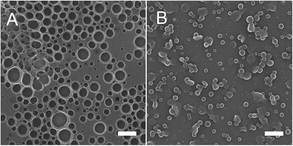 | ||
| Fig. 1 (A) Porous film obtained by spin-coating a CS2 solution of PFO under humid flow. (B) Polymer beads obtained by spin-coating in the same conditions from a THF solution of the same material. The scale bar is 500 nm. | ||
This result clearly shows that, if water vapor is used as the templating nonsolvent, a solvent fully miscible with water is needed in order to promote the formation of beads instead of porous film. Therefore, with the aim of keeping as simple as possible the fabrication conditions, we chose THF/water vapor as the primary solvent/nonsolvent couple for our experiments.
As regards the polymeric material, it is desirable for a patterning technique to be applicable to a wide range of materials. This point can be generally considered as a limit for the practical application of BFs, because sometimes subtle differences in the chemical structure of the polymers used can make a big difference in the quality of the films. We applied our RBF procedure to THF solutions of different polymers, either commercial or synthesized in our laboratory, by replicating the same working conditions, and we obtained micrometric beads with almost all materials, hence attesting the high versatility of the technique (see Scheme S1 and Table S1 in ESI†). Interestingly, we could prepare fluorescent microspheres with conjugated polymers, with polymers terminated with a fluorescent dye and even by simply mixing a non-fluorescent polymer with a fluorescent dye. It is worth to notice that the list of materials from which the RBFs were successfully attempted comprises polymers widely employed in optoelectronic devices such as PFO and polythiophene (P3HT), including two state-of-the-art materials for organic photovoltaic like PTB7 and PCDTBT. If we consider that the polymer particles spontaneously form during the few seconds of the spin-coating run and remain on the substrate at the end of the process, this opens the way to intriguing application in optoelectronic, such as the possibility of making pixel structured micro-OLEDs for displays.
Fig. 2 shows some representative examples of different materials processed with our RBF procedure, resulting in different shape objects. In the first row, SEM micrographs of commercial PS and PMMA are reported in panel A and B, respectively. PS is structured in the form of spheres and mostly hemispheres of 100–400 nm in diameter. Interestingly, most of the hemispheres are oriented with the flat side looking up, suggesting that the particles are formed during the film formation rather than in the solution bulk. The shape of PMMA particles is somewhat fancy. The riddled disks shown in panel B let us think that for this material an intermediate condition between standard BF and RBF formation has occurred. Such button-like structures may find interesting potential applications in the separation field; at this stage of the study, anyway, we did not investigate them further. The second row in Fig. 2 shows structures of a commercial poly(9,9-dioctylfluorene-alt-benzothiadiazole) (F8BT). The fluorescence microscopy view (panel C) clearly indicates the green-yellow emission of the particles, while the magnified SEM micrograph (panel D) shows beads of ≈200 nm connected in clusters. The three examples above confirm that this technique is compatible with easily affordable commercial polymers; hence it can in principle be exploited in any laboratory by a simple procedure. As example of red emitter, a perylene diimide derivative synthesized in our laboratory was employed (redpery). Because of the low viscosity of its THF solutions, as expected for a small molecule based dye, the RBF process did not afford any particles (see Fig. S2 in ESI†). However, when we blended the dye with PS in 1![[thin space (1/6-em)]](https://www.rsc.org/images/entities/char_2009.gif) :20 weight ratio, fluorescent spherical beads of 2–300 nm in diameter, were readily obtained (panels E and F). These emitting microspheres can be regarded as the proof of concept that, by following the same strategy, any fluorescent dye can in principle be incorporated into PS spheres. In the last row of Fig. 2, structures obtained with a blue emitting commercial PFO are displayed. Once again, the RBF process produced bright emitting particles in the form of both single spheres and clusters.
:20 weight ratio, fluorescent spherical beads of 2–300 nm in diameter, were readily obtained (panels E and F). These emitting microspheres can be regarded as the proof of concept that, by following the same strategy, any fluorescent dye can in principle be incorporated into PS spheres. In the last row of Fig. 2, structures obtained with a blue emitting commercial PFO are displayed. Once again, the RBF process produced bright emitting particles in the form of both single spheres and clusters.
 | ||
| Fig. 2 SEM micrographs (A, B, D, F, H) and fluorescence microscopy images (C, E, G) of micrometric polymer beads prepared by spin-coating under humid nitrogen flow THF solutions (2 mg mL−1) of different materials: PS (A), PMMA (B), F8BT (C and D), PS/redpery 20:1 (w/w) (E and F), PFO (G and H). The scale bar for fluorescence microscopy images is 10 μm. | ||
The above displayed results demonstrate the straightforwardness and versatility of the technique, which can be applied to different kind of materials, with similar success. Compared to conventional BFs, the here developed RBF procedure is much less sensitive to the molecular structure of the polymer used; specifically, it does not require the presence of polar groups, nor a particular polydispersity control.
When other water miscible solvents, such as acetone and methyl ethyl ketone, were tested, we observed microsphere formation only in particular working conditions. THF, on the other hand, worked with most of the materials tested. Spin-coating on glass substrate afforded similar results as silicon, suggesting that substrate contribution to RBFs is negligible.
Even though this process does not generally produce monodispersed particles, the particle size can be tuned to some extent by adjusting the polymer concentration of the THF solution. As shown in Fig. 3, the optimal concentration for particle formation normally ranges from 0.5 to 5 mg mL−1, resulting in average size between 0.02 and 0.5 μm2, respectively. By ideally considering spherical shapes, corresponding diameters of 150–800 nm are calculated. At higher concentration, the deposition results in a mixture of particles and network-like polymer film. Lowering the concentration too much makes the beads rather far away from each other and their shape becomes more and more flattened. The spinning rate does not particularly affect the particle shape, or their diameter. Similar results in fact were obtained by setting the spin-coater rate at 1000, 3000 and 5000 rpm. The amount of humidity, on the other hand, mainly affects the particle size and their clustering tendency, but not their shape. By adjusting the R.H. of the nitrogen flow from 9 to 80%, we observed that 2 mg mL−1 THF solutions of commercial PFO produce particles starting from R.H. 16%, and their average size increases with the increase of R.H., from 0.06 to 0.12 μm2. The maximum size is reached for R.H. 50%; further humidity increment does not produce larger particles. In addition, particles obtained at R.H. 80% are aggregated in bigger clusters than those produced at R.H. 16% (see Fig. S1 in ESI†).
 | ||
| Fig. 3 (A–D) SEM micrographs of commercial PFO spin-coated from THF solution at different concentrations: 20 (A), 10 (B), 5 (C) and 2 (D) mg mL−1. Scale bar is 10 μm for frame A and 1 μm for frames (B–D). In the insets, particle size distribution is shown. (E) Variation of average particle size with concentration of PFO. | ||
The mechanism of microsphere formation has been extensively discussed in some previous articles. According to Xiong and coworkers, during solvent evaporation the polymer solution is separated into liquid microdroplets by the condensation of the nonsolvent, so that the microdroplets of polymer solution act as templates dispersed in the continuous nonsolvent phase. After all liquids are evaporated, the microdroplets of polymer solution turn into microspheres.16 Let's transfer this basic explanation to our RBF condition, by taking into account that we use a spin-coating instead of a drop casting deposition, and that our solvent/nonsolvent couple is THF/water instead of toluene/mixture of methanol and water. We can imagine that, as soon as the spinning starts and the moist nitrogen flow is applied, water condensation quickly enriches THF with water, which induces the polymer solution to separate into microdroplets. After solvent evaporates and most of the free solvents are spinned off the rotating substrate, only solid polymer particles remain on it. On the other hand the general rule, resulting from the same reference study, that microspheres instead of porous films are obtained only when the surface tension of polymer solution is at least 1.5 mN m−1 higher than that of the condensed nonsolvent, does not apply to our system. In all our experiments, in fact, microparticles are formed even though the surface tension of the condensed nonsolvent (72 mN m−1 for water at 20 °C) is much higher than that expected for the polymer solution at any concentration (26 mN m−1 for pure THF).21 As widely established by the literature, both BF and RBF processes are complex thermodynamic, kinetic and entropic phenomenon; in order to address them thoroughly, many other factors including viscosity, surface energy, interfacial tensions between the liquids and van der Waals interactions between liquid and substrate, should be taken into account, which is out of the scope of this communication.
In order to test further the potential of the technique, the effect of multiple depositions on the same substrate was studied. Because THF is a good solvent for all the polymers used, one could expect that a second deposition of THF solution on preformed particles would simply wash away the first layer and replace it with the second material. When we studied these films by microscopy, the scenario was completely different.
Fig. 4 shows the particles resulting from sequential spin-coating depositions of a blue (PFO), a green (PS-bodipy) and a red (PS/redpery 20:1) emitting polymer, in this order. As evidenced by the fluorescence microscopy views of the same portion of the sample, each single particle looks bright when excited with three different excitation filters,22 resulting in blue, green and red colors. The white ovals on the pictures have been drawn to visualize the same group of particles in each picture, evidencing that the same particles are mixtures of the three emitting materials. The SEM micrograph of this film, shown in panel D, reveals that the particle shape resembles more discs and domes than microspheres. Some of the particles have a flat cylindrical shape, which suggests that a stratification of the materials may occur during the sequential spin-coating runs. As displayed in panel E, after the third deposition, we could detect the photoluminescence (PL) emission associated with the three single emitting polymers. In particular, emission peaks centered at 440, 465, 543 and 610 nm, ascribable to the PL contribution coming from PFO (first two peaks), PS-bodipy and redpery emission, respectively, were recorded. Consequently, by properly regulating the amount of each material in the spin-coating solutions, we were able to achieve white emission with Commission Internationale de l'Eclairage (CIE) chromaticity coordinates of (0.38, 0.38) (see ESI, Fig. S4†). A comparable result can be obtained by spinning in a single deposition a THF mixed solution of the three materials. In other words, besides corroborating the adaptability of RBFs, we found out a simple way to cover a substrate with patterns of different materials, mixing different PL colors, by using solutions of single materials.
 | ||
| Fig. 4 (A–C) Fluorescence microscopy images of polymer beads obtained by sequential deposition of PFO, PS-bodipy and PS/redpery 20:1 (w/w). The same portion of film is shown by using UV (A), blue (B) and green (C) filters. (D) SEM micrograph showing a tilted view of the same film. (E) PL spectra of the same film (bottom) and of the three nanoparticle films prepared with the corresponding single materials (top). Spectra were shifted along the y axis to aid the comparison. | ||
In summary, we have developed a convenient fabrication strategy for shaping micrometric polymer beads on a substrate, based on the naturally occurring phenomenon of RBFs. With the aim of proposing a universal approach which unlike traditional BFs is not affected by too many parameters, we have shown that a number of different polymers can be deposited in form of micrometric beads by applying a fast and facile fabrication process based on spin-coating. In particular, bright single color emitting particles and multicolor emitting particles can be obtained by single and sequential deposition, respectively. This ongoing work, which can be extended to other polymer systems, may find intriguing application in the development of a new generation of optoelectronic devices manufactured by self-assembly approach.
Experimental
PFO, F8BT were purchased from American Dye Source. PTB7, PCDTBT, PS and PMMA were purchased from Aldrich. PFO-THP, PFO-NBr, PS-pery, PS-bodipy, PS-P3HT and redpery were prepared according to already published procedures.23–28To prepare the polymer beads, the selected material is dissolved in THF at concentration between 0.5 and 10 mg mL−1. As soon as 50 μL of this solution is deposited on a 1 × 1 cm2 silicon substrate, a nitrogen humid flow of 500 L h−1 at 80% R.H. is applied and the spin-coating run (3000 rpm) is started. The water condensation triggered by the fast evaporation of THF causes the polymer to organize into micrometric beads instead of a continuous film, according to the RBF phenomenon. Typically the polymer beads formation is complete within ≈5 s; the remaining runtime helps the full evaporation of water. When the same process was performed on glass, microscopy coverslips were employed. In the sequential deposition, THF solutions of different materials are spin-coated sequentially on the same substrate. As the first deposition is complete, the second one is performed immediately on it, without removing the sample from the spin-coater. In the sequential deposition of three emitting materials, in order to detect all the PL signals, the concentrations were regulated by taking account of the energy transfer occurring between the materials with overlapping emission/absorption bands. Specifically, to obtain the spectrum reported in Fig. 4E (bottom), concentration were regulated as follows: 5 mg mL−1 for PFO (first deposition); 10 mg mL−1 for PS-bodipy (second deposition); 1 mg mL−1 for PS/redpery 20:1 (third deposition).
Acknowledgements
This work has been supported by Accordo Quadro between Regione Lombardia and CNR – Cluster Project ‘Energy’ no. 17348, and by Progetto Cariplo Edonhist no. 2012-0844.Notes and references
- C.-Y. Ma, Y.-W. Zhong, J. Li, C.-K. Chen, J.-L. Gong, S.-Y. Xie, L. Li and Z. Ma, Chem. Mater., 2010, 22, 2367–2374 CrossRef CAS.
- H. Yabu and M. Shimomura, Langmuir, 2005, 21, 1709–1711 CrossRef CAS PubMed.
- F. Galeotti, W. Mróz, G. Scavia and C. Botta, Org. Electron., 2013, 14, 212–218 CrossRef CAS PubMed.
- F. Galeotti, F. Trespidi, G. Timò and M. Pasini, ACS Appl. Mater. Interfaces, 2014, 6, 5827–5834 CAS.
- Y. Ou, C.-J. Lv, W. Yu, Z.-W. Mao, L.-S. Wan and Z.-K. Xu, ACS Appl. Mater. Interfaces, 2014, 6, 22400–22407 CAS.
- L.-S. Wan, J.-W. Li, B.-B. Ke and Z.-K. Xu, J. Am. Chem. Soc., 2011, 134, 95–98 CrossRef PubMed.
- H. M. Ma, P. C. Gao, Y. Zhang, D. W. Fan, G. B. Li, B. Du and Q. Wei, RSC Adv., 2013, 3, 25291–25295 RSC.
- H. L. Cong, J. L. Wang, B. Yu and J. G. Tang, Soft Matter, 2012, 8, 8835–8839 RSC.
- M. Du, P. Zhu, X. Yan, Y. Su, W. Song and J. Li, Chem.–Eur. J., 2011, 17, 4238–4245 CrossRef CAS PubMed.
- D. Beattie, K. H. Wong, C. Williams, L. A. Poole-Warren, T. P. Davis, C. Barner-Kowollik and M. H. Stenzel, Biomacromolecules, 2006, 7, 1072–1082 CrossRef CAS PubMed.
- A. S. de León, A. del Campo, A. L. Cortajarena, M. Fernández-García, A. Muñoz-Bonilla and J. Rodríguez-Hernández, Biomacromolecules, 2014, 15, 3338–3348 CrossRef PubMed.
- M. Pisco, F. Galeotti, G. Quero, A. Iadicicco, M. Giordano and A. Cusano, ACS Photonics, 2014, 1, 917–927 CrossRef CAS.
- B. W. Lim and M. C. Suh, Nanoscale, 2014, 6, 14446–14452 RSC.
- M. B. Avinash, E. Verheggen, C. Schmuck and T. Govindaraju, Angew. Chem., Int. Ed., 2012, 51, 10324–10328 CrossRef CAS PubMed.
- L. S. Wan, L. W. Zhu, Y. Ou and Z. K. Xu, Chem. Commun., 2014, 50, 4024–4039 RSC.
- X. Xiong, W. Zou, Z. Yu, J. Duan, X. Liu, S. Fan and H. Zhou, Macromolecules, 2009, 42, 9351–9356 CrossRef CAS.
- V. D. Deepak and S. K. Asha, J. Polym. Sci., Part A: Polym. Chem., 2008, 46, 1278–1288 CrossRef CAS PubMed.
- A. S. de León, A. Muñoz-Bonilla, M. Fernández-García and J. Rodríguez-Hernández, J. Polym. Sci., Part A: Polym. Chem., 2012, 50, 851–859 CrossRef PubMed.
- E. Ferrari, P. Fabbri and F. Pilati, Langmuir, 2011, 27, 1874–1881 CrossRef CAS PubMed.
- J.-P. Gao, W. Wu, L. Rong, G.-L. Mao, Y.-N. Ning, Q.-L. Zhao, J. Huang and Z. Ma, Eur. Polym. J., 2014, 59, 171–179 CrossRef CAS PubMed.
- J. J. Jasper, J. Phys. Chem. Ref. Data, 1972, 1, 841–1010 CrossRef CAS PubMed.
- Fluorescence microscopy images were taken with three different excitation filters, UV: 356 nm excitation bandpass and 420 nm filter cut, Blue: 470 nm bandpass and 515 nm filter cut, Green: 535 nm bandpass and 610 nm filter cut.
- A. Bolognesi, F. Galeotti, U. Giovanella, F. Bertini and S. Yunus, Langmuir, 2009, 25, 5333–5338 CrossRef CAS PubMed.
- C. Chi, A. Mikhailovsky and G. C. Bazan, J. Am. Chem. Soc., 2007, 129, 11134–11145 CrossRef CAS PubMed.
- E. Kozma, W. Mróz and F. Galeotti, Dyes Pigm., 2015, 114, 138–143 CrossRef CAS PubMed.
- F. Galeotti, V. Calabrese, M. Cavazzini, S. Quici, C. Poleunis, S. Yunus and A. Bolognesi, Chem. Mater., 2010, 22, 2764–2769 CrossRef CAS.
- X. Yu, K. Xiao, J. Chen, N. V. Lavrik, K. Hong, B. G. Sumpter and D. B. Geohegan, ACS Nano, 2011, 5, 3559–3567 CrossRef CAS PubMed.
- E. Kozma, D. Kotowski, S. Luzzati, M. Catellani, F. Bertini, A. Famulari and G. Raos, RSC Adv., 2013, 3, 9185–9188 RSC.
Footnote |
| † Electronic supplementary information (ESI) available: Instruments and methods, chemical structures and description of all materials, fluorescence microscopy and AFM images, PL spectra and CIE chromaticity coordinates plots. See DOI: 10.1039/c5ra05118e |
| This journal is © The Royal Society of Chemistry 2015 |