

Generation of nitrogen-doped photoluminescent carbonaceous nanodots via the hydrothermal treatment of fish scales for the detection of hypochlorite
Guoqin Wu,
Miao Feng and
Hongbing Zhan*
College of Materials Science and Engineering, Fuzhou University, Fuzhou, 350116, China. E-mail: hbzhan@fzu.edu.cn; Tel: +86 591 22866539
First published on 12th May 2015
Abstract
Preparing nitrogen-doped (N-doped) photoluminescent carbonaceous nanodots (C-dots) from the recycling/utilisation of nitrogen-rich carbonaceous waste has received considerable research interest. In this work, by using fish scales, an abundant food industry waste, as a starting material, we have developed a green, cheap and convenient approach for the preparation of N-doped photoluminescent C-dots, which represents a great potential for large-scale production. The as-synthesized C-dots are highly soluble, regularly sphere shaped and homogeneously sized with an average diameter of 2 nm. X-ray photoelectron spectroscopy results demonstrate that the nitrogen content can reach 14.6%, which is higher than that of most N-doped C-dots reported previously. The C-dots present a narrow photoluminescence emission band (400–490 nm) with a high quantum yield up to 17%, owing to the fluorescence enhancement effect of nitrogen doping. Besides, their fluorescence can be sensitively quenched by the addition of hypochlorite (ClO−), making them a promising sensing platform for ClO−.
1. Introduction
Owing to their superiority in water solubility, chemical inertness, low toxicity, ease of functionalization and resistance to photobleaching, carbonaceous nanodots (C-dots) have inspired intensive research efforts in recent years.1,2 Particularly, considerable research efforts have been focused on the doping of C-dots with nitrogen and it has been demonstrated that nitrogen doping can significantly enhance their properties and expand their novel applications.3–7 Several methods have been developed for the preparation of nitrogen-doped (N-doped) C-dots.3–17 Among which, hydrothermal synthesis is the most attractive for being simple, inexpensive, and green.8–17 Many nitrogen-containing natural products (grass, silk, etc.),8–12 biomass derivatives (citric acid, folic acid, glucose, etc.)13–15 and artificial compounds16,17 have been used as precursors for hydrothermal treatments to obtain N-doped C-dots.Along with the uprising global awareness of creating a sustainable community via waste minimization, the choice of recycling and reusing waste for the generation of useful, efficient materials for tomorrow's nanotech applications is immensely preferred by providing an elegant mechanism to sequester atmospheric CO2 and completing a holistic approach and material benefit at the same time.18–24 From this view point, the recycling/utilisation of nitrogen-rich carbonaceous waste especially food industry waste as starting materials for synthesis of N-doped C-dots would be highly advantageous.
Reactive oxygen species (ROS) control a wide range of physiological functions. Hypochlorite (ClO−), one of the most important ROS, acts as a dominant microbicidal mediator in the natural immune system. High level of ClO− is also widely used to treat food surfaces and water supplies, and this composes a potential health hazard to humans and animals. Thus, it is important to develop simple and effective methods for the detection of ClO− in both living cells and environment. Amongst various techniques, fluorescent probes are the most promising candidate for this respect owing to their capability, like high sensitivity, simplicity of implementation, short response time and offering application methods for not only in vitro assays but also in vivo imaging studies.25–29
Herein, we reported a simple and green method for the generation of photoluminescent N-doped C-dots via hydrothermal treatment of fish scales (Fig. 1) for the detection of ClO−. Fish scales are essentially waste of fish industry. They represent an inexpensive, accessible source of nitrogen containing biomass that does not directly impact the food chain. Nitrogen containing polysaccharide, chitin (poly-b(1/4)-N-acetyl-D-glucosamine), which is the main organic component of fish scales, provides an efficient precursor for the N-doped C-dots.
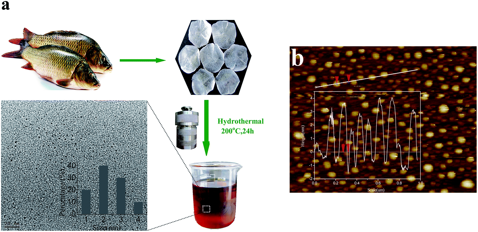 | ||
| Fig. 1 (a) Schematic illustration of the formation of C-dots from hydrothermal treatment of fish scales and TEM image of the as-prepared C-Dots. Inset: the corresponding particle size distribution histogram. (b) AFM image of the C-Dots. Inset: height profile along the line. | ||
2. Experimental section
2.1. Materials
Ca(NO3)2, CuCl2, FeCl3, Hg(NO3)2, Mg(NO3)2, Pb(NO3)2, Zn(OH)2, K2Cr2O7 and KMnO4 were purchased from Beijing Chemical Corp. Quinine sulphate, NaClO, KH2PO4 and K2HPO4 were purchased from Aladdin Ltd. (Shanghai, China). All chemicals were of analytical grade and were used as received without further purification. NaClO solution (10%) was prepared with Milli-Qultrapure water (Millipore, ≥18 MΩ cm). Milli-Qultrapure water was used throughout the experiments. The detection buffer was phosphate buffered saline (PBS, 0.2 M, pH 7.0).2.2. Preparation of N-doped CDs
N-doped CDs were prepared by hydrothermal treatment of fish scales. The fish scales used in this study were collected from Grass carp and washed thoroughly with cold water prior to lab use. The clean ‘wet’ fish scales were dried in oven at 60 °C for 3 h to get ‘dry’ fish scales for use. In a typical synthesis, ‘dry’ fish scales (2 g) were added into deionised H2O (30 mL). Then the mixture was transferred into a 50 mL Teflon lined autoclave and heated at 200 °C for a period of 24 h. The N-doped CDs were collected by removing the solid deposition and the large dots though filtration over a 220 nm pore size PTFE membranes. The C-dots thus obtained were dispersed in water for further characterizations and use.2.3. Determination of quantum yield
Determination of the quantum yield (QY) of all the samples was accomplished by comparison of the wavelength integrated intensity of sample to that of the standard quinine sulfate. The QY was calculated using| Φ = Φs[(IAsn2)/(IsAns2)] |
2.4. Detection of ClO−
All fluorescence spectra were recorded at room temperature in PBS (0.2 M, pH 7.0). The following metal ions were chosen to evaluate the influence of metal ions on fluorescence of C-dots, and assess the selectivity of ClO− based on fluorescence variation: Mg2+, Zn2+, Ca2+, Cu2+, Pb2+, Hg2+, Fe3+, Cr2O72+ and MnO4− (1 mM for each). For selectivity of the C-dots toward different ions, the concentration of the chosen ions was 10 mM. In a typical run, C-dots (40 μL) dispersion was added into PBS (3 mL), followed by the addition of a calculated amount of different ions. The PL spectra were recorded after standing for 5 min at room temperature. The quenching effect of ClO− on the fluorescence of C-dots was conducted as follows: 40 μL of C-dots dispersion and 3.0 mL of PBS were mixed. Then, a series of ClO− solutions with different concentrations were added into the proceeding solutions.2.5. Characterizations
Ultraviolet-visible (UV-vis) spectra were obtained with a Shimadzu UV-2450 spectrophotometer. Fluorescent emission spectra were recorded on a HJY FM-4-Tcspc spectrofluorometer (Horiba Jobin Yvon, Japan). Transmission electron microscopy (TEM) images were obtained on a JEM-2010 field emission transmission electron microscope (JEOL, Tokyo, Japan) with an accelerating voltage of 200 kV. The sample for TEM characterization was prepared by placing a drop of colloidal solution on carbon-coated copper grid and dried at room temperature. The height distribution of the obtained C-dots was characterized by atomic force microscopy (AFM, Bruker Dimension Icon) by using ScanAsyst mode. X-ray photoelectron spectroscopy (XPS, ESCALAB 250) were used to analyze the composition of the as-prepared materials using Mg as the exciting source. Fourier-transform infrared (FTIR) spectra were obtained using a Nicolet 5700 spectrometer, spectrum was recorded from 4000 to 400 cm−1 using 12 scans at a resolution of 4 cm−1. Powder X-ray diffraction (XRD) patterns were obtained with an XPert-PMD diffractometer, using Cu Kα radiation (λ = 0.15405 nm, 40 kV, 100 mA).3. Results and discussion
TEM image (Fig. 1a) and AFM image (Fig. 1b) show that the obtained C-dots are regularly sphere shaped and homogeneously sized. They are highly water-soluble and can be kept for several months without any observed aggregation.The structure and components of the resultant C-dots were characterized by XPS, XRD and FTIR spectroscopy. The XPS survey spectrum (Fig. 2a) shows three peaks at 284.0, 400.0, and 530.6 eV, which are attributed to C 1s, N 1s, and O 1s, respectively. The corresponding content of each element was displayed in the inset of Fig. 2a. It can be seen that the nitrogen content in our C-dot samples can reach 14.6%, which is higher than that of other N-doped C-dots reported previously.8–17 The spectrum of C 1s (Fig. 2b) can be deconvoluted into three single peaks that correspond to C–C (284.4 eV), C–N (285.5 eV), and C![[double bond, length as m-dash]](https://www.rsc.org/images/entities/char_e001.gif) N/CO (288.0 eV) functional groups. The N 1s spectrum (Fig. 2c) shows two peaks at 399.7 and 400.8 eV, which are attributed to C–N–C and N–(C)3 bands, respectively. The O 1s spectrum (Fig. 2d) further confirms the observations with the characteristic oxygen states of CO (530.7 eV).30–32 The XRD pattern of the C-dots (Fig. 3a) gives a broad peak around 2θ = 23.4–24.6°, revealing an amorphous carbon phase, which is attributed to the introduction of nitrogen- and oxygen-containing groups.33,34 Besides, the weak peak at 2θ = 35.9° implies the partial graphitization of the C-dots. FT-IR spectrum (Fig. 3b) displays two significant bands at 3424 cm−1 (O–H stretching vibration) and 1655 cm−1 (CO stretching vibration). Besides, the bands at 3247 cm−1 and 1510 cm−1 could be attributed to the vibration of N–H. The two bands at 1330 cm−1 and 1410 cm−1 can be identified as –CH2–. The bands at 1455 cm−1 and 1113 cm−1 are characteristic of the amide III C–N stretch and C–O, respectively.34–37 All these results indicate that the as-prepared C-dots possess abundant nitrogen and oxygen containing groups on their surface.
N/CO (288.0 eV) functional groups. The N 1s spectrum (Fig. 2c) shows two peaks at 399.7 and 400.8 eV, which are attributed to C–N–C and N–(C)3 bands, respectively. The O 1s spectrum (Fig. 2d) further confirms the observations with the characteristic oxygen states of CO (530.7 eV).30–32 The XRD pattern of the C-dots (Fig. 3a) gives a broad peak around 2θ = 23.4–24.6°, revealing an amorphous carbon phase, which is attributed to the introduction of nitrogen- and oxygen-containing groups.33,34 Besides, the weak peak at 2θ = 35.9° implies the partial graphitization of the C-dots. FT-IR spectrum (Fig. 3b) displays two significant bands at 3424 cm−1 (O–H stretching vibration) and 1655 cm−1 (CO stretching vibration). Besides, the bands at 3247 cm−1 and 1510 cm−1 could be attributed to the vibration of N–H. The two bands at 1330 cm−1 and 1410 cm−1 can be identified as –CH2–. The bands at 1455 cm−1 and 1113 cm−1 are characteristic of the amide III C–N stretch and C–O, respectively.34–37 All these results indicate that the as-prepared C-dots possess abundant nitrogen and oxygen containing groups on their surface.
 | ||
| Fig. 2 (a) XPS full scan spectrum, and (b) C 1s, (c) N 1s, and (d) O 1s spectra. | ||
 | ||
| Fig. 3 (a) XRD pattern, (b) FT-IR spectrum of the C-dots. | ||
To study the optical properties of the C-dots, UV-vis absorption and PL studies were carried out in detail. Fig. 4a illustrates that the C-dots in aqueous solution has two typical UV/Vis peaks at 266 and 316 nm. While the former corresponds to the typical absorption of an aromatic π system or the n–π* transition of the carbonyl, the latter is due to the trapping of excited state energy by the surface states.11–13,34–36 When excited at 360 nm, a broad emission centered at 430 nm was observed in the emission spectrum. The QY was determined to be 17.08% using quinine sulfate as a reference. The PL effect is so strong that, under the illumination of UV (360 nm) light, even at a very low concentration, the C-dots still give very bright violet-blue luminescence (inset of Fig. 4a). Both the radiative recombination of excitons and the functional groups on the surface of the C-dot may be responsible for the PL emission.11–13,34–36 Moreover, the PL spectra also display the characteristic feature that the maximum emission moves to a longer wavelength as the excitation wavelength increased. As shown in Fig. 4b, the strongest emission peak shifts from 414 nm to 462 nm as the excitation alters from 310 nm to 400 nm. This phenomenon has been widely observed in the luminescent carbon nanomaterials, which is assumed to be caused by the optical selection of different surface defect states near the Fermi level and the different size distribution of C-dots.34–38
 | ||
| Fig. 4 (a) UV-vis absorption, excitation and emission spectra of the C-dots. Inset: the optical image under daylight and UV light (365 nm), respectively. (b) PL emission spectra for the C-dots dispersed in water at excitation wavelengths progressively increasing from 310 nm to 400 nm. | ||
To evaluate the selectivity of this sensing system, we examined the PL intensity changes in the presence of representative metal ions under the same conditions, including Mg2+, Zn2+, Ca2+, Cu2+, Pb2+, Hg2+, Fe3+, Cr2O72+ and MnO4− (1 mM for each), as shown in Fig. 5a. It is seen that a much lower PL was observed for C-dots upon addition of ClO−. In contrast, no tremendous decrease was observed by addition of other ions into the C-dots dispersion. Fig. 5b shows the PL spectral evolution of the C-dots aqueous solution upon addition of different concentrations of ClO− (0–10 mM). Fig. 5b inset shows dependence of F/F0 on the concentrations of ClO−, where F0 and F are C-dots fluorescence intensities at 430 nm in the absence and presence of ClO−, respectively. Both reveal that the PL intensity of the mixture is sensitive to ClO− concentration and decreases with the increase of ClO− concentration. We attribute this fluorescence quenching to the effective and selective photoinduced electron transfer from the N atom to ClO−.
 | ||
| Fig. 5 (a) The difference in PL intensity of C-dots dispersion between the blank and solutions containing different ions (1 mM for each). (b) PL emission spectra of the C-dots dispersion in the presence of different concentrations of ClO− (from top to bottom: 0, 0.1, 0.2, 0.3, 0.4, 0.5, 0.6, 0.7, 0.8, 0.9, 1, 2, 3, 4, 5, 6, 7, 8, 9, 10 mM, excitation at 360 nm). Inset shows the dependence of F/F0 on the concentrations of ClO− within the range of 0–10 mM. | ||
4. Conclusions
In conclusion, we have developed a green, cheap and convenient approach for the preparation of C-dots. By using fish scales, an abundant food industry waste, as staring materials, this approach represents a great potential for large-scale production. The obtained C-dots are nitrogen-rich and possess abundant heteroatoms groups on the surface. They are strongly fluorescent and the fluorescence can be sensitively quenched by the addition of ClO−, making them a sensing system for ClO−.Acknowledgements
This research was supported by the National Natural Science Foundation of China (nos 51172045 and 51402051).References
- S. N. Baker and G. A. Baker, Luminescent Carbon Nanodots: emergent nanolights, Angew. Chem., Int. Ed., 2010, 49, 6726–6744 CrossRef CAS PubMed.
- H. Li, Z. Kang, Y. Liu and S. T. Lee, Carbon Nanodots: synthesis, properties and applications, J. Mater. Chem., 2012, 22, 24230–24253 RSC.
- W. Wei, C. Xu, L. Wu, J. Wang, J. Ren and X. Qu, Non-enzymatic-browning-reaction: A versatile route for production of Nitrogen-Doped carbon dots with tunable multicolor luminescent display, Sci. Rep., 2014, 4, 3564 Search PubMed.
- H. Zhang, Y. Li, X. Liu, P. Liu, Y. Wang and T. An, et al. Determination of iodide via direct fluorescence quenching at Nitrogen-Doped carbon quantum dot fluorophores, Environ. Sci. Technol. Lett., 2014, 1, 87–91 CrossRef CAS.
- C. Wang, X. Wu, X. Li, W. Wang, L. Wang and M. Gu, et al. Upconversion fluorescent carbon nanodots enriched with nitrogen for light harvesting, J. Mater. Chem., 2012, 22, 15522–15525 RSC.
- W. S. Kwon, S. G. Do, J. Lee, S. Y. Hwang, J. K. Kim and S. W. Rhee, Freestanding luminescent films of nitrogen-rich carbon nanodots toward large-scale phosphor-based white-light-emitting devices, Chem. Mater., 2013, 25, 1893–1899 CrossRef CAS.
- Y. Xu, M. Wu, Y. Liu, X. Z. Feng, X. B. Yin and X. W. He, et al. Nitrogen-Doped Carbon Dots: A facile and general preparation method, photoluminescence investigation, and imaging applications, Chem.–Eur. J., 2013, 19, 2276–2283 CrossRef CAS PubMed.
- S. Liu, J. Tian, L. Wang, Y. Zhang, X. Qin and Y. Luo, et al. Hydrothermal Treatment of Grass: A low-cost, green route to Nitrogen-Doped, carbon-rich, photoluminescent polymer nanodots as an effective fluorescent sensing platform for label-free detection of Cu(II) Ions, Adv. Mater., 2012, 24, 2037–2041 CrossRef CAS PubMed.
- W. Li, Z. Zhang, B. Kong, S. Feng, J. Wang and L. Wang, et al. Simple and green synthesis of Nitrogen-Doped photoluminescent carbonaceous nanospheres for bioimaging, Angew. Chem., Int. Ed., 2013, 52, 8151–8155 CrossRef CAS PubMed.
- H. Zhang, Y. Wang, D. Wang, Y. Li, X. Liu and P. Liu, et al. Hydrothermal transformation of dried grass into graphitic carbon-based high performance electrocatalyst for oxygen reduction reaction, Small, 2014, 10, 3371–3378 CrossRef CAS PubMed.
- Z. Wu, P. Zhang, M. Gao, C. Liu, W. Wang and F. Cheng, et al. One-pot hydrothermal synthesis of highly luminescent nitrogen-doped amphoteric carbon dots for bioimaging from Bombyx mori silk - natural proteins, J. Mater. Chem. B, 2013, 1, 2868–2873 RSC.
- Y. Sha, J. Lou, S. Bai, D. Wu, B. Liu and Y. Ling, Hydrothermal synthesis of nitrogen-containing carbon nanodots as the high-efficient sensor for copper(II) ions, Mater. Res. Bull., 2012, 48, 1728–1731 CrossRef PubMed.
- Y. Dong, H. Pang, H. Yang, C. Guo, J. Shao and Y. Chi, et al. Carbon-based dots co-doped with nitrogen and sulfur for high quantum yield and excitation-independent emission, Angew. Chem., Int, Ed., 2013, 52, 7800–7804 CrossRef CAS PubMed.
- R. Zhang and W. Chen, Nitrogen-doped carbon quantum dots: Facile synthesis and application as a “turn-off” fluorescent probe for detection of Hg2+ ions, Biosens. Bioelectron., 2014, 55, 83–90 CrossRef CAS PubMed.
- T. Lai, E. Zheng, L. Chen, X. Wang, L. Kong and C. You, et al. Hybrid carbon source for producing nitrogen-doped polymer nanodots: one-pot hydrothermal synthesis, fluorescence enhancement and highly selective detection of Fe(III), Nanoscale, 2013, 5, 8015–8021 RSC.
- Y. Q. Zhang, D. K. Ma, Y. Zhuang, X. Zhang, W. Chen and L. L. Hong, et al. One-pot synthesis of N-doped carbon dots with tunable luminescence properties, J. Mater. Chem., 2012, 22, 16714–16718 RSC.
- C. L. Li, C. M. Ou, C. C. Huang, W. C. Wu, Y. P. Chen and T. E. Lin, et al. Carbon dots prepared from ginger exhibiting efficient inhibition of human hepatocellular carcinoma cells, J. Mater. Chem. B, 2014, 48, 1728–1731 Search PubMed.
- R. J. White, M. Antonietti and M. M. Titirici, Naturally inspired nitrogen doped porous carbon, J. Mater. Chem., 2009, 19, 8645–8650 RSC.
- J. H. Clark, V. Budarin, F. E. I. Deswarte, J. J. E. Hardy, F. M. Kerton and A. J. Hunt, et al. Green chemistry and the biorefinery: a partnership for a sustainable future, Green Chem., 2006, 8, 853–860 RSC.
- S. Y. Park, H. U. Lee, E. S. Park, S. C. Lee, J. W. Lee and S. W. Jeong, et al. Photoluminescent green carbon nanodots from food-waste-derived sources: large-scale synthesis, properties, and biomedical applications, ACS Appl. Mater. Interfaces, 2014, 6, 3365–3370 CAS.
- H. U. Lee, S. Y. Park, E. S. Park, B. Son, S. C. Lee and J. W. Lee, et al. Photoluminescent carbon nanotags from harmful cyanobacteria for drug delivery and imaging in cancer cells, Sci. Rep., 2014, 4, 4665 Search PubMed.
- X. W. Tan, A. N. B. Romainor, S. F. Chin and S. M. Ng, Carbon dots production via pyrolysis of sago waste as potential probe for metal ions sensing, J. Anal. Appl. Pyrolysis, 2014, 105, 157–165 CrossRef CAS PubMed.
- P. C. Hsu, Z. Y. Shih, C. H. Lee and H. T. Chang, Synthesis and analytical applications of photoluminescent carbon nanodots, Green Chem., 2014, 14, 917–920 RSC.
- J. Zhou, Z. Sheng, H. Han, M. Zou and C. Li, Facile synthesis of fluorescent carbon dots using watermelon peel as a carbon source, Mater. Lett., 2012, 66, 222–224 CrossRef CAS PubMed.
- S. Goswami, A. P. Manna, S. Paul, C. K. Quah and H. K. Fun, Rapid and ratiometric detection of hypochlorite with real application in tap water: molecules to low cost devices (TLC sticks), Chem. Commun., 2013, 49, 11656–11658 RSC.
- S. Goswami, S. Paul and A. Manna, Highly reactive (<1 min) ratiometric “naked eye” detection of hypochlorite with real application in tap water, Dalton Trans., 2013, 42, 10097–10101 RSC.
- B. Zhu, Y. Xu, W. Liu, C. Shao, H. Wu and H. Jiang, et al. A highly selective colorimetric probe for fast and sensitive detectionof hypochlorite in absolute aqueous solution, Sens. Actuators, B, 2014, 191, 473–478 CrossRef CAS PubMed.
- Y. Koide, Y. Urano, K. Hanaoka, T. Terai and T. Nagano, Development of an Si-rhodamine-based far-red to near-infrared fluorescence probe selective forhypochlorous acid and its applications for biological imaging, J. Am. Chem. Soc., 2011, 133, 5680–5682 CrossRef CAS PubMed.
- T. I. Kim, S. Park, Y. Choi and Y. Kim, A BODIPY-based probe for the selective detection of hypochlorous acid in living cells, Chem.–Asian J., 2011, 6, 1358–1361 CrossRef CAS PubMed.
- W. Lu, X. Qin, S. Liu, G. Chang and Y. Zhang, et al. Economical, green synthesis of fluorescent carbon nanoparticles and their use as probes for rapid, sensitive, and selective detection of mercury(II) ions, Anal. Chem., 2012, 84, 5351–5357 CrossRef CAS PubMed.
- L. Zhou, B. He and J. Huang, Amphibious fluorescent carbon dots: one-step green synthesis and application for light-emitting polymer nanocomposites, Chem. Commun., 2013, 49, 8078–8080 RSC.
- Y. Deng, D. Zhao, X. Chen, F. Wang, H. Song and D. Shen, Long lifetime pure organic phosphorescence based on water soluble carbon dots, Chem. Commun., 2013, 49, 5751–5753 RSC.
- Q. Liang, W. Ma, Y. Shi, Z. Li and X. Yang, Easy synthesis of highly fluorescent carbon quantum dots from gelatin and their luminescent properties and applications, Carbon, 2013, 60, 421–428 CrossRef CAS PubMed.
- P. Hsu and H. Chang, Synthesis of high-quality carbon nanodots from hydrophilic compounds: role of functional groups, Chem. Commun., 2012, 48, 3984–3986 RSC.
- Z. Qian, X. Shan, L. Chai, J. Ma, J. Chen and H. Feng, Si-Doped Carbon Quantum Dots: A facile and general preparation strategy, bioimaging application, and multifunctional sensor, ACS Appl. Mater. Interfaces, 2014, 6, 6797–6805 CAS.
- Y. Li, Y. Zhao, H. Cheng, Y. Hu, G. Shi and L. Dai, et al. Nitrogen-Doped graphene quantum dots with oxygen-rich functional groups, J. Am. Chem. Soc., 2012, 134, 15–18 CrossRef CAS PubMed.
- Z. Qian, J. Ma, X. Shan, H. Feng, L. Shao and J. Chen, Highly luminescent N-Doped carbon quantum dots as an effective multifunctional fluorescence sensing platform, Chem.–Eur. J., 2014, 20, 2254–2263 CrossRef CAS PubMed.
- S. Sahu, B. Behera, T. K. Maiti and S. Mohapatra, Simple one-step synthesis of highly luminescent carbon dots from orange juice: application as excellent bio-imaging agents, Chem. Commun., 2012, 48, 8835–8837 RSC.
| This journal is © The Royal Society of Chemistry 2015 |