DOI:
10.1039/C5RA04726A
(Paper)
RSC Adv., 2015,
5, 34243-34250
Monodisperse erythrocyte-sized and acid-soluble chitosan microspheres prepared via electrospraying
Received
17th March 2015
, Accepted 8th April 2015
First published on 8th April 2015
Abstract
Monodisperse erythrocyte-sized and acid-soluble chitosan microspheres are successfully prepared by an electrospraying method for the first time. Effects of the physical properties of the polymer solution and the condition parameters of the electrospraying process on the size and size distribution of the chitosan microspheres are systematically studied, to optimize the conditions for preparation of chitosan microspheres with our specific requirements. The microsphere size is mainly controlled by the viscosity of the spray liquid, and the microsphere monodispersity mainly depends on the electric conductivity of the spray liquid, the flow rate and the needle size. Under the optimized conditions, monodisperse chitosan microspheres with an average diameter of 6.4 μm, which is similar to the erythrocyte size, are prepared with a narrow size distribution (CV < 3%). Due to the use of terephthalaldehyde as cross-linker via formation of Schiff base bonds, the prepared chitosan microspheres can maintain structural integrity and show green fluorescence in a neutral medium, but display rapid acid-triggered decomposition. The prepared erythrocyte-sized and acid-soluble chitosan microspheres are highly attractive as promising substitutes of blood samples for calibration of hematology analyzers and flow cytometers.
1. Introduction
Polymer microspheres with good biocompatibility, uniform size, large specific surface area and unique functions have received considerable attention in biomedical and biochemical fields for drug delivery,1–3 tissue engineering,4,5 enzyme immobilization,6,7 bioseparation,8,9 bioassay and diagnosis.10,11 Among them, monodisperse polymer microspheres with a size range of 5–8 μm, which have same size as human erythrocytes, are highly potential as the standard samples replacing the human erythrocyte samples for the calibration of hematology analyzers and flow cytometers.12–14 The polymer microsphere samples can avoid a series of annoying things coming from the blood samples, such as troublesome processing procedure, poor stability, bio-hazard, limited shelf-life, lot-to-lot variation and high cost. Furthermore, polymer microspheres with chemical-triggered decomposition properties can be easily removed without residue, which can improve the accuracy of the instrument to be used again. Therefore, development of monodisperse polymer microspheres with erythrocyte-like size and chemical-triggered decomposition properties is of both scientific and commercial interests for hematology applications.
Chitosan, a natural cationic polysaccharide, is of great interest in biomedical and pharmaceutical fields owing to its excellent biocompatibility, biological activity, and biodegradability.15,16 In our previous works, we have found that chitosan hydrogels prepared using terephthalaldehyde as the cross-linker by forming Schiff base bonds exhibited great pH-dependent stability.17 In neutral medium, the cross-linked chitosan hydrogels are stable, and they can maintain their original shape and structural integrity. While in acidic environment at low pH value, the Schiff base bonds become instable and the cross-linked chitosan hydrogels are decomposed finally. In addition, the cross-linked chitosan containing Schiff base bonds display autofluorescent properties without the need to conjugate any external fluorochromes.17,18 Therefore, monodisperse chitosan microspheres cross-linked by terephthalaldehyde could serve as ideal substitutes of human erythrocytes for calibration of hematology analyzers and flow cytometers. Shirasu porous glass (SPG) membrane emulsification technology is a promising method to prepare monodisperse chitosan microspheres.18–20 Microfluidic emulsification is also a rapidly developing method to controllably prepare monodisperse chitosan microspheres.17,21,22 However, these two methods still have certain limitations. The SPG membranes are required hydrophobic modification before use for fabrication of chitosan microspheres. For microfluidic technology, it is difficult to achieve mass production of products. And more importantly, the size of chitosan microspheres prepared by microfluidic technology is usually too large to serve as the substitutes of human erythrocytes for calibration in hematology applications. In recent years, electrospraying technology, also known as electrohydrodynamic atomization, has been developed rapidly to promote the industrialized production of polymer particles with a wide variety of morphology.23,24 Compared with conventional mechanical spraying systems, the electrospraying setup is quite simple and low-cost, and the operation is easy and the preparation conditions are mild. More significantly, the size distribution of the generated droplets is usually narrow and droplet size can be flexibly controlled, which provides a viable and facile way to prepare monodisperse chitosan-based microspheres.25–34 However, all of previously reported chitosan-based microspheres prepared by electrospraying had no acid-solubility and most chitosan-based microspheres were hundreds of micrometers in size.25–30 Up to now, monodisperse chitosan microspheres with both erythrocyte-like size and acid-solubility have not been reported yet.

In this study, monodisperse erythrocyte-sized and acid-soluble chitosan microspheres are fabricated by electrospraying technology for the first time. Monodisperse water-in-oil (W/O) emulsions, which are generated by homogeneous dispersion of electrosprayed chitosan droplets into the collection solution, are served as the reaction templates. Effects of the solution properties and the condition parameters of electrospraying process on the size and size distribution of the prepared chitosan microspheres are systematically studied to optimize the conditions for preparation of monodisperse erythrocyte-sized chitosan microspheres.
2. Experimental
2.1. Materials
Chitosan with a deacetylation level of 85% (Mw = 5000, Jinan Haidebei Marine Bioengineering), terephthalaldehyde (Sinopharm Chemical Reagents) as the cross-linker, and polyglycerol polyricinoleate (PGPR 90) (Danisco) as the emulsifier are used as received without further purification. All other chemicals are of analytical grade and purchased from Chengdu Kelong Chemical Reagents. Deionized water, with a pH value of about 5.8, is obtained from a Millipore Milli-Q water purification-system and used throughout the experiments.
2.2. Properties of chitosan solutions
Chitosan aqueous solution is used as the spray liquid for fabricating the chitosan microspheres. The pH value of chitosan aqueous solutions is adjusted to 6.3 by adding sodium hydroxide. The viscosity and electrical conductivity of chitosan aqueous solutions with different chitosan concentrations are measured at different environment temperatures. The viscosity is determined by a rotational viscometer (NDJ-79, Shanghai Sendi Scientific Apparatus) at a spindle speed of 750 rpm. The electrical conductivity is measured by a conductivity meter (SevenMultiTM, Mettler-Toledo). The viscosity and electrical conductivity for each chitosan aqueous solution at each condition are measured three times to obtain the arithmetical average values.
2.3. Preparation of chitosan microspheres
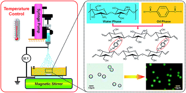
Monodisperse chitosan microspheres are fabricated by a monoaxial electrospraying setup (SS-2534H, Ucalery Beijing), as schematically shown in Fig. 1. Briefly, the electrospraying setup mainly consists of a syringe pump (TS2-60, Baoding Longer Precision Pump), a 10 mL plastic syringe fitted with a stainless steel needle as the capillary nozzle, a high-voltage power supply, and a grounded magnetic stirrer (85-1, Shanghai Meiyingpu Instrument).
 |
| Fig. 1 Schematic illustration of the electrospraying setup and preparation process of cross-linked chitosan microspheres. | |
To fabricate chitosan microspheres, chitosan solution is loaded into the syringe and pumped through the needle. The flow rate is precisely controlled by the syringe pump. The syringe with its needle is positioned vertically, and the positive electrode of the high voltage is connected to the stainless steel needle. A glass chamber filled with the collection solution is used as the collector and placed on the magnetic stirrer, which is located directly below the needle. To generate well-dispersed monodisperse W/O emulsions, the collection solution is constantly stirred at a low rotational speed (about 200 rpm) during the electrospraying process and cross-linking reaction. The reaction system is left overnight to make sure the chitosan in the water phase is cross-linked completely. All preparations are carried out in the electrospraying chamber, and the operation temperature inside the chamber is controlled from 25 to 50 °C, and the humidity is maintained between 20% and 30% RH. The resultant chitosan microspheres are washed using isopropanol to remove the organic solution and finally dispersed into deionized water.
2.4. Characterization of chitosan microspheres
The morphology of chitosan microspheres dispersed in collection solution is observed by a fluorescent microscope (DM 4000, Leica) under an I3 filter set (BP 450-490, Leica). The size distribution of chitosan microspheres is determined by using an automatic analytic software (Tiger 3000, Chongqing Xinminfeng Instruments) on the basis of obtained optical micrographs. The average diameter of each microsphere sample is calculated from the measured results of more than 100 particles.
Acid-induced dissolution experiment of chitosan microspheres is performed in a transparent glass container at 25 °C. At first, a certain amount of chitosan microspheres equilibrated in a small amount of deionized water in the container. To change the ambient solution into an acidic medium, the deionized water around the microspheres is replaced with excess phosphate buffer solution (0.01 mol L−1, pH 3.5) rapidly. Ionic strength of the buffer solution is adjusted to 0.1 mol L−1 by adding sodium chloride. The acid-induced dissolution behavior of chitosan microspheres are monitored by a confocal laser scanning microscope (CLSM, SP5-II, Leica) in real time and recorded by a CCD camera. The green fluorescent channel of CLSM is excited at 488 nm.
3. Results and discussion
Electrospraying, also called electrohydrodynamic atomization, is a unique one-step technique of liquid atomization by the use of high voltage.20,21 Relied on the electrostatic force, the spray liquid is deformed and broken up into fine charged droplets at the nano- to micro-scale. Although electrospraying is accepted as a technique that can produce particles with monodisperse size distributions and reproducible morphologies by controlling the electrospraying parameters, production of particles with very specific requirements still remains challenging due to many variables involved in the process as well as the complex interaction and interdependence.20 Therefore, in this work, to prepare monodisperse erythrocyte-sized and acid-soluble chitosan microspheres, effects of the solution properties and the condition parameters of electrospraying process on the size and size distribution of chitosan microspheres are studied systematically.
3.1. Setting of applied voltage and jet distance
Because the aim of our work is to prepare monodisperse erythrocyte-sized and acid-soluble chitosan microspheres, the primary pre-requisite is to establish the stable cone-jet mode, which is necessary for formation of monodisperse electrosprayed droplets.20,35 For a given electrospraying polymer solution, the cone-jet mode could be established only within certain range of applied voltage.35 In our work, stable cone-jet mode is observed at the tip of the needle when the high voltage is applied between 5 and 6 kV. Outside this voltage range, different instable electrospraying modes are observed and the obtained droplets are polydisperse. When the applied voltage is lower than 5 kV, the jet-breaking mode is turned into a pulsating mode, with a discontinuous liquid emission from the liquid meniscus. While above the upper limit of this voltage range, various types of instable jet-breaking modes like multi-jet mode are observed as the applied voltage is gradually increased.
Furthermore, because in our work the chitosan microspheres are prepared through the cross-linking reaction instead of solvent evaporation, the jet distance between the needle tip and surface of the collection solution is found to have little effect on the formation of chitosan microspheres. Therefore, the jet distance is fixed at 5 cm as electrode gap distance throughout our study, which is the minimum safe distance of our electrospraying setup.
3.2. Effect of the collection solution
In this work, chitosan microspheres cross-linked by terephthalaldehyde are synthesized by using monodisperse W/O emulsions as the templates, which are generated by homogeneous dispersion of electrosprayed chitosan droplets into the collection solution. As shown in Fig. 1, oil-soluble terephthalaldehyde is dissolved in the collection solution, and diffuses across the oil–water interface to the aqueous phase of W/O emulsions to react with the amino groups of the chitosan. Monodisperse chitosan microspheres are formed finally when the cross-linking reaction is sufficient and complete inside the W/O emulsions. In this case, the electrosprayed chitosan droplets should enter into the collection solution to form chitosan microspheres through cross-linking reaction. Furthermore, if the electrosprayed droplets cannot quickly enter into the collection solution, they may tend to coalesce with the newly formed and incoming droplets on the surface of the collection solution. Therefore, a mixture of toluene and n-hexanol with volume ratio of 1
:
1 is used as the collection solution to optimize the viscosity and density. For example, the viscosity of collection solution at 25 °C is 2.62 mPa s, and the density of the collection solution is about 0.846 g cm−3 measured by a glass hydrometer, which is much less than that of the chitosan solution (1.01 g cm−3). So, the chitosan droplets can enter into the collection solution immediately after they reach the surface of the collection solution. Besides terephthalaldehyde, PGPR 90 and N,N,N′,N′-tetramethylethylenediamine (TMEDA) are also added into the collection solution to stabilize the W/O emulsions, in which PGPR 90 is used as emulsifier to stabilize the W/O emulsions and TMEDA is an alkaline agent to prevent flocculation and coalescence of emulsion droplets by speeding up cross-linking rate. In the collection solution, the concentrations of terephthalaldehyde, PGPR 90 and TMEDA are 2.0 wt%, 6.0 wt% and 0.6% v/v, respectively.
3.3. Effect of the chitosan solution
It has been reported that low viscosity of the polymer solution is one of the key factors to prepare monodisperse particles with several micrometers in size.36 Therefore, chitosan with lower molecular weight is chosen in this work to prepare the spray liquid with low viscosity, which is likely to be used for obtaining erythrocyte-sized chitosan microspheres. Moreover, in the electrospraying process, the viscosity and electrical conductivity of the spray liquid also greatly affect the size and size distribution of the sprayed droplets. The polymer solutions with different chitosan concentrations have different viscosities and electrical conductivities. Therefore, the effect of chitosan concentration on the microsphere preparation is also studied in this work. In the experiments, the chitosan concentrations are varied from 1 wt% to 4 wt% with an increment of 1 wt%, and other processing parameters are kept at constant. As mentioned above, the high voltage is applied in the range of 5–6 kV and the jet distance is fixed at 5 cm. The flow rate of the spray liquid is kept at 110 μL h−1 and the preparation temperature is 45 °C. A 32-gauge (32G) needle with inner diameter of 104 μm is used in the experiments.
The viscosity and electrical conductivity of chitosan solutions with different chitosan concentrations are measured at 45 °C, and the results are listed in Table 1. With the increase of the chitosan concentration, both viscosity and electrical conductivity of chitosan solution increase.
Table 1 Physical properties of chitosan solutions with different chitosan concentrations measured at 45 °C
Chitosan concentration (wt%) |
1 |
2 |
3 |
4 |
Viscosity (mPa s) |
1.10 |
1.13 |
1.48 |
1.68 |
Electrical conductivity (mS cm−1) |
3.83 |
6.18 |
8.45 |
10.26 |
Fig. 2a–d shows the fluorescence micrographs of chitosan microspheres obtained with different chitosan concentrations. Due to the Schiff base bonds, these chitosan microspheres cross-linked by terephthalaldehyde display obvious green fluorescence. All prepared chitosan microspheres exhibit good dispersion and spherical shape, but the size and size distribution of the prepared chitosan microspheres are different with the change of chitosan concentration. As shown in Fig. 2e, the sizes of all prepared chitosan microspheres are in the range of 6–10 μm, which increase slightly with increasing the chitosan concentration in the test range. Such a slight size increase is mainly caused by the viscosity increase of chitosan solution. The higher the liquid viscosity is, the larger the droplets form through the jet break-up process, and thus the larger the chitosan microspheres are.
 |
| Fig. 2 Fluorescent micrographs of chitosan microspheres prepared with chitosan concentrations of 1 wt% (a), 2 wt% (b), 3 wt% (c), 4 wt% (d), and their corresponding sizes (e) and size distributions (f). All scale bars are 10 μm. | |
The size monodispersities of chitosan microspheres prepared at various chitosan concentrations are quite different. In essence, the size distribution of chitosan microspheres is mainly affected by the electrical conductivity of spray liquid. A polydispersity index called the coefficient of variation (CV), which is defined as the ratio of the standard deviation of the size distribution to its arithmetic mean, is used to accurately evaluate the size monodispersity of the microspheres.
|
 | (1) |
where,
di is diameter of the
ith microsphere,
![[d with combining macron]](https://www.rsc.org/images/entities/i_char_0064_0304.gif)
is arithmetic average diameter of microspheres, and
n is the total number of microspheres counted (
n > 100). Obviously, the smaller the CV value is, the better the monodispersity of microspheres is.
As shown in Fig. 2f, the CV value of chitosan microspheres decreases first and then increases with the increase of chitosan concentration. When the chitosan concentration is 1 wt%, the size distribution of chitosan microspheres is very wide and the CV value is larger than 10%. The electrical conductivity of spray liquid directly affects the jet break-up process. It has been reported that the stable cone-jet mode can be maintained only when the electrical conductivity and flow rate of spray liquid having a good matching.35 Chitosan solution with low chitosan concentration has small electrical conductivity, in this case it is difficult to obtain cone-jet mode at a lower flow rate. As a result, secondary and satellite droplets are formed and the resultant chitosan microspheres are quite polydisperse. Electrospraying using 2 wt% chitosan solution at flow rate of 110 μL h−1 is observed to generate stable cone-jet mode in this study, so that the resultant chitosan microspheres present the best monodisperse size distribution as shown in Fig. 2b and the CV value is only 2.7%. However, when the electrical conductivity of the chitosan solution further increases, the cone-jet mode becomes unstable and the size monodispersity of chitosan microspheres is affected. Hence, chitosan concentration of 2 wt% is chosen as the optimized value for carrying out subsequent studies.
3.4. Effect of operation temperature
Although the chitosan microspheres are not prepared through solvent evaporation method in this work, the physical properties of chitosan solution can be affected by the operation temperature. Therefore, the influence of operation temperature on the microsphere preparation is also studied here. To ensure the study accuracy, the temperature of chitosan solution and environmental temperature inside the electrospraying chamber are kept the same, which are varied from 25 to 50 °C with an increment of 5 °C. Other processing parameters are kept at constant, in which the chitosan concentration of the spray liquid is 2 wt% and its flow rate is 110 μL h−1, and the needle gauge is 32G.
The viscosity and electrical conductivity of chitosan solutions measured at different operation temperatures are listed in Table 2. When the operation temperature is varied from 25 to 50 °C, the viscosity of chitosan solution decreases while the electrical conductivity increases. Fig. 3a–f shows the fluorescence micrographs of chitosan microspheres obtained at different operation temperatures, and the comparisons of the size and size distribution are shown in Fig. 3g and h. Similarly, all chitosan microspheres display obvious green fluorescence and most of them exhibit good spherical shape. The average size of prepared chitosan microspheres decreases with the increase of the operation temperature due to the change of the viscosity of chitosan solution. When the operation temperature is in the range from 25 to 35 °C, the average sizes of chitosan microspheres are larger than 12 μm, and the size distributions of chitosan microspheres formed in this temperature range are quite wide, with the CV values larger than 13%. For example, in the same batch of microsphere samples prepared at 30 °C (Fig. 3b), some microspheres are even larger than 17 μm and some are less than 9 μm. Such a large size and polydispersity are still mainly caused by the high viscosity and low electrical conductivity of chitosan solution at low temperature. As the operation temperature is increased further, the microsphere size is reduced to less than 10 μm, and correspondingly the size monodispersity is improved very well. However, further increase of the temperature to 50 °C makes the electrosprayed droplets dehydrated, which results in chitosan microspheres formed with irregular shape (Fig. 3f).
Table 2 Physical properties of 2 wt% chitosan solutions measured at different temperatures
Temperature (°C) |
25 |
30 |
35 |
40 |
45 |
50 |
Viscosity (mPa s) |
1.55 |
1.48 |
1.35 |
1.23 |
1.13 |
1.01 |
Electrical conductivity (mS cm−1) |
5.66 |
5.72 |
5.77 |
5.98 |
6.18 |
6.22 |
 |
| Fig. 3 Fluorescent micrographs of chitosan microspheres prepared at operation temperatures of 25 °C (a), 30 °C (b), 35 °C (c), 40 °C (d), 45 °C (e), 50 °C (f) and their corresponding sizes (g) and size distributions (h). All scale bars are 10 μm. | |
Actually, the results of effects of chitosan concentration and operation temperature reveal the fact that the viscosity and electrical conductivity of chitosan spray liquid play very important roles in the formation of sprayed droplets and resultant chitosan microspheres. The liquid viscosity greatly affects the size of sprayed droplets, and electrospraying solution with lower viscosity can form smaller droplets and microspheres. The electrical conductivity of spray liquid has a significant impact on the generation of stable cone-jet mode, which is a key to obtain monodisperse chitosan microspheres.
3.5. Effect of the flow rate
In electrospraying, the flow rate of polymer solution is a key processing parameter to control the formation of spayed droplets. The effects of the flow rate on the size and size distribution of obtained chitosan microspheres are studied and evaluated in a variation from 90 to 140 μL h−1. The chitosan concentration, operation temperature and needle gauge are kept constant at 2 wt%, 45 °C and 32G, respectively.
From the fluorescence micrographs of chitosan microspheres prepared at different flow rates, it can be seen that the size change is not so remarkable (Fig. 4a–f). The average sizes of chitosan microspheres obtained at different flow rates are in the range of 6–10 μm (Fig. 4g). Especially, the average size of chitosan microspheres obtained at 110 μL h−1 is 6.4 μm, which is closest to the erythrocyte size. However, the size monodispersity of the chitosan microspheres are quite different (Fig. 4h), which verifies again that the stable cone-jet mode can be maintained only when the flow rate and given spray liquid have a good matching as mentioned above.
 |
| Fig. 4 Fluorescence micrographs of chitosan microspheres prepared at flow rates of 90 μL h−1 (a), 100 μL h−1 (b), 110 μL h−1 (c), 120 μL h−1 (d), 130 μL h−1 (e), 140 μL h−1 (f), and their corresponding sizes (g) and size distributions (h). All scale bars are 10 μm. | |
It has been reported that every certain liquid has a minimum flow rate.35 When the liquid flow rate is near to this minimum flow rate, the liquid jet can be broken up into monodisperse droplets in the cone-jet mode due to axisymmetric instabilities. For the polymer solution with 2 wt% chitosan concentration, the flow rate of 110 μL h−1 is the optimum value to obtain the stable cone-jet mode. Fortunately, the chitosan microspheres prepared using 2 wt% chitosan solution at flow rate of 110 μL h−1 not only have the erythrocyte-like size, but also achieve the best monodispersity as shown in Fig. 4. However, the stable cone-jet mode cannot exist when the flow rate is below 110 μL h−1, as a result the size distribution of sprayed droplets becomes wider and the formed microspheres are polydisperse. On the other hand, the liquid cone at the needle tip becomes larger with the increase of the flow rate, and then the sprayed droplets also become larger. Furthermore, at higher flow rates, the jet break-up process is influenced by lateral or azimuthal instabilities of the jet, which can trigger the formation of a number of satellites or secondary droplets between main droplets.35,37 Therefore, as shown in Fig. 4d–f, chitosan microspheres prepared at higher flow rates have both many large particles and small particles, and the size distributions are quite wide.
3.6. Effect of needle size
The inner diameter of a needle is commonly expressed in gauge (G). To find out a needle gauge with proper inner diameter to prepare monodisperse erythrocyte-size chitosan microspheres, the effect of needle size on the microsphere preparation is also evaluated here. Four needle gauges including 32G, 30G, 30G′ and 28G are used in this work, and the actually measured inner diameter are 104, 138, 149 and 205 μm, respectively. The chitosan concentration, operation temperature and flow rate for this study are kept constant at 2 wt%, 45 °C and 110 μL h−1, respectively. From the fluorescence micrographs it can be obviously found out that, as the needle size increases, the size of obtained chitosan microspheres increase slightly and the size distribution becomes wider (Fig. 5a–d).
 |
| Fig. 5 Fluorescent micrographs of chitosan microspheres prepared with needle inner diameters of 104 μm (a), 138 μm (b), 149 μm (c), 205 μm (d) and their corresponding sizes (e) and size distributions (f). All scale bars are 10 μm. | |
With the given polymer solution and at the given flow rate, the spray liquid can flow more freely and a sputtering phenomenon is observed when using larger size of needle, which causes the obtained microspheres with larger size and poor monodispersity (Fig. 5b–d). The needle with the smallest inner diameter of 104 μm can generate a narrow size distribution (Fig. 5a). It is worth noting that when the needle diameter changes from 138 to 205 μm, there is no significant change in microsphere size, the average diameters of microspheres only increase from 10 to 12 μm; however, the CV values obviously increase (Fig. 5e and f). The results suggest that the needle size mainly affect on the monodispersity of microspheres rather than size of microspheres.
On the basis of above results, it can be concluded that the size of chitosan microsphere is mainly controlled by the viscosity of spray liquid, and the microsphere monodispersity mainly depends on electric conductivity and flow rate of spray liquid. Actually, the viscosity and electric conductivity of spray liquid are affected by the chitosan concentration and the operation temperature. Among these four factors, the chitosan concentration is the most effective variable in the production. So, when preparing monodisperse erythrocyte-sized chitosan microspheres, the chitosan concentration is firstly to be screened and optimized.
3.7. Acid-induced dissolution of chitosan microspheres
As mentioned above, erythrocyte-size chitosan microspheres with high monodispersity are successfully prepared is this work. To estimate the acid-soluble capability of the microspheres, the decomposition processes of chitosan microspheres in the pH of 3.5 is studied. Microspheres are firstly immersed in a small amount of deionized water. Then, the deionized water around the microspheres is replaced with excess phosphate buffer solution with pH 3.5 rapidly. The acid-induced dissolution behavior of erythrocyte-size chitosan microspheres is observed under CLSM microscope. Once in the environment of pH 3.5 buffer solution, chitosan microspheres swell immediately at first, and then a rapid and complete decomposition is achieved within 60 s, as shown in Fig. 6. The cross-linked network of the chitosan microspheres is dependent on the formation of Schiff base bonds between amino groups of chitosan and aldehyde groups of terephthalaldehyde. The Schiff base bonds between chitosan and terephthalaldehyde possess an interesting pH-responsive stability. In acidic solution, Schiff base bonds are broken and decomposed. The acid-induced swelling behavior of chitosan microspheres is mainly caused by the decrease of cross-linking effect and the electrostatic repulsion among protonated free amino groups in chitosan polymer chains in acidic medium.38,39 Therefore, the green fluorescence mainly from Schiff base bonds is gradually lost as show in Fig. 6. The final dissolution of chitosan microspheres in acidic solution is also a result of acid-induced hydrolysis of Schiff base bonds between chitosan and terephthalaldehyde.
 |
| Fig. 6 CLSM microscope snapshots of the acid-triggered decomposition process of chitosan microspheres, in which pH 3.5 buffer solution is added at t = 0 s. All scale bars are 10 μm. | |
The above results show that, the chitosan microspheres prepared in our work have both satisfactory erythrocyte-like size and excellent acid-soluble capability, which are highly attractive for serving as the substitutes of human erythrocytes for the calibration of hematology analyzers and flow cytometers.
4. Conclusions
In this paper, monodisperse erythrocyte-sized and acid-soluble chitosan microspheres are successfully prepared via electrospraying technology. Chitosan aqueous solution is used as the spray liquid and a mixture of toluene and n-hexanol is used as the collection solution. By optimizing the solution and operation parameters, chitosan microspheres with average diameter of 6.4 μm and narrow size distribution (CV < 3%) are prepared. The optimized process condition parameters for preparation of the desired monodisperse erythrocyte-sized chitosan microspheres are as follows: 2 wt% chitosan aqueous solution as the spray liquid, operation temperature of 45 °C, flow rate of 110 μL h−1, and 32G needle with inner diameter of 104 μm. The size of chitosan microspheres is mainly controlled by the viscosity of spray liquid. The size monodispersity of microspheres shows heavy dependence on the electric conductivity, the flow rate and the needle size. Besides the similar size range with human erythrocytes and narrow size distribution, the prepared chitosan microspheres also display excellent autofluorescent and acid-induced dissolution properties. Acid-induced decomposition properties make the chitosan microspheres easy-to-be removed without residue, which can improve the reuse accuracy of the instrument. Autofluorescent properties make the chitosan microspheres easy-to-be detected without any necessity to conjugate external fluorochromes. Due to these advantages and properties, our chitosan microspheres are more suitable as the substitutes of human erythrocytes in the calibration of hematology analyzers and flow cytometers than other polymeric microspheres, and they are also highly attractive in many biological and biomedical applications.
Acknowledgements
The authors gratefully acknowledge support from the National Natural Science Foundation of China (21322605, 21276002, 91434202), the Training Program of Sichuan Province Distinguished Youth Academic and Technology Leaders (2013JQ0035) and the State Key Laboratory of Polymer Materials Engineering (sklpme2014-3-02).
Notes and references
- J. M. Anderson and M. S. Shive, Adv. Drug Delivery Rev., 2012, 64, 72 CrossRef PubMed.
- S. R. Mao, C. Q. Guo, Y. Shi and L. C. Li, Expert Opin. Drug Delivery, 2012, 9, 1161 CrossRef CAS PubMed.
- S. J. Park, H. S. Lim, Y. M. Lee and K. D. Suh, RSC Adv., 2015, 5, 10081 RSC.
- W. Huang, X. L. Li, X. T. Shi and C. Lai, Biomater. Sci., 2014, 2, 1145 RSC.
- W. K. Xu, L. Y. Wang, Y. Ling, K. Wei and S. Z. Zhong, RSC Adv., 2014, 4, 13495 RSC.
- Y. T. Zhang, Y. K. Yang, W. F. Ma, J. Guo, Y. Lin and C. C. Wang, ACS Appl. Mater. Interfaces, 2013, 5, 2626 CAS.
- Z. Liu, H. S. Wang, B. Li, C. Liu, Y. J. Jiang, G. Yu and X. D. Mu, J. Mater. Chem., 2012, 22, 15085 RSC.
- J. Huang, P. Su, J. W. Wu and Y. Yang, RSC Adv., 2014, 4, 58514 RSC.
- Z. Zhang, J. H. Li, J. Q. Fu and L. X. Chen, RSC Adv., 2014, 4, 20677 RSC.
- G. Bazin and X. X. Zhu, Prog. Polym. Sci., 2013, 38, 406 CrossRef CAS PubMed.
- W. F. Ma, Y. Zhang, W. L. Miao, C. Zhang, M. Yu, Y. T. Zhang, J. Guo, H. J. Lu and C. C. Wang, RSC Adv., 2014, 4, 42957 RSC.
- A. Schwartz, G. E. Marti, R. Poon, J. W. Gratama and E. Fernández-Repollet, Cytometry, 1998, 33, 106 CrossRef CAS.
- A. Schwartz, H. Rey and P. Rico, US Pat., 5 837 547, 1998.
- D. Zelmanoric, W. Moskalski, R. DeClercq, F. Louwet, E. Kuckert and W. Podszun, US Pat., 6 074 879, 2000.
- A. Anitha, S. Sowmya, P. T. S. Kumar, S. Deepthi, K. P. Chennazhi, H. Ehrlich, M. Tsurkan and R. Jayakumar, Prog. Polym. Sci., 2014, 39, 1644 CrossRef CAS PubMed.
- M. Larssona, W. C. Huanga, M. H. Hsiaoa, Y. J. Wanga, M. Nydénb, S. H. Chiouc and D. M. Liua, Prog. Polym. Sci., 2013, 38, 1307 CrossRef PubMed.
- L. Liu, J. P. Yang, X. J. Ju, R. Xie, Y. M. Liu, W. Wang, J. J. Zhang, C. H. Niu and L. Y. Chu, Soft Matter, 2011, 7, 4821 RSC.
- W. Wei, L. Y. Wang, L. Yuan, Q. Wei and G. H. Ma, Adv. Funct. Mater., 2007, 17, 3153 CrossRef CAS PubMed.
- K. Akamatsu, Y. Ikeuchi, A. Nakao and S. Nakao, J. Colloid Interface Sci., 2012, 371, 46 CrossRef CAS PubMed.
- K. Akamatsu, D. Kaneko, T. Sugawara, R. Kikuchi and S. Nakao, Ind. Eng. Chem. Res., 2010, 49, 3236 CrossRef CAS.
- H. Zhao, J. H. Xu, T. Wang and G. S. Luo, Lab Chip, 2014, 14, 1901 RSC.
- X. M. Xu, J. H. Xu, H. C. Wu and G. S. Luo, RSC Adv., 2014, 4, 37142 RSC.
- N. Bock, T. R. Dargaville and M. A. Woodruff, Prog. Polym. Sci., 2012, 37, 1510 CrossRef CAS PubMed.
- A. Sosnik, J. Biomed. Nanotechnol., 2014, 10, 2200 CrossRef CAS PubMed.
- S. M. Kuo, G. C. C. Niu, S. J. Chang, C. H. Kuo and M. S. Bair, J. Appl. Polym. Sci., 2004, 94, 2150 CrossRef CAS PubMed.
- S. J. Chang, G. C. C. Niu, S. M. Kuo and S. F. Chen, J. Biomed. Mater. Res., Part A, 2007, 81, 554 CrossRef PubMed.
- X. H. Peng, L. N. Zhang and J. F. Kennedy, Carbohydr. Polym., 2006, 65, 288 CrossRef CAS PubMed.
- Y. J. Maeng, S. W. Choi, H. O. Kim and J. H. Kim, J. Biomed. Mater. Res., Part A, 2010, 92, 869 Search PubMed.
- D. T. Vu, X. Li and C. Wang, Sci. China: Chem., 2013, 56, 678 CrossRef CAS PubMed.
- C. Y. Wang, C. H. Yang, K. S. Huang, C. S. Yeh, A. H. J. Wang and C. H. Chen, J. Mater. Chem. B, 2013, 1, 2205 RSC.
- S. L. Zhang and K. Kawakami, Int. J. Pharm., 2010, 397, 211 CrossRef CAS PubMed.
- N. Arya, S. Chakraborty, N. Dube and D. S. Katti, J. Biomed. Mater. Res., Part B, 2009, 88, 17 CrossRef PubMed.
- T. Pengpong, P. Sangvanich, K. Sirilertmukul and N. Muangsin, Eur. J. Pharm. Biopharm., 2014, 86, 487 CrossRef CAS PubMed.
- P. Suvannasara, K. Siralertmukul and N. Muangsin, Int. J. Biol. Macromol., 2014, 64, 240 CrossRef CAS PubMed.
- R. P. A. Hartman, D. J. Brunner, D. M. A. Camelot, J. C. M. Marijnissen and B. Scarlett, J. Aerosol Sci., 2000, 31, 65 CrossRef CAS.
- K. Pancholi, N. Ahras, E. Stride and M. Edirisinghe, J. Mater. Sci.: Mater. Med., 2009, 20, 917 CrossRef CAS PubMed.
- M. Enayati, M. W. Chang, F. Bragman, M. Edirisinghe and E. Stride, Colloids Surf., A, 2011, 382, 154 CrossRef CAS PubMed.
- K. Chaturvedi, K. Ganguly, M. N. Nadagouda and T. M. Aminabhavi, J. Controlled Release, 2013, 165, 129 CrossRef CAS PubMed.
- C. Alvarez-Lorenzo, B. Blanco-Fernandez, A. M. Puga and A. Concheiro, Adv. Drug Delivery Rev., 2013, 65, 1148 CrossRef CAS PubMed.
|
This journal is © The Royal Society of Chemistry 2015 |
Click here to see how this site uses Cookies. View our privacy policy here.