A simple two-step silane-based (bio-) receptor molecule immobilization without additional binding site passivation

A. Gangab, G. Gaberneta, L. D. Rennerac, L. Baraban*a and G. Cuniberti*ab
aInstitute for Materials Science and Max Bergmann Center of Biomaterials TU Dresden, 01062 Dresden, Germany. E-mail: larysa.baraban@nano.tu-dresden.de; g.cuniberti@tu-dresden.de
bCenter for Advancing Electronics Dresden, TU Dresden, 01062 Dresden, Germany
cLeibniz Institute of Polymer Research Dresden and the Max-Bergmann Center of Biomaterials, 01069 Dresden, Germany

Received 13th March 2015 , Accepted 9th April 2015

First published on 13th April 2015


Abstract

We present a simple method for immobilizing (bio-) receptor molecules on glass surfaces using the silane 3-(triethoxysilyl)propylsuccinic anhydride (TESPSA). Its succinic anhydride functionality enables the covalent binding of amino-terminated molecules in a ring opening reaction under formation of an amide bond. We demonstrate proof-of-concept using fluorescence microscopy that antibodies immobilized with the developed method maintain their specificity for their fluorescently labelled analytes, and that no additional binding site passivation is required. Therefore, the presented method significantly facilitates the functionalization of surfaces for a wide range of biosensor systems.

One of the most important aims in biosensor research is the fabrication of surfaces that allow a specific interaction with the environment. This step is required for well-established sample screening technologies like protein and DNA microarrays or surface plasmon resonance (SPR-) based sensors,1–3 as well as for more recently developed techniques relying on the use of nanomaterials and offering a high sensitivity like silicon nanowire or graphene-based sensors.4,5 A high specificity for a desired target analyte can be obtained by immobilizing selectively-binding receptor molecules on the sensor surface. Depending on the substrate material, various surface modification approaches can be applied to achieve covalent immobilizations of the receptor molecules – e.g. thiol-based chemistry on gold surfaces,3,6 or silane-based chemistry on silica surfaces, with the goal to arrange the linker molecules as self-assembled monolayers.4,7–11 Several molecule binding strategies were investigated for silanes. Various groups proposed the use of 3-aminopropyltriethoxysilane (APTES) as starting material to obtain amino group-terminated functional layers.7,8 Subsequently, succinic anhydride and in the following step N-(3-dimethylaminopropyl)-N′-ethylcarbodiimide (EDC) and N-hydroxysuccinimide (NHS) can be used to covalently bind molecules providing an amino group for a stable bond formation.7 By applying cross-linker molecules such as glutaraldehyde it is possible to establish the bond between amino-terminated surfaces and amino-terminated molecules reduced by one processing step.8 The immobilization effort can be further reduced by initially modifying the surface with silanes of different functionalities, e.g. aldehydes (using 3-trimethoxysilyl)propyl aldehyde),4,9 isocyanates (using 3-(triethoxysilyl)propyl isocyanate),10 or epoxies (using (3-glycidoxypropyltrimethoxy-silane).11 In the latter cases,4,9–11 no additional linker reactant is required for attaching amino-terminated molecules, because they readily form covalent bonds with the surface. However, in all the referenced procedures1,4,7–11 it is necessary to deactivate the prepared binding sites on the respective surfaces after the final receptor immobilization. A commonly used molecule to block the covalent attachment of unspecific molecules during subsequent experiments is ethanolamine.4,7–11 In summary, the preparation of surfaces for sensing applications usually comprises at least three steps: (i) the linker deposition, (ii) the binding of receptor molecules and (iii) the passivation of excess binding sites.

Here, we propose a receptor binding approach, which allows the covalent binding of amino-terminated molecules on oxide surfaces without the subsequent binding site passivation step. By means of fluorescence microscopy investigations using different kinds of specific and unspecific antibodies we provide proof-of-concept that the specificity of receptor molecules immobilized via the presented method is maintained and that unspecific binding to the surface is suppressed.

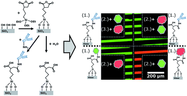
In our experiments, we used 3-triethoxysilylpropyl succinic anhydride (TESPSA; abcr GmbH, Germany) which allowed the immobilization of silanes with succinic anhydride functionalities on oxide surfaces (see Fig. 1A). It has been demonstrated that such anhydride groups readily react with amines in a ring opening reaction forming an amide bond.12,13 With this procedure, antibodies can be covalently bound in a single preparation step (see Fig. 1B).12,13 Additionally, when exposed to water the anhydride group gets hydrolysed and two carboxy groups are generated (see Fig. 1C) inhibiting the subsequent covalent binding in the presence of amines.13


image file: c5ra04469c-f1.tif
Fig. 1 Schematic drawings of (A) the surface functionalization using 3-triethoxysilylpropyl succinic anhydride (TESPSA), (B) the covalent attachment of amines via a succinic anhydride functionality ring opening reaction leading to an amide bond formation, (C) the hydrolysis of the succinic anhydride functionalities resulting in the generation of two carboxyl groups, and (D) the silanization procedure performed in an desiccator with an IR-lamp as an external heat source next to it to evaporate the TESPSA and bring it into contact with the glass surface to induce the silanization reaction. (E) Fluorescence microscopy image showing a glass slide of which one half was dipped into water prior to the immobilization of fluorescence labelled oligonucleotides. The visible fluorescence intensity edge goes along the middle line of the glass slide.

Consequently, using a TESPSA layer to attach receptor molecules to oxide surfaces makes the additional binding site passivation dispensable. Hence, the surface functionalization process is reduced by one step. Therefore, this procedure will facilitate the surface preparation for many sensory devices (Fig. 1).

To test the applicability of TESPSA it was deposited in stripe patterns on glass substrates and immersed with antibody molecules. We investigated (i) the attachment of the antibodies to the surface and (ii) the specificity against their target molecules with fluorescence microscopy. The experimental setup enabled us to simultaneously obtain information about the surface passivation properties to resist unspecific binding.

We used microscopy-grade glass slides as substrates (Paul Marienfeld GmbH, Germany). Prior to the silane deposition, the glass slides were consecutively cleaned in acetone, isopropanol and deionized water, dried under a stream of nitrogen and plasma treated (air plasma; Plasma Prep II, SPI supplies, USA). In the next step, the glass slides were placed in a desiccator with an open container of TESPSA on the bottom (Fig. 1D). The silane was evaporated by applying a vacuum of approx. 50 mbar and via an additional heat input using an externally positioned IR-lamp to induce the silanization reaction at the glass surface for at least 4 h (see Fig. 1D). Subsequently, the glass slides were rinsed in isopropanol and dried under a stream of nitrogen to remove any unbound silane molecules from the surface. We observed a contact angle of approx. 52° using sessile drop contact angle measurements. This contact angle result is approx. twice as high as reported by Lee et al.,14 maybe due to different silanization regimes or to the fact that silicon wafers with an oxide surface were used as substrates in the latter case.

Prior to the immobilization of the desired amine molecules, the samples were cured and annealed in a furnace at 120 °C for at least 1.5 h to remove any water from the surface and to dehydrate all succinic anhydride functionalities.13,15 In an initial test we submerged one half of an annealed glass slide into a water bath for 1.5 h to investigate the effect of water on the amine binding capabilities. Subsequently, the sample was entirely dipped into a 10 mM PBS solution (pH 7.4) containing 0.1 mM fluorescently labeled amino-terminated oligonucleotides (Eurofins MWG Operon, Germany) for 30 min. After submerging, the glass slide was rinsed with deionized water and dried under a stream of nitrogen. A typical fluorescence microscopy image (Axiovert 200M microscope, Carl Zeiss Jena GmbH, Germany) is shown in Fig. 1E. We observed a sharp edge along the centerline of the glass slide at which the fluorescence intensity strongly increases. This edge is a result of the hydrolysis of the succinic anhydride functionalities on the right-hand side and the annealed functionalities on the left-hand side prior to the oligonucleotide immobilization. On the left-hand side the binding affinity towards the amino-terminated oligonucleotides was much higher, resulting in the visible fluorescence contrast. The result shown in Fig. 1E proves the concept of the presented procedure (Fig. 1A–C).

We tested the influence of the described amine binding strategy on the behavior of immobilized receptor molecules by depositing the silanes in a stripe pattern following a similar procedure as described by Shah et al.16 Briefly: (i) after the intensive cleaning procedure of the glass slides (as described above), the surface was covered with the protein repellent polyethylene glycol silane (mPEG-silane, MW 350; Nanocs Inc., New York, NY, USA)17 in a 2 vol% ethanol solution for 45 min, (ii) the repellent surfaces were rinsed with isopropanol and dried under a stream of nitrogen, (iii) we deposited a stripe-patterned photoresist film (AZ 6632, MicroChemicals GmbH, Germany) on top of the mPEG layer (contact angle approx. 35°). (iv) The samples were plasma treated a second time for 15 min to remove the mPEG layer from unexposed stripe areas, to obtain a bare glass surface, (v) followed by TESPSA deposition. (vi) We removed the photoresist with acetone and isopropanol from the surface, resulting in an alternating pattern of stripes with amine-binding functionalities and stripes with protein repellent properties (see Fig. 2A). (vii) Finally, the glass slides were dried and annealed in a furnace for at least 1.5 h at 120 °C.


image file: c5ra04469c-f2.tif
Fig. 2 Schematic drawing of (A) the surface pattern consisting of stripes formed from TESPSA next to stripes derived from the deposition of mPEG-silanes, (B) the experimental layout of the microfluidic channels to run four experiments in parallel on one substrate, (C) the receptor analyte combination – a receptor without fluorescence label is immobilized on the surface and specifically attracts the green fluorescently-labelled analyte but not the red analyte. (D) Merged fluorescence microscopy images of green and red channel – in four microfluidic channels (indicated by grey dotted lines). Receptor and analyte molecules were incubated with the surface in different orders as schematically depicted next to the channels.

On top of the prepared glass, we assembled a polydimethylsiloxane (PDMS) based microfluidic channel to load the desired receptor molecules onto the surface, as described elsewhere.18 The design consists of four channels arranged in a cross like manner (see Fig. 2B) to run and microscopically observe four independent experiments simultaneously.

The composition of the receptor–analyte system used for testing the applicability of our immobilization strategy is schematically shown in Fig. 2C. We used the following biomolecules: a donkey anti-rabbit IgG as the receptor molecule (IgGreceptor); a rabbit anti-mouse IgG (DyLight 488 fluorescence label, λemission = 518 nm) as the corresponding target analyte (IgGtarget_green); a donkey anti-goat IgG (DyLight 594 fluorescence label, λemission = 617 nm) as a control analyte (IgGcontrol_red). All IgG were ordered from Fisher Scientific GmbH, Germany.

The conducted experimental sequences are schematically summarized in Fig. 2D for the four different channels. The observed results are shown as merged images consisting of false colored green and red fluorescence images. In each microfluidic channel, we subsequently loaded different antibody solutions (0.01 mg ml−1 IgG in 10 mM PBS buffer, pH 7.4) over the surface for 30 min, followed by an intermediate rinsing step using 10 mM PBS-Tween (pH 7.4, 0.05% Tween20) between the applications of two subsequent antibody solutions. We used the following antibodies for the different channels:

Channel 1: (1) IgGreceptor, (2) IgGtarget_green, (3) IgGcontrol_red

Channel 2: (1) IgGreceptor, (2) IgGcontrol_red, (3) IgGtarget_green

Channel 3: (1) IgGtarget_green, (2) IgGcontrol_red

Channel 4: (1) IgGcontrol_red, (2) IgGtarget_green

The highest fluorescence signals were observed in all cases along the identical stripes (Fig. 2D). At other parts of the channel with different patterns, we confirmed that the dark areas were covered with photoresist during the second plasma treatment (see ESI, Fig. S1). Due to their repellent effect on the antibodies, we conclude that the dark areas were still coated with mPEG-silane.

In channel 4, we only observed a red fluorescence signal, which is due to the immobilization of IgGcontrol_red. The subsequent incubation with IgGtarget_green did not result in additional unspecific fluorescence emission, proving the additional binding site passivation dispensable (see also ESI, Fig. S2). All excess anhydride groups, i.e. all excess binding sites, are hydrolyzed – and therefore passivated – by the water in the antibody (IgGcontrol_red) solution before the incubation with the analyte (IgGtarget_green) solution.

In channel 3, we mainly detected a green fluorescence signal as a result of the direct immobilization of IgGtarget_green. The subsequent incubation with IgGcontrol_red led to minor unspecific interactions (see ESI, Fig. S2), suggesting a high affinity of the IgGcontrol_red to adhere to the antibody covered surfaces. We observed a similar behavior of the IgGcontrol_red in channel 1 and 2, where also weak unspecific interactions occurred. However, we mainly detected a strong green fluorescence signal in channel 1 and 2, indicating that the immobilization sites, namely the attached IgGreceptor, specifically attract the IgGtarget_green.

The established covalent bonds were irreversible, even after multiple subsequent cleaning steps the fluorescent signal remained constant.

We believe that the receptor immobilization strategy presented here will strongly facilitate the task to develop sensory devices with high specificity towards selected analyte species. Particularly, our finding that the utilization of TESPSA, instead of other silanes,4,7–11 can reduce the need for an additional binding site passivation makes the developed approach attractive for other applications in various scientific fields.

Acknowledgements

This work was funded by the European Union (ERDF) and the Free State of Saxony via the ESF project 100098212 InnoMedTec. The authors thank Ralf Zimmermann and Carsten Werner (Leibniz Institute of Polymer Research Dresden, Germany) for access and assistance with contact angle measurements.

Notes and references

  1. G. MacBeath and S. L. Schreiber, Science, 2000, 289, 1760 CAS.
  2. M. Schena, D. Shalon, R. W. Davis and P. Brown, Science, 1995, 270, 467 CAS.
  3. J. Homola, S. S. Yee and G. Gauglitz, Sens. Actuators, B, 1999, 54, 3 CrossRef CAS.
  4. G. Zheng, F. Patolsky, Y. Cui, W. U. Wang and C. M. Lieber, Nat. Biotechnol., 2005, 23, 1294 CrossRef CAS PubMed.
  5. C. Shan, H. Yang, J. Song, D. Han, A. Ivaska and L. Niu, Anal. Chem., 2009, 81, 2378 CrossRef CAS PubMed.
  6. C. D. Bain, E. B. Troughton, Y.-T. Tao, J. Evall, G. M. Whitesides and R. G. Nuzzo, J. Am. Chem. Soc., 1989, 111, 321 CrossRef CAS.
  7. L. Römhildt, C. Pahlke, F. Zörgiebel, H.-G. Braun, J. Opitz, L. Baraban and G. Cuniberti, ACS Appl. Mater. Interfaces, 2013, 5, 12029 Search PubMed.
  8. J. H. Chua, R.-E. Chee, A. Agarwal, S. M. Wong and G.-J. Zhang, Anal. Chem., 2009, 81, 6266 CrossRef CAS PubMed.
  9. F. Patolsky, G. Zheng and C. M. Lieber, Nat. Protoc., 2006, 1, 1711 CrossRef CAS PubMed.
  10. J. Escorihuela, M. J. Bañuls, J. G. Castelló, V. Toccafondo, J. García-Rupérez, R. Puchades and Á. Maquieira, Anal. Bioanal. Chem., 2012, 404, 2831 CrossRef CAS PubMed.
  11. G. Festag, A. Steinbrück, A. Wolff, A. Csaki, R. Möller and W. Fritzsche, J. Fluoresc., 2005, 15, 161 CrossRef CAS PubMed.
  12. R. G. Chapman, E. Ostuni, L. Yan and G. M. Whitesides, Langmuir, 2000, 16, 6927 CrossRef CAS.
  13. T. Pompe, S. Zschoche, N. Herold, K. Salchert, M.-F. Gouzy, C. Sperling and C. Werner, Biomacromolecules, 2003, 4, 1072 CrossRef CAS PubMed.
  14. M. H. Lee, D. Boettiger, P. Ducheyne and R. J. Composto, Silanes and Other Coupling Agents, 2007, 4, 163 Search PubMed.
  15. T. Pompe, L. Renner, M. Grimmer, N. Herold and C. Werner, Macromol. Biosci., 2005, 5, 890 CrossRef CAS PubMed.
  16. S. S. Shah, M. C. Howland, L.-J. Chen, J. Silangcruz, S. V. Verkhoturov, E. A. Schweikert, A. N. Parikh and A. Revzin, ACS Appl. Mater. Interfaces, 2009, 1, 2592 CAS.
  17. J. Blümmel, N. Perschmann, D. Aydin, J. Drinjakovic, T. Surrey, M. López-García, H. Kessler and J. P. Spatz, Biomaterials, 2007, 28, 4739 CrossRef PubMed.
  18. A. Gang, N. Haustein, L. Baraban and G. Cuniberti, RSC Adv., 2015, 5, 11806 RSC.

Footnote

Electronic supplementary information (ESI) available. See DOI: 10.1039/c5ra04469c

This journal is © The Royal Society of Chemistry 2015
Click here to see how this site uses Cookies. View our privacy policy here.