PVdF/graphene oxide hybrid membranes via electrospinning for water treatment applications
Wongi Janga,
Jaehan Yuna,
Kyungsu Jeonb and
Hongsik Byun*a
aDepartment of Chemical Engineering, Keimyung University, Deagu, 704-701, Korea. E-mail: hsbyun@kmu.ac.kr
bNano Characterization & Analysis Service Team, Convergence Practical Application Center, Daegu 704-801, Korea
First published on 19th May 2015
Abstract
Membrane materials based on poly (vinylidene fluoride) (PVdF) have received great attention recently due to their outstanding mechanical property and chemical resistance. However, this material can easily cause a membrane fouling problem due to its hydrophobic nature. This paper describes how to overcome this problematic issue by incorporating hydrophilic graphene oxide (GO) into PVdF-based membranes. Herein, PVdF nanofiber membranes loaded with GO were prepared via an electrospinning method and the hybrid membranes were characterized for water treatment applications. Graphene oxide sheets were initially prepared by the Hummer's method. The pore property of the PVdF/GO hybrid nanofiber membrane for microfiltration (MF) applications was controlled by systematically increasing the number of nanofiber layers and thermal treatment. These resulting materials were characterized by SEM, FT-IR, UV-Vis, Raman spectroscopy, and tensometer. The overall results showed the reliable formation of the hybrid membranes which possessed controlled pore-diameters (∼0.2 micron) and narrow distribution. Based on contact angle tests, these PVdF/GO nanofiber hybrid membranes exhibited very hydrophilic characteristics. In addition, the hybrid membrane showed high pure water flux results up to 3 times and outstanding flux decline with 0.1 g L−1 Kaolin solutions compared to a neat PVdF nanofiber membrane. Based on all these results, it can be speculated that the incorporation of GO into PVdF could also improve the antifouling ability of the membrane system and will allow for their use as a water-treatment membrane.
Introduction
Synthetic polymer membranes including poly (vinylidene fluoride) (PVdF), Poly (sulfone) (PSf), poly (ether sulfone) (PES), poly (ethylene) (PE), and poly (propylene) (PP) have been widely used in water treatment processes due to their outstanding physical and chemical properties.1,2 However, these materials often exhibit hydrophobic properties causing some critical problems including the flux and rejection decline, and organic fouling.3–6 In order to resolve some of these issues, various approaches have been developed to render hydrophilic properties to these polymeric materials by introducing hydrophilic monomers by low energy plasma7 and irradiation,8 as well as by blending hydrophilic organic and inorganic materials.9,10Recently, nano-scale metal oxide particles,11 carbon nanotubes,12 and graphene13,14 have been utilized to improve these polymer-based membrane properties with increased permeability, selectivity, and anti-fouling effect. In particular, graphene as an additive has many advantages for water purification due to its high surface area (2630 m2 g−1) and chemical stability.15,16 As exfoliated graphene oxide (GO) can possess various hydrophilic functional groups such as carboxyl, epoxyl, and hydroxyl groups,17 GO-incorporated membranes have been tested in waste-water treatment systems to remove heavy metal ions (e.g., Pb, Cd, and As ions).15,18,19
Electrospinning technique is a relatively new approach to manufacture nanofiber-based membranes for microfiltration (MF) and ultrafiltration (UF) systems.20 It has been introduced to prepare a non-woven nanofibrous web or membrane by electrostatic charge to the polymer solution jet. This technique offers many advantages such as a high surface area-to-volume ratio, outstanding selectivity of polymer materials, and easy incorporation of various functional groups. In addition, this technology can reduce the environmental treatment costs as the membrane manufacturing process doesn't require the use of non-solvent bath.21–23
Herein, GO was prepared by the modified Hummer's method and was hybridized into an electrospun poly (vinylidene fluoride) (PVdF) membrane to prepare hybrid membrane for microfiltration (MF) application. We systematically controlled the pore property of the hybrid membrane, and investigated the changes of water flux and surface modifications of the nanofiber membrane as a function of GO content.
Experimental section
Materials
Materials used to synthesize graphene oxide were graphite flake (Bay city, Michigan 48706), sulfuric acid (98%), potassium permanganate (Sigma Aldrich), sodium nitrate (NaNO3, ≥99.0%, Sigma Aldrich), hydrogen peroxide (H2O2, 35%, Samchen Co. Ltd. in South Korea). Materials used to manufacture hybrid nanofiber membrane were poly (vinylidene fluoride) (PVdF, Arkema, Kynar 761), N,N-dimethyl formamide (Duksan Pure Chemical Co. Ltd., Korea, >99.0%), acetone (Duksan Pure Chemical Co. Ltd.). The distilled water was purified through a Millipore system (∼18 MΩ cm). All of the chemicals and reagents were used as received without further purification.Synthesis of graphene oxide (GO) and GO paper
GO was synthesized by the oxidation of natural graphite flake using the modified Hummer's methods.24 Specifically, 2 g of graphite flake and 1.52 g of NaNO3 were mixed with sulfuric acid (67.6 mL) under stirring at room temperature, after which 9 g of KMnO4 were gradually added into the mixture. The resulting mixture was put into an ice-bath and stirred for 5 hours under 10 °C. The mixture was then additionally stirred at room temperature for 5 days and eventually turned into a brownish paste. The brown paste compound was further fully dissolved by 5 vol% of sulfuric acid and stirred for 3 hours, followed by the addition of 5 mL of H2O2 into the compound. The color changed to bright yellow. A mixture of 4 vol% of H2SO4 and 1.5 vol% of H2O2 solution was added to this bright yellow solution to oxidize graphite flakes. The oxidized graphite was washed several times with distilled water and centrifuged (Hanil Science Industrial Co., Ltd., FLETA 5) at 4000 rpm for 10 min until the pH of the top solution became neutral. To obtain the GO powder, vacuum drying was performed for 2 day. Exfoliation of graphite oxide to GO was achieved using a tip sonicator (Sonic VCX-750, Sonics & Materials, Inc.) with 1 mg mL−1 of graphite oxide solution for 1 hour. Then, the final GO paper was obtain by filtering the graphene oxide solution with a vacuum filtration system with 0.45 μm PVdF filter (Millipore Co., Ltd.).Preparation of PVdF/GO hybrid nanofiber membrane
The PVdF (PNMs) and PVdF/GO hybrid nanofiber membranes (PGNMs) were prepared by the electrospinning method after the preparation of an electrospinning solution consisting of 20 wt% of PVdF powder and the 0.1–0.4 wt% of GO in DMF and Acetone under sonication for 1 hour. The composition of the solution is listed in Table 1. The prepared solution was then filled into a 5 mL syringe with a 22 gauge needle. The syringe was positioned vertically for 30 min. By pushing the end of syringe plunger, the air was completely removed. The ejection speed was controlled with a KDS100 (KD Scientific Inc.), and the voltage supply equipment used was a CPS 60K02VIT (Chungpa EMT Co., Ltd.). The following electrospinning conditions were used: flow rate 0.6 mL h−1, voltage 15 kV, TCD (tip to collector distance) 10 cm, duration 6 h, and relative humidity 20–40%. To improve physical property and to control pore diameter, the PGNMs were thermally treated in a dry-oven at 120 °C for 24 hours after stacking PGNM's layers and placing between glass plates. After peeling off the membrane from the glass plates, the membrane was rinsed with methanol and distilled water to remove any residues.| Sample code | PVdF (wt%) | GO (wt%) | DMF (wt%) | Acetone (wt%) |
|---|---|---|---|---|
| PNM | 20.0 | 0.0 | 64 | 16 |
| PGNM01 | 19.9 | 0.1 | 64 | 16 |
| PGNM02 | 19.8 | 0.2 | 64 | 16 |
| PGNM03 | 19.7 | 0.3 | 64 | 16 |
| PGNM04 | 19.6 | 0.4 | 64 | 16 |
Characterization of synthesized GO and PVdF/GO hybrid nanofiber membranes
The surface morphology of GO was observed by STEM (Scanning Transmission Electron Microscope, Hitachi, HD-2300). The GO solution was prepared to 0.1 mg mL−1 density in ethanol by dispersion for about 10 minutes with an ultra-sonicator, and the STEM samples were prepared by completely drying of 1–2 drops of this GO solution on the TEM grid with infrared. In this experiment 400 mesh carbon was used for grid and the acceleration voltage was 200 keV.
The SEM and EDS (Hitachi, S-4800) analysis were performed to observe the surface morphology of the PNMs and PGNMs, and to confirm the presence of GO in nanofiber. These membranes were completely dried in a vacuum oven at room temperature for 1 hour and the osmium (Os) was coated for 5 second on the membranes by using a vacuum sputter. Raman spectroscopy (WITec project, alpha 300 R) was used to determine whether the introduction of GO in the nanofiber membrane was successful or not. The sample was made to a 1 cm × 1 cm size and mounted on a glass substrate, then the laser beam was focused on the center of the membrane surface.
To analyze the porosity of membranes, the prepared nanofiber membranes (5 cm × 5 cm) were soaked in n-butanol (Junsei Chemical Co. Ltd.) at room temperature for 2 hours. The membranes were taken out from the solvent and wiped with Kimwipes to remove excess n-butanol from the surface. The mass of these wet membranes (Wwet) was measured. To determine the mass of dry membranes (Wdry) and volume (Vdry), the wet membranes were dried in the oven at 100 °C for 24 hours. The average water uptake values were determined based on five measurements. The porosity was then determined by the following eqn (1).
| Porosity (%) = (Wwet − Wdry)/(ρbVdry)100% | (1) |
| Water flux (kg m−2 h−1) = mx/ΔtAx | (2) |
 | ||
| Fig. 1 Schematic diagram of a dead-end-cell device.25 | ||
 | (3) |
Results and discussion
Morphology and structure of synthesized GO and PVdF/GO hybrid nanofiber membranes
The synthesized GO was analyzed by FT-IR (Fig. 2). From Fig. 2, it was clearly observed that a peak appeared at 1630 cm−1 for the stretching vibration of remaining sp2 character of C![[double bond, length as m-dash]](https://www.rsc.org/images/entities/char_e001.gif) C bond. The peak in the range of 3000–3700 cm−1 is attributed to the –OH stretching vibration. The peak at 1720 cm−1 is corresponding to the carboxyl group (C
C bond. The peak in the range of 3000–3700 cm−1 is attributed to the –OH stretching vibration. The peak at 1720 cm−1 is corresponding to the carboxyl group (C![[double bond, length as m-dash]](https://www.rsc.org/images/entities/char_e001.gif) O stretching vibration); 1050 cm−1 peak is the vibrational absorption peak for the C–O–C group. This shows that under our experimental conditions, the synthesized GO possesses many hydrophilic functional groups including –OH, –COOH, –C
O stretching vibration); 1050 cm−1 peak is the vibrational absorption peak for the C–O–C group. This shows that under our experimental conditions, the synthesized GO possesses many hydrophilic functional groups including –OH, –COOH, –C![[double bond, length as m-dash]](https://www.rsc.org/images/entities/char_e001.gif) O, –CH(O)CH–.
O, –CH(O)CH–.
The STEM analysis was carried out to examine the surface morphology of synthetic GO. The winkle shape of the GO unique features was observed from the STEM images (Fig. 3(e)). Further, it was shown that the GO sheets were multi-layered, which was expected since the GO layers were not completely exfoliated during the sample preparation process due to the relatively short time of ultrasonic dispersion. Thus, it can be expected that the GO will be introduced between the nanofiber in a multilayer structure, not less than 1 nm of single layer structure.
 | ||
| Fig. 3 Optical images of prepared nanofiber membranes; (a) PNM, (b) PGNM01, (c) PGNM02, (d) PGNM03, and (e) PGNM04. | ||
The surface morphology of the neat PVdF nanofiber membrane and PVdF/GO hybrid nanofiber membranes was observed by SEM analysis. The Fig. 4(a)–(e) images exhibit the surface morphology of nano-sized fibers with network-like porous structures. The average diameter of nanofiber was measured to be between 600–700 nm. However, a two-dimensional structure of GO couldn't be observed for all of PGNM samples. But it was found that the colour of PGNM samples was gradually changed from white to brownish colour with increase of the GO content. In order to confirm the existence of GO in the nanofiber the EDS was taken and the results showed small amount of oxygen atom peaks (1.0–1.7%) due to the small amount of GO in nanofiber (Fig. 5).
 | ||
| Fig. 4 STEM, and SEM images of prepared nanofiber membrane; (a) PNM, (b) PGNM01, (c) PGNM02, (d) PGNM03, (e) PGNM04, and (f) GO. | ||
In order to confirm the existence of GO in the nanofiber membrane, Raman spectroscopy analysis was carried out. This technique has been known as an efficient and quick method for determining the structure of graphene derivatives.
The Raman spectra in Fig. 6 show the D-band peak at ∼1,352 cm−1 and G-band peaks at ∼1,601 cm−1 in all of GO sheets and PGNM samples unlike the PNM samples. It is well known that the G-band corresponds to the first-order scattering of the E2g mode observed for sp2 carbon domains, and the pronounced D band is associated with structural defects, amorphous carbon, or edges that can break the symmetry and selection rule.16 As PGNMs also clearly exhibit the D- and G-band peaks. From the Raman spectroscopy results it could be concluded that GO was successfully incorporated with the PVdF nanofiber membrane.
Pore size and porosity analysis of PVdF/GO hybrid nanofiber membrane
It was observed that neat PVdF membrane had a bubble point of 0.4 μm and mean pore size of 0.26 μm. By increasing the content of GO in PVdF nanofiber membranes, the pore size of PGNMs gradually decreased. The largest pore size of PGNM04 containing 0.4 wt% of GO was tested to be ∼0.2 μm. The pore properties of a series of membrane samples are summarized in Table 2. Although the membrane pore sizes somewhat decreased as a function of GO content, overall porosity of the membranes were measured to be about 35%, and the thickness was in a range of 55–65 μm. It is speculated that the GO, a two-dimensional material, is not like other nano-fillers such as a nano-clay which affected the overall porosity of membranes.| Sample code | Biggest pore diameter (nm) | Smallest pore diameter (nm) | Avg. pore diameter (nm) | Porosity (%) | Thickness (μm) |
|---|---|---|---|---|---|
| PNM | 402.7 | 144.9 | 259.2 | 35 | 55–64 |
| PGNM01 | 316.5 | 135.2 | 218.8 | 38 | 54–62 |
| PGNM02 | 256.4 | 133.4 | 203.5 | 35 | 55–65 |
| PGNM03 | 241.2 | 132.9 | 185.0 | 34 | 57–62 |
| PGNM04 | 200.6 | 118.6 | 167.1 | 32 | 58–61 |
Mechanical properties of PVdF/GO hybrid nanofiber membranes
Fig. 7 shows the mechanical properties of various hybrid nanofiber membranes as a function of GO content. As the amount of GO in nanofiber membrane increased, the tensile strength gradually increased. This is presumably caused by a strong hydrogen bond interaction between the GO and PVdF nanofiber. Thus, all the samples exhibited enhanced mechanical properties higher than 280 kgf cm−2. These results could be explained by the high number of complex physical bonds in the nanofiber itself.Contact angle analysis of PVdF/GO hybrid nanofiber membranes
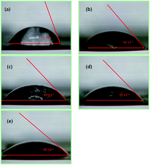
As GO possesses hydrophilic carboxyl, hydroxyl, and epoxide groups, it was speculated that the hydrophobic PVdF could exhibit hydrophilic property upon introduction of small amount of GO. With systematic increase of the GO content in the nanofiber membrane, the water flux was obviously improved and the contact angle of hybrid membranes gradually decreased from 70° to 40° which are shown in Fig. 8. | ||
| Fig. 8 Contact angle results of prepared nanofiber membranes; (a) PNMs, (b) PGNM01, (c) PGNM02, (d) PGNM03, and (e) PGNM04. | ||
Pure water flux of PVdF/GO hybrid nanofiber membranes
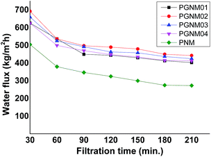
According to other research, it is known that water molecules can be easily drawn to the inside of the membrane with a hydrophilic surface, hence the flux of a membrane can be increased by enhancing hydrophilicity of the membrane.12 The pure water flux results were shown in Fig. 9. Although the PVdF membrane has the largest pore-diameter among all samples, the pure water flux was very low due to the hydrophobic nature. Under the 1 bar pressure condition, the water flux value of neat PVdF membrane was lower than 200 kg m−2 h−1. However, when adding GO into PVdF nanofiber, all of the PVdF/GO hybrid nanofiber membranes showed 3 times higher values of pure water flux than that of neat PVdF membrane due to its hydrophilic nature. Meanwhile, PGNM02 sample had the highest value of water flux among all of the samples. However, the water flux values of PGNM04 were lower than PGNM02 and PGNM03 despite of having the lowest contact angles (Fig. 7). This result is in accordance with the pore-properties of PGNMs and it is expected that the membrane pores are gradually blocked by the aggregation of GO.Turbidity change and flux decline results of PVdF/GO hybrid nanofiber membranes
For the rejection of turbidity test, 0.1 g L−1 Kaolin solution (160 NTU) was used, where standard distill water showed a turbidity of 0.16 NTU. Filtrate rejection rate and flux decline results of our membranes were shown in Table 3 and Fig. 10, respectively.| Sample code | After 30 min rejection (%) | 60 min (%) | 120 min (%) | 180 min (%) |
|---|---|---|---|---|
| PNM | 99.89 | 99.90 | 99.89 | 99.89 |
| PGNM01 | 99.90 | 99.90 | 99.90 | 99.90 |
| PGNM02 | 99.90 | 99.89 | 99.89 | 99.90 |
| PGNM03 | 99.90 | 99.90 | 99.90 | 99.89 |
| PGNM04 | 99.90 | 99.89 | 99.90 | 99.90 |
During filtration with the kaolin solution, it was expected that the pore of membrane could be gradually blocked, resulting in the decrease of flux. As the neat PVdF nanofiber membrane was hydrophobic, it could be easily polluted by kaolin grains compared to PGNM samples. The flux obviously declined after 90 min of filtration. The rejection value of all samples was measured to be 99.9% during filtration tests with 0.1 g L−1 Kaolin feed solution. But the flux decline was improved with increase of GO in the nanofiber membrane, indicating the PVdF/GO hybrid nanofiber membrane could lower the membrane fouling which is one of the general problems with polymeric membrane. It can thus be concluded that the PVdF/GO hybrid nanofiber membrane can be used for the water treatment application as a microfiltration membrane.
GO elution test of PVdF/GO hybrid nanofiber membranes during filtration
From the Fig. 11 it was observed that GO showed maximum absorption peak at ∼237 nm attributable to π–π* transition of the atomic C–C bonds and shoulder peak at ∼300 nm due to n-π* transitions of aromatic C–C bonds.26 The filtrated water did not show the absorption peaks of both ∼237 nm and ∼300 nm, thus, it was confirmed that the GO in the nanofiber matrix wasn't eluted. | ||
| Fig. 11 UV-Vis spectra results of various concentrated GO and filtrate of PGNM02 after filtration for 7 day at 1 bar pressure. | ||
Conclusions
In this study, we focused on the preparation of porous PVdF/GO hybrid nanofiber membranes (PGNMs) for possible water-treatment applications. The formation of PGNMs was completed via the electrospinning method using a solution containing a mixture of PVdF powder (20 wt%) and exfoliated graphene oxide (0.1 to 0.4 wt%) in dimethylformamide (64 wt%) and acetone (16 wt%). The resulting PGNMs had improved mechanical properties upon thermal treatment. The pore diameter of PGNMs was systematically controlled by simply increasing the number of PGNMs layers. The prepared membrane and nanomaterials (GO) were characterized by SEM, FT-IR, UV-Vis spectra, Raman analysis and tensometer. These results showed that the pore-diameter was controlled to be 0.2 μm with narrow distribution. Based on contact angle tests these prepared PVdF/GO hybrid nanofiber membranes exhibited hydrophilic characteristics. In addition, the PGNMs showed pure water flux results up to 3 times higher, and outstanding flux decline with 0.1 g L−1 Kaolin solutions compared to neat PVdF nanofiber membrane. Thus it is expected that the antifouling ability of membranes can be improved by adding GO. Based on the results of GO elution test, it was confirmed that the GO in the nanofiber matrix wasn't eluted. It was concluded that the PVdF/GO hybrid nanofiber membranes can be utilized for water-treatment.Acknowledgements
This subject is supported by Korean Ministry of Environment as “The Eco-Innovation project (Global-Top project), code number 2014001080001”.References
- Y. Yang, H. X. Zhang, P. Wang, Q. Z. Zheng and J. Li, J. Membr. Sci., 2007, 288, 231–238 CrossRef CAS PubMed.
- J. N. Shen, H. M. Ruan, L. G. Wu and C. J. Gao, Chem. Eng. J., 2011, 168, 1272–1278 CrossRef CAS PubMed.
- I. S. Chang, P. L. Clech, B. Jefferson and S. Judd, J. Environ. Eng., 2002, 125, 1018–1029 CrossRef.
- J. R. Du, S. Peldszus, P. M. Huck and X. S. Feng, Water Res., 2009, 43, 4559–4568 CrossRef CAS PubMed.
- W. Zhou, A. Bahi, Y. Li, H. J. Yang and F. Ko, RSC Adv., 2013, 3, 11614–11620 RSC.
- T. He, W. Zhou, A. Bahi, H. J. Yang and F. Ko, Chem. Eng. J., 2014, 252, 327–336 CrossRef CAS PubMed.
- L. Zou, I. Vidalis, D. Steele, A. Michelmore, S. P. Low and J. Q. J. C. Verberk, J. Membr. Sci., 2011, 369, 420–428 CrossRef CAS PubMed.
- D. Rana and T. Matsuura, Chem. Rev., 2010, 110, 2448–2471 CrossRef CAS PubMed.
- S. Boributh, A. Chanachai and R. Jiraratananon, J. Membr. Sci., 2009, 342, 97–104 CrossRef CAS PubMed.
- F. Shi, Y. Ma, J. Ma, P. Wang and W. Sun, J. Membr. Sci., 2013, 427, 259–269 CrossRef CAS PubMed.
- A. Razmjou, J. Mansouri and V. Chen, J. Membr. Sci., 2011, 378, 73–84 CrossRef CAS PubMed.
- B. Zhang, L. Liu, S. Xie, F. Shen, H. Yan, H. Wu, Y. Wan, M. Yu, H. Ma, L. Li and J. Li, RSC Adv., 2014, 4, 16561–16566 RSC.
- C. A. Crock, A. R. Rogensues, W. Shan and V. V. Tarabara, Water Res., 2013, 47, 3984–3996 CrossRef CAS PubMed.
- J. W. Lee, H. R. Chae, Y. J. Won, K. B. Lee, C. H. Lee, H. H. Lee, I. C. Kim and J. M. Lee, J. Membr. Sci., 2013, 448, 223–230 CrossRef CAS PubMed.
- H. Jabeen, K. C. Kemp and V. Chandra, J. Environ. Manage., 2014, 130, 429–435 CrossRef PubMed.
- M. Hu and B. Mi, Environ. Sci. Technol., 2013, 47, 3715–3723 CrossRef CAS PubMed.
- S. Stankovich, D. A. Dikin, R. D. Piner, K. A. Kohlhass, A. Kleinhammes, Y. Y. Jia, Y. Wu, S. B. T. Nguyen and R. S. Ruoff, Carbon, 2007, 45, 1558–1565 CrossRef CAS PubMed.
- V. Chandra, J. S. Park, Y. Chun, J. W. Lee, I. C. Hwang and K. S. Kim, ACS Nano, 2010, 4, 3979–3986 CrossRef CAS PubMed.
- G. Zhao, J. Li, X. Ren, C. Chen and X. Wang, Environ. Sci. Technol., 2011, 25, 10454–10462 CrossRef PubMed.
- C. Feng, K. C. Khulbe, T. Matuura, S. Tabe and A. F. Ismail, Sep. Purif. Technol., 2013, 102, 118–135 CrossRef CAS PubMed.
- S. Ramakrishna, K. Fujihara, W. E. Teo, T. Yong, Z. Ma and R. Ramaseshan, Mater. Today, 2006, 9, 40–50 CrossRef CAS.
- N. Chanunpanich, B. S. Lee and H. S. Byun, Macromol. Res., 2008, 16, 212–217 CrossRef CAS.
- N. Daels, S. D. Vrieze, I. Sampers, B. Decostere, P. Westbroek, A. Dumoulin, P. Dejans, K. D. Clerck and S. W. H. Van Hulle, Desalination, 2011, 275, 285–290 CrossRef CAS PubMed.
- W. D. Chen, H. M. Jung, W. G. Jang, B. P. Hong and H. S. Byun, Adv. Mater. Res., 2013, 726–731, 1715–1719 CrossRef.
- W. G. Jang, K. S. Jeon and H. S. Byun, Desalin. Water Treat., 2013, 51, 5283–5288 CrossRef CAS PubMed.
- L. Shahriary and A. A. Athawale, International Journal of Renewable Energy and Environmental Engineering, 2014, 2(1), 58–63 Search PubMed.
| This journal is © The Royal Society of Chemistry 2015 |


