DOI:
10.1039/C5RA04359J
(Paper)
RSC Adv., 2015,
5, 38100-38110
Facile fabrication of a flower-like CuO/Cu(OH)2 nanorod film with tunable wetting transition and excellent stability†
Received
12th March 2015
, Accepted 13th April 2015
First published on 13th April 2015
Abstract
A flower-like CuO/Cu(OH)2 nanorod film on a brass substrate has been synthesized for the first time, with a tunable wetting transition and excellent stability. We devised a facile etching method utilizing an electrolyte containing K2S2O8 and KOH in this study. The growth process and the surface morphology of the flower-like CuO/Cu(OH)2 nanorods are well documented. Moreover, the surface wetting behavior is reversible between superhydrophilic and superhydrophobic. The as-prepared superhydrophilic surface can be converted to superhydrophobic by modification with myristic acid and can be changed back to superhydrophilic after annealing at 200 °C for 6 min. The wetting transition can be cycled more than 30 times and takes less than 10 min per cycle. We also discussed the wetting transition mechanism based on the surface composition analysis and the relevant theoretical model and investigated the abrasion resistance and flush resistance. For the primary application, we propose that a water-drop collecting device based on a large-scale and complex superhydrophobic surface would show an excellent performance.
1. Introduction
In the recent years, superhydrophobic surfaces (with a water contact angle larger than 150°) and superhydrophilic surfaces (with a water contact angle less than 5°), which are two important forms of the solid surfaces, have attracted extensive attention due to their potential applications.1–7 Furthermore, smart surfaces with tunable transitions between superhydrophobicity and superhydrophilicity have been investigated by many researchers8–11 due to their numerous industrial applications such as antifogging surfaces,12 self-cleaning coatings,13–15 oil–water separation,16 microfluid manipulation17 and biotechnology.18 Many methods are employed to fabricate these smart surfaces such as anodic oxidation, electrodeposition, plasma etching, dip coating, hydrothermal processing, electrospinning, sol–gel processing, and laser irradiation.19–26 However, these surfaces are fabricated on expensive substrates and demand strict reaction conditions, which greatly limit their applications.
Our group has committed to study controllable wetting behavior for a few years.27–29 In our previous study, controllable superhydrophobic and superhydrophilic surfaces have been fabricated on brass substrates (an alloy of Cu and Zn) by an alternating current (AC) etching method in an acidic solution.29 Brass is widely used in industrial applications due to its good performance and low price. AC etching is also facile and suitable. However, the prepared pine-cone structure was attained by the decomposition of Zn without further reaction, which made the products uneven and large-sized (about a hundred nanometers to several micrometers). The large-sized structure performs badly in abrasion resistance and flush resistance tests, which greatly limits its potential applications.
In this study, an easy and suitable alternating current (AC) etching method is employed to fabricate a surface that is tunable between superhydrophobicity and superhydrophilicity on a brass substrate in an alkaline solution with an etching duration of 15 min at 20 V AC. According to the results of the contact angle test, this surface is superhydrophilic. After modification by myristic acid (CH3-(CH2)12-COOH) for 30 s, the superhydrophilic surface transforms to superhydrophobic. After annealing at 200 °C for 6 min, the surface returned to superhydrophilic again. Furthermore, this modification and annealing process can be cycled more than 30 times while maintaining good performance. Moreover, the entire wetting transition cycle takes less than 10 min. Unlike Wang's method,29 the fabrication process in this study contains two steps: decomposition of Zn and oxidation of the remaining Cu. Further oxidation of Cu results in uniform and smaller sized nanorods, which greatly improves the stability of the prepared surface and extend its potential applications. It is mainly because the decomposition reaction is strong and uncontrollable, while the oxidation process is more gentle and specific. As a preliminary application, a water drop collecting device based on superhydrophobic surface is proposed and a high performance is expected.
2. Experimental details
2.1 Materials
KOH, K2S2O8, acetone, ethanol and myristic acid (CH3-(CH2)12-COOH) were of analytical grade and did not require further treatment. Brass substrates containing 64 wt% Cu and 36 wt% Zn and an aqueous electrolyte solution containing KOH and K2S2O8 were used. Before etching, the brass substrate was ultrasonically cleaned in acetone, polished with sandpaper and sequentially rinsed with deionized water and ethanol.
2.2 Fabrication of the flower-like CuO/Cu(OH)2 nanorods
At the beginning of the AC etching, two polished brass substrates were immersed in the prepared aqueous solution as electrodes. Then, an AC voltage (20 V and 50 Hz) was applied to the two electrodes for a series of time periods of 1 min, 5 min, 10 min, 15 min, 20 min and 30 min. The electrolyte solution contained 0.025 M K2S2O8 and 1.0 M KOH and the etching temperature was maintained at 50 °C by a water bath. The agitation rate was 600 rpm. Finally, the two brass substrates were taken out and rinsed with deionized water and ethanol in sequence.
2.3 Wetting transition cycle
To fabricate the superhydrophobic surface, 0.48 g myristic acid was dissolved in 500 mL ethanol to modify the surface of the obtained brass substrate. The prepared substrate was immersed in the solution for 30 s at room temperature. After removal from the solution, the prepared substrate was dried at room temperature and then a superhydrophobic surface was obtained.
After annealing at 200 °C for several minutes, the superhydrophobic surface converted rapidly into the superhydrophilic surface, and the appropriate annealing time was determined. By repeating the previous modification step, the annealed surface returned to superhydrophobicity again. Further annealing can also make it superhydrophilic. This transition cycle can be repeated more than 30 times while maintaining a stable wetting behavior.
2.4 Characterizations
A scanning electron microscope (SEM, JSM-7500F, JEOL Ltd., Japan) was used to observe the surface morphology of the obtained flower-like nanorods. Before observation, the samples were sputter-coated with Pt under vacuum to enhance electrical conduction. The crystallographic structure was investigated using an X-ray diffractometer (XRD, ARL XTRA, Thermo Electron Co., Switzerland) with Cu K radiation at a scan speed of 6° min−1 in the 2θ range from 5° to 90°. To study the surface composition, an X-ray photoelectron spectrometer (XPS; VG Scientific ESCA-LAB 250) with 200 W Al Kα radiation was used. Transmission electron microscopy (TEM; JEM-2100M) was used to evaluate the microstructure of the prepared substrate. The digital images were obtained by a digital camera (Olympus E-PL1, Japan). The contact angles were measured using a contact angle meter (DSA 20, Krüss Instruments GmbH) at five different positions for each surface. The volume of an individual droplet in all measurements was about 6 μL.
3. Results and discussion
3.1 Formation of the flower-like CuO/Cu(OH)2 nanorods
To better observe the formation of flower-like CuO/Cu(OH)2 nanorods, SEM images of the growth process are shown in Fig. 1. Fig. 1(A) shows the bare brass substrate before AC etching. It is rough with clear scratches due to the mechanical polishing. The XRD pattern in Fig. 2(D)(a) shows that the brass substrate is composed of 64 wt% Cu and 36 wt% Zn. When the AC etching time was increased to 1 min, the surface morphology of the brass substrate changed dramatically. Particles ranging from hundreds of nanometers to several micrometers were observed on the surface due to the release of the Zn, as shown in Fig. 1(B). Zn would be preferentially oxidized to Zn2+ because it is more active than Cu.30 As the etching time was increased to 5 min, more Zn decomposed. In addition, some small crystals with a size of a few nanometers can be observed on the remaining particles, which were the first generation of CuO/Cu(OH)2, as shown in Fig. 1(C). The irregular particles with first generated CuO/Cu(OH)2 disappeared when the etching time increased to 10 min due to the further decomposition of the Zn. The solution also became turbid with the decomposition of Zn. Crystals with a 200 nm diameter lay on the surface, which can be confirmed as mainly Cu by EDS, as shown in Fig. 1(D) (the peak on the right corresponds to Pt sputter-coated initially). These crystals were stable and provided appropriate place for CuO/Cu(OH)2 to grow. Small nanorods, which are the prototypes of the flower-like nanorods, can be observed in Fig. 1(D). Fig. 1(E) and (F) show the surface morphology and a high-resolution image of the brass substrate after etching for 15 min. The prepared CuO/Cu(OH)2 nanorods with a diameter of about 20 nm and a length of about 200 nm lay uniformly on the surface. The prepared CuO/Cu(OH)2 nanorods marked by a white square in Fig. 1(F) presented a flower-like and uniform morphology. Fig. 1(G) shows the surface morphology after etching for 20 min. Some of the prepared nanorod film was destroyed with the further decomposition of Zn, while some of the nanorods were left on the surface (marked by a white arrow in Fig. 1(G)). New nanorods were generated on the newly exposed Cu and the surface morphology became uneven with mixed structures. As the etching time was increased to 30 min, more nanorods broke away from the surface, as shown in Fig. 1(H). At this point, the brass substrate surface completely lost its flower-like morphology. Moreover, the AC etching time of 15 min is appropriate (the effects of the different concentrations of KOH and K2S2O8 on the morphology of the prepared nanorods are also shown in ESI Fig. S1 and S2.† A contrast experiment without the AC current, under other experimental conditions, was carried out and the result is shown in ESI Fig. S3†).
 |
| | Fig. 1 SEM images of the surface morphology with different AC etching durations: (A) 0 s (bare brass substrate); (B) 1 min; (C) 5 min; (D) 10 min; (E) & (F) 15 min; (G) 20 min and (H) 30 min; insets are the EDS images of the corresponding surfaces. | |
 |
| | Fig. 2 (A) TEM image of the prepared CuO/Cu(OH)2 crystals; (B) HRTEM image of the region marked with the white arrow in (A); (C) the electron diffraction pattern of the area marked by the white arrow in (A) (white is for Cu(OH)2 and red is for CuO); (D) XRD patterns of the (a) untreated brass substrate and (b) the AC etching treated substrate. Inset is the high-resolution image of XRD peaks match with CuO/Cu(OH)2. | |
Fig. 2(A) shows the TEM image of the prepared nanorods. The area of the rectangle is about 400 nm × 600 nm and it contains tens of nanorods and nanoparticles. The nanorods with diameters of about 20 nm and lengths of about 200 nm can be observed clearly in the TEM image, which confirmed the SEM image shown earlier (ESI Fig. S4†). Fig. 2(B) is the high-resolution TEM image of the region marked by a white arrow in Fig. 2(A), showing a lattice fringe of 0.252 nm, which is attributed to the CuO (002) lattice spacing. Fig. 2(C) is the electron diffraction pattern of the area marked by the white arrow in Fig. 2(A), which shows various index facets of the marked area. Lattice spacings of 0.252 nm, 0.232 nm and 0.150 nm can be observed, which correspond to the (002), (111) and (113) planes, respectively, of the CuO phase (PDF 45-0937). Moreover, we can also find the lattice spacings of 0.263 nm, 0.208 nm, 0.193 nm and 0.172 nm, which correspond to Cu(OH)2 (PDF 35-0505) phases of (002), (131), (112) and (132), respectively. Fig. 2(D) shows the XRD patterns of the pure brass substrates and the substrates after AC etching treatment for 15 min. As shown in Fig. 2(D)(a), the peaks at 42.3°, 49.3°, 72.2° and 87.5° match well with the diffraction peaks of Cu0.64Zn0.36 (PDF no. 50-1333), which indicates that the brass substrate is composed of 64 wt% Cu and 36 wt% Zn. The emergence of peaks at 35.5° and 38.7° in Fig. 2(D)(b) indicates that CuO (PDF no. 45-0937) appears on the brass substrate surface after AC etching for 15 min. Furthermore, the existence of peaks at 31.5°, 34.1°, 35.9°, 43.5° and 53.3° match well with the diffraction peaks of Cu(OH)2 (PDF = 35-0505), which indicates that Cu(OH)2 appears after the AC etching. In addition, we can also find significant peaks of Cu0.64Zn0.36 in Fig. 2(D)(b), which indicates that the CuO/Cu(OH)2 film is thin and this is consistent with the SEM analysis results. The results of TEM and XRD analyses further confirm the existence of CuO/Cu(OH)2.
From what has been discussed above, we conclude that the formation of the flower-like CuO/Cu(OH)2 nanorods proceeds as follows:31
| | |
Instant anode: Zn + 4OH− − 2e− → Zn(OH)42−
| (1) |
| | |
Instant cathode: 2H2O + 2e− → 2OH− + H2↑
| (2) |
| | |
Secondary reaction: Cu + S2O82− + 2OH− → Cu(OH)2↓ + 2SO42−
| (3) |
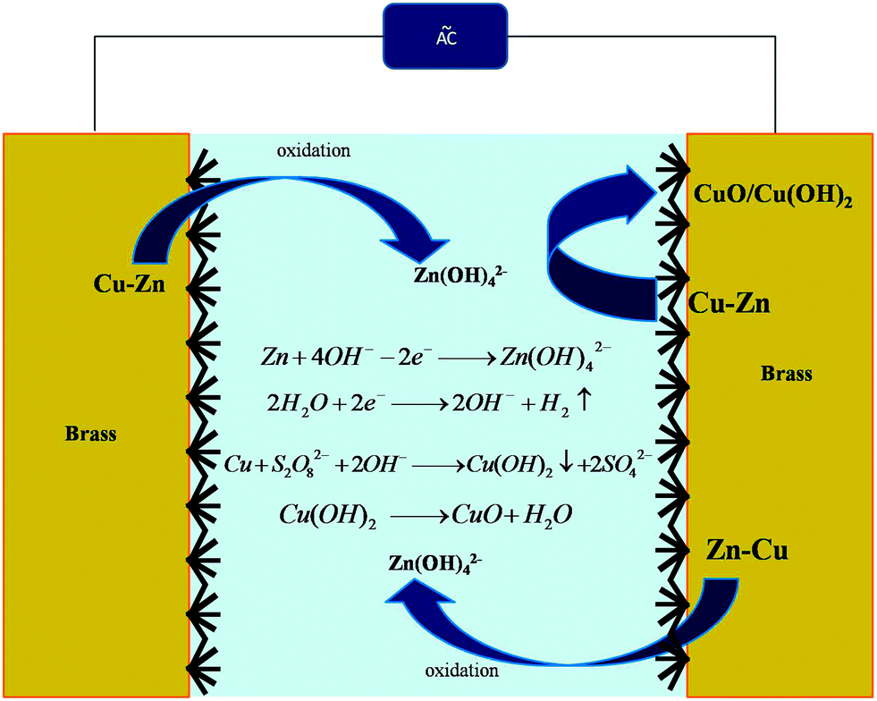
Fig. 3 shows the mechanism of the AC etching of the brass substrate in a solution containing KOH and K2S2O8. During AC etching, the two brass substrates serve as the anode and cathode alternately and both the brass substrates become covered with flower-like CuO/Cu(OH)2 nanorods, i.e. we can fabricate two products at the same time.
 |
| | Fig. 3 Mechanism of AC etching of the brass substrate in a solution containing KOH and K2S2O8. | |
At the beginning of AC etching, Zn in the brass substrate reacted with OH−, as it is an amphoteric metal and more active than Cu,30 as shown in eqn (1). This process is called “dezincification”. Zn gradually dissolved in the solution and Cu was left on the surface of the substrate. This “dezincification” process follows the same method as in our previous study,29 while the products are different due to the different reaction conditions. In this study, after “dezincification”, the remaining Cu can be easily oxidized by S2O82− in the presence of OH− and turn into Cu(OH)2,11 as shown in eqn (3). Cu(OH)2 is not stable and can decompose into CuO and H2O under high temperatures, as shown in eqn (4). The AC etching process released a great deal of thermal energy and further accelerated the decomposition of Cu(OH)2. Thus, the newly generated nanorods are a mixture of CuO and Cu(OH)2. Some of the remaining Cu grains with newly generated CuO/Cu(OH)2 on them would dissolve simultaneously with Zn32 and get mixed into the solution, which made the solution turbid, as described above. The new CuO/Cu(OH)2 would grow on the newly exposed Cu grains. This process would continue until the remaining Cu grains can stay on the surface, as shown in Fig. 1(D). This process takes about 15 min. Then the newly-generated CuO/Cu(OH)2 can grow into uniform nanorods with flower-like structures, as shown in Fig. 1(E) and (F). The prepared CuO/Cu(OH)2 nanorods are more uniform and smaller than the pine-cone structures discussed in our previous study as the oxidation process is milder than the decomposition process. As time increases, more Zn grains are dissolved in the solution and the generated CuO/Cu(OH)2 nanorod film is destroyed, as shown in Fig. 1(G) and (H). We can also find the remaining CuO/Cu(OH)2 nanorods in Fig. 1(G) (marked by the white arrow), which further validated our speculation.
3.2 Wetting transition between superhydrophobicity and superhydrophilicity
The as-prepared surface was superhydrophilic with a contact angle less than 3°, as shown in Fig. 4(A). After immersing in the myristic acid ethanol solution for 30 s and drying at room temperature for several min, the surface converted to superhydrophobic, as shown in Fig. 4(B). By comparing the SEM images before and after modification shown in Fig. 4(A) and (B), we can find that the surface morphology shows no apparent change and is still composed of flower-like nanorods. After annealing at 200 °C for several minutes, the contact angle returned to less than 3° without any apparent change in surface morphology, as shown in Fig. 4(C). This indicates that both the modification and annealing do not change the surface morphology and the change of wetting behavior is mainly due to the modification by the myristic acid and annealing. Myristic acid combined with the surface during the immersing process and the “combination” was damaged during annealing, which caused the wetting behavior to change between superhydrophilicity and superhydrophobicity, as shown in Fig. 4(D) (the digital images of the CAs of the modified surfaces with different AC etching durations are shown in ESI Fig. S5.† Digital images of the process of modified surface contact with the water droplet are shown in ESI Fig. S6†).
 |
| | Fig. 4 The digital CA images and the SEM images of the prepared flower-like CuO/Cu(OH)2 nanorod surface (A) before and (B) after modification by myristic acid, and (C) after annealing at 200 °C; (D) the schematic of the wetting behavior transition. | |
It is known that chemical composition and microstructure are the key factors influencing the wetting behavior of a solid surface.33,34 The phenomenon described above indicates that the superhydrophobicity of the substrate is governed by both the flower-like nano structure and the low surface energy materials. Contrast experiments measuring CAs on blank brass substrates and modified brass substrates were also carried out and the result is shown in Fig. 5. The blank brass is hydrophilic with a CA of 68° ± 1.0° and the modified blank brass is hydrophobic with a CA of 95° ± 1.2°. The blank brass is hydrophilic because the hydroxyl group is absorbed on the metal surface. After modification with myristic acid, the CAs increased as myristic acid combines with the surface and stops the hydroxyl group from absorbing onto the metal surface. Fig. 5(C) and (D) show that the prepared CuO/Cu(OH)2 nanorod film substrate is superhydrophilic with a CA of 0° and the modified substrate is superhydrophobic with a CA of 162 ± 2.1°; this can also be attributed to the combination of myristic acid on the surface. According to previous study,29,35–37 the combination between CuO/Cu(OH)2 and myristic acid can be shown as follows:
| | |
CuO + 2CH3(CH2)12COOH ↔ Cu[CH3(CH2)12COO]2 + H2O
| (5) |
| | |
Cu(OH)2 + 2CH3(CH2)12COOH ↔ Cu[CH3(CH2)12COO]2 + 2H2O
| (6) |
 |
| | Fig. 5 The digital CA images of the (A) blank brass substrate; (B) modified brass substrate; (C) prepared nanorod film substrate and (D) modified nanorod film substrate. | |
CuO and Cu(OH)2 are prone to react with myristic acid in the ethanol solution,11,29,36 thus the myristic acid group will react with CuO/Cu(OH)2 and form Cu[CH3(CH2)12COO]2 on the surface of the prepared CuO/Cu(OH)2 nanorod film. The modified surface is superhydrophobic as Cu[CH3(CH2)12COO]2 is a low surface energy material with long-chain aliphatic groups. During the annealing process, the long-chain aliphatic groups are damaged and the surface becomes superhydrophilic. Moreover, the “combination” between myristic acid and the pure brass substrate in Fig. 5(B) can also be attributed to the formation of Cu[CH3(CH2)12COO]2 and Zn[CH3(CH2)12COO]2 in similar reactions.29,36,37
According to the Cassie–Baxter model38 shown as follows:
where
θr represents the contact angle on the rough surface,
θ represents the contact angle on the smooth surface, and
f1 and
f2 are the fractions of the solid surface and air trapped in grooves when in contact with water, respectively, it can be easily shown that
f1 plus
f2 is 1, as shown in
eqn (8). According to the result of the contact angle test, the
θr after modification was 162° ± 2.1° and the CA of the modified smooth brass substrate surface was 95° ± 1.2° (shown in
Fig. 5(B)), which can be considered as
θ.
39 By substituting them into
eqn (7), the values of
f1 and
f2 can be calculated to be 0.0537 and 0.9463, respectively. The results mean that air occupies about 94.63% of the contact area when the modified surface is in contact with the water droplet. This is because the 3D capillary effect of the prepared CuO nano-structure provides more space for trapping air, which greatly increases the air/liquid interfaces, as shown in
Fig. 4(D). The modification of the low surface energy materials decreases the interaction force to further improve the superhydrophobicity of the prepared surface.
In addition, the entire transition process can be concluded as follows: a rough surface made from high energy materials tends to be hydrophilic and a rough surface made from low energy materials tends to be hydrophobic. The as-prepared nanorod film is superhydrophilic as it is a high surface energy material and the hydroxyl group can be easily absorbed on the surface. After modification with myristic acid, CuO/Cu(OH)2 reacts with myristic acid and produces Cu[CH3(CH2)12COO]2, which is a low surface energy material with a long-chain aliphatic group. The nanorod film with a hierarchical nano-structure and Cu[CH3(CH2)12COO]2 on it shows superhydrophobic property. After annealing at 200 °C, the long-chain aliphatic group decomposed and the surface becomes superhydrophilic. The re-modification process can form new long-chain aliphatic groups and the surface becomes superhydrophobic again. The modification and annealing process does not affect the morphology of the prepared surface, thus this wetting transition can be cycled many times.
As stated above, the wettability of the solid surface depends on the surface morphology and low surface energy materials. In this study, superhydrophobicity was achieved by the modification of the prepared flower-like CuO/Cu(OH)2 nanorods with myristic acid. To remove the low surface energy materials, the modified substrate was annealed at 200 °C. To find the appropriate time of annealing, the contact angles of the prepared surface after different annealing times were measured, as shown in Fig. 6(A). The results illustrate that as annealing time was prolonged, the contact angles of the prepared surface decreased and after annealing for 6 min, the contact angle decreased to zero, i.e., the superhydrophobic surface became a superhydrophilic surface after annealing and 6 min is long enough for this transition. If the annealed substrate is again immersed in myristic acid ethanol solution for 30 s, the surface will revert to superhydrophobicity after drying at room temperature. In further experiments, this wettability change was cycled several times to confirm its stability. The experimental results shown in Fig. 6(B) indicate good reversibility of the wettability. In fact, after 30 cycles, the prepared surface can retain excellent reversibility in this experiment, which is longer than other surfaces reported in the literature.4,10,29,39–44 This can be attributed to the smaller size of the nanorods, which are not easily damaged and make the surface more stable. It is obvious that the longer the wettability cycling lasts, the wider its application will be. In other words, the new prepared surface has great potential in practical applications. In addition, each cycle of the wettability change in this study takes less than 10 min, which is much shorter than that previously reported.
 |
| | Fig. 6 (A) The contact angles of the surface after different annealing times (average of 5 different positions); (B) cycles between superhydrophobicity and superhydrophilicity of the prepared surface after modification and annealing. | |
3.3 Component analysis of the surfaces
As stated above, we know that the modification by myristic acid and annealing do not change the surface morphology, and the change in wetting behavior is mainly because of the absorbance and release of myristic acid on the surface. To confirm this viewpoint, X-ray photoelectron (XP) spectra of the prepared surfaces before modification with myristic acid (the blank sample), after modification with myristic acid (the modified sample) and after annealing at 200 °C (the annealed sample) were carried out and are shown in Fig. 7, which can further illustrate the surface composition of the three different states. The results of XPS survey spectra in Fig. 7(a) indicate that Cu, O and C can be detected on the three surfaces. Fig. 7(b) shows the high-resolution of Cu 2p regions, in which we can find a typical Cu 2p spectrum. The blank sample and modified sample present obvious peaks at 933.4 and 934.4 eV, which correspond to CuO and Cu(OH)2, respectively. The Cu 2p intensity for the blank sample is higher than for the modified sample. After annealing, the increase in peak intensity at 933.7 eV indicates an increase in CuO. The result demonstrates that the Cu(OH)2 is further decomposed during the thermal treatment. High-resolution spectra of O 1s are also shown in Fig. 7(c). The blank sample contains the main peaks of CuO at 529.4 eV and Cu(OH)2 at 531.7 eV, indicating that the prepared surfaces are mainly composed of CuO and Cu(OH)2. An obvious peak at 533.3 eV appears after modification, corresponding to O–C![[double bond, length as m-dash]](https://www.rsc.org/images/entities/char_e001.gif) O, which indicates that myristic acid was adsorbed on the blank sample during the modification and produced Cu[CH3(CH2)12COO]2 as stated above. After annealing, the peak representing O–CO can also be found, which indicates that some Cu[CH3(CH2)12COO]2 remained after thermal treatment. A high-resolution spectrum of O 1s of the annealed sample in Fig. 7(c) shows that the main peaks of CuO at 529.4 eV increased after annealing, which further confirms the decomposition of Cu(OH)2. Fig. 7(d) shows the high-resolution XPS spectra of the C 1s. After modification, obvious peaks at 288.5 and 284.6 eV appear on the graph, corresponding to O–CO and C–C, which are attributed to the existence of Cu[CH3(CH2)12COO]2. After annealing, the C 1s peak intensity declines significantly, which is due to the Cu[CH3(CH2)12COO]2 decomposing during the thermal treatment. Comparing the modified sample and the annealed sample indicates that although most of Cu[CH3(CH2)12COO]2 is decomposed in the thermal treatment, some peaks of C 1s can also be observed, which is consistent with the O 1s analysis.
O, which indicates that myristic acid was adsorbed on the blank sample during the modification and produced Cu[CH3(CH2)12COO]2 as stated above. After annealing, the peak representing O–CO can also be found, which indicates that some Cu[CH3(CH2)12COO]2 remained after thermal treatment. A high-resolution spectrum of O 1s of the annealed sample in Fig. 7(c) shows that the main peaks of CuO at 529.4 eV increased after annealing, which further confirms the decomposition of Cu(OH)2. Fig. 7(d) shows the high-resolution XPS spectra of the C 1s. After modification, obvious peaks at 288.5 and 284.6 eV appear on the graph, corresponding to O–CO and C–C, which are attributed to the existence of Cu[CH3(CH2)12COO]2. After annealing, the C 1s peak intensity declines significantly, which is due to the Cu[CH3(CH2)12COO]2 decomposing during the thermal treatment. Comparing the modified sample and the annealed sample indicates that although most of Cu[CH3(CH2)12COO]2 is decomposed in the thermal treatment, some peaks of C 1s can also be observed, which is consistent with the O 1s analysis.
 |
| | Fig. 7 (a) XPS survey spectra and high-resolution spectra of (b) Cu 2p regions, (c) O 1s regions and (d) C 1s regions. (1), (2) and (3) Correspond to the blank sample, the modified sample and the annealed sample, respectively. | |
Table 1 presents the surface composition of the blank sample, the modified sample and the annealed sample detected by XPS. The increase of C from 33.74% to 76.47% after modification by myristic acid implies that an integral aliphatic group forms on the blank sample surface during the modification process. After annealing at 200 °C, the amount of C reduces from 76.47% to 32.33%, which indicates the decomposition of the long-chain aliphatic group. This is consistent with the results of XPS analysis.
Table 1 Surface atomic content of C, O, and Cu detected by XPS on a blank sample, a modified sample and an annealed sample
| At% |
C |
O |
Cu |
| Blank sample |
33.74 |
43.93 |
12.42 |
| Modified sample |
76.47 |
16.02 |
4.86 |
| Annealed sample |
32.33 |
37.41 |
25.73 |
By further analysis we know that after annealing, Cu[CH3(CH2)12COO]2 decomposed but the residual carbon can also adhere to the surface or just stay on the surface by physical adsorption.29,45 Moreover, from the CAs test results in Fig. 6(B) it can be concluded that the wetting behavior of the prepared surface is less influenced by the residual carbon. This is mainly because most of the long-chain aliphatic group has decomposed due to the thermal treatment and a new integral long-chain aliphatic group will be introduced onto the surface after re-modification.
3.4 Abrasion resistance test and flush resistance test
To further observe the abrasion resistance of the prepared superhydrophobic surface, a scratch test was carried out with 800# SiC sandpapers, which served as an abrasive surface.46 The prepared superhydrophobic surface to be tested was faced with the rough side of the sandpaper. 50 g load was applied to the superhydrophobic surface and then the surface was moved 10 cm on the sandpaper longitudinally and transversely, which was defined as an abrasion cycle. Table 2 shows the changes in the surface morphology and contact angle after 2 abrasion cycles. The water contact angle changed slightly within 15 cycles and then decreased gradually. The sample, which had undergone 20 abrasion cycles, was re-modified by immersing in a myristic acid ethanol solution and the CAs were measured to be 146 ± 1.9°, which indicates that the micro/nano structure of the superhydrophobic surface was slightly damaged during the abrasion test. Moreover, water droplets could easily roll off at a small sliding angle after the surface was scratched repeatedly. This indicates that the prepared superhydrophobic surfaces possessed good abrasion resistance (schematic illustration is shown in ESI Fig. S7†).
Table 2 Contact angles and rolling angles of the prepared superhydrophobic surface after different cycles of abrasion test
| Cycle of abrasion test/time |
Contact angle/degree |
Rolling angle/degree |
| 0 |
162 ± 2.1 |
1 ± 0.2 |
| 2 |
160 ± 2.0 |
1 ± 0.3 |
| 5 |
159 ± 1.9 |
3 ± 0.3 |
| 10 |
159 ± 2.1 |
3 ± 0.4 |
| 15 |
158 ± 2.3 |
5 ± 0.5 |
| 20 |
146 ± 1.9 |
22 ± 1.2 |
| Re-modification |
146 ± 2.1 |
21 ± 1.1 |
A flush test was also carried out to further verify the stability of the superhydrophobic surface. The prepared surface was flushed with deionized water from 50 cm above the surface at a speed of 5 mL per second.47 After a continuous test lasting for several hours, the sample was moved away to test the CAs at the place where water was flushed to evaluate the flush resistance. The results in Table 3 show that after a continuous flush test of about 6 hours, the CAs of the prepared surface remain as high as 155 ± 2.1°, which indicates that the surface superhydrophobicity decreased during the flush test. In spite of this, the surface still has excellent superhydrophobicity. After a continuous flush test of 12 hours, the superhydrophobicity is destroyed completely as the modified myristic acid has been flushed away. However, after simple immersion in a myristic acid ethanol solution for 30 s followed by drying, the surface regained its superhydrophobicity. It also remained superhydrophobic after more than 6 hours of flushing, which indicates that the flush test of 12 hours just flushed away the modified myristic acid and did not damage the nanostructures of the prepared surface. The results of the flush test indicate that the prepared superhydrophobic surface has good resistance to flushing (schematic illustration is shown in ESI Fig. S8†).
Table 3 Contact angles and rolling angles of the prepared superhydrophobic surface after different times of flush test
| Time of flush test/hour |
Contact angle/degree |
Rolling angle/degree |
| 0 |
162 ± 2.1 |
1 ± 0.2 |
| 3 |
161 ± 1.9 |
2 ± 0.3 |
| 6 |
155 ± 2.1 |
5 ± 0.5 |
| 12 |
0 |
— |
| Re-modification |
162 ± 2.1 |
1 ± 0.2 |
| 3 |
160 ± 1.9 |
2 ± 0.3 |
| 6 |
156 ± 2.0 |
5 ± 0.5 |
3.5 Further application potential
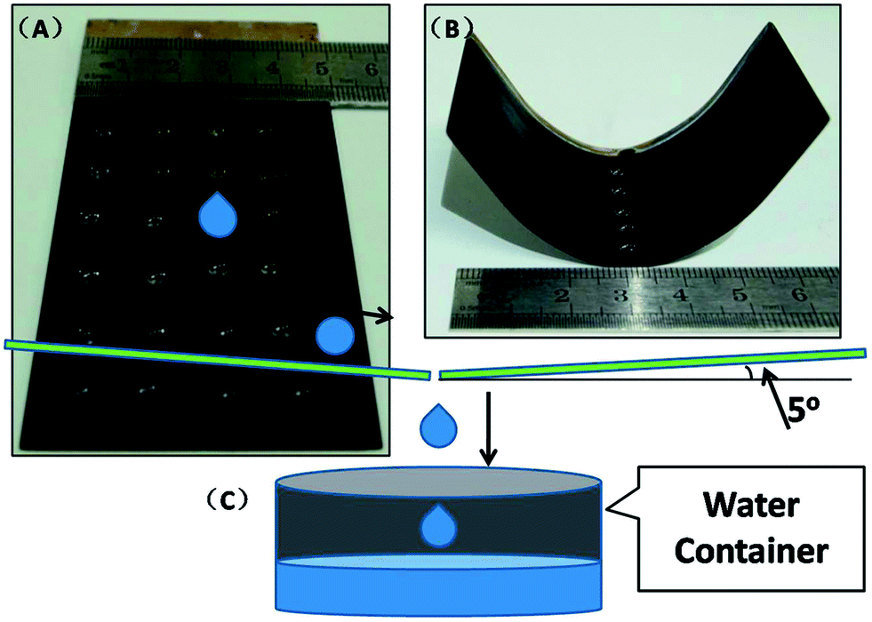
Superhydrophobic materials are always required to be in the form of large and complex shapes in practical industrial applications.48–50 To meet this requirement, the AC etching method is also employed to fabricate large materials with complex shapes. As shown in Fig. 8(A) and (B), we fabricated a superhydrophobic surface on a large, curved brass substrate (100 mm × 50 mm × 2 mm) with all the parameters set up as described above. We can see many water droplets (10 μL) presenting round shapes with large CAs on the prepared surface, which indicates that the AC etching method of fabricating a superhydrophobic surface can be successfully applied to large substrates with complex shapes.
 |
| | Fig. 8 (A) Digital image of prepared superhydrophobic brass substrate; (B) digital image of large scale superhydrophobic brass substrate with a curved shape; (C) the schematic of the suggested device: a water drop collecting device based on the prepared large-scale superhydrophobic surface. | |
On the basis of the large complex shape superhydrophobic surface, a water drop collecting device is proposed, constructed as per the schematic shown in Fig. 8. The main working part is built from the prepared superhydrophobic substrate with a tilt of about 5° with the superhydrophobic surface facing upwards, as shown in Fig. 8(A). If a water drop falls on the surface of the collecting device, it will roll along the tilt direction due to the small rolling angle, which is less than 3°. The entire working process has been demonstrated in Fig. 8(C): a water droplet falls on the superhydrophobic region and then rolls to the lower region due to the small rolling angle. Finally, water droplets will collect together at the centre and flow into a container. This water drop collecting device should have a good performance and a long service life due to the excellent performance of the prepared superhydrophobic surface in abrasion resistance and flush resistance tests, and it will be very useful for collecting water without the need for energy in arid areas.51
4. Conclusion
We demonstrate the fabrication of a flower-like CuO/Cu(OH)2 nanorod film on a brass substrate using a 15 min and 20 V alternating current etching method in K2S2O8 and KOH aqueous solution. The flower-like nanorods, which are about 20 nm in diameter and 200 nm in length, stand on the brass substrate neatly and evenly, producing good abrasion resistance and flush resistance. The superhydrophobicity is achieved by myristic acid modification. After annealing at 200 °C for 6 min, the superhydrophobic surface becomes superhydrophilic. This tunable wetting transition can be realized by myristic acid modification and annealing for more than 30 cycles with a short time of 10 min for each cycle. As a preliminary application, it is proposed that the prepared surface is used to construct a water drop collecting device. The prepared surface is expected to have potential applications in water-collecting devices, antifogging surfaces and self-cleaning coatings.
Acknowledgements
The authors are very grateful for the support from the National Natural Science Foundation of China (Grant no. 51401011).
References
- G. Caputo, B. Cortese, C. Nobile, M. Salerno, R. Cingolani, G. Gigli, P. D. Cozzoli and A. Athanassiou, Reversibly Light-Switchable Wettability of Hybrid Organic/Inorganic Surfaces With Dual Micro/Nanoscale Roughness, Adv. Funct. Mater., 2009, 19, 1149–1157 CrossRef CAS PubMed.
- H. Y. Erbil, A. L. Demirel, Y. Avci and O. Mert, Transformation of a simple plastic into a superhydrophobic surface, Science, 2003, 299, 1377–1380 CrossRef CAS PubMed.
- X. J. Feng, J. Zhai and L. Jiang, The fabrication and switchable superhydrophobicity of TiO2 nanorod films, Angew. Chem., Int. Ed., 2005, 44, 5115–5118 CrossRef CAS PubMed.
- S. Nishimoto and B. Bhushan, Bioinspired self-cleaning surfaces with superhydrophobicity, superoleophobicity, and superhydrophilicity, RSC Adv., 2013, 3, 671–690 RSC.
- J. Heikenfeld and M. Dhindsa, Electrowetting on superhydrophobic surfaces: present status and prospects, J. Adhes. Sci. Technol., 2008, 22, 319–334 CrossRef CAS PubMed.
- P. Roach, N. J. Shirtcliffe and M. I. Newton, Progress in superhydrophobic surface development, Soft Matter, 2008, 4, 224–240 RSC.
- X. M. Li, D. Reinhoudt and M. Crego-Calama, What do we need for a superhydrophobic surface? A review on the recent progress in the preparation of superhydrophobic surfaces, Chem. Rev., 2007, 36, 1350–1368 RSC.
- R. B. Pernites, R. R. Ponnapati and R. C. Advincula, Superhydrophobic–superoleophilic polythiophene films with tunable wetting and electrochromism, Adv. Mater., 2011, 23, 3207–3213 CrossRef CAS PubMed.
- X. F. Hao, L. P. Du, H. G. Cai, C. Y. Zhang, X. Q. Zhang and H. X. Zhang, Preparation of superhydrophobic cross-linked syndiotactic 1,2-polybutadiene membranes by electrospinning, J. Nanosci. Nanotechnol., 2012, 12, 8077–8080 CrossRef CAS PubMed.
- G. Wang and T. Y. Zhang, Easy Route to the Wettability Cycling of Copper Surface between Superhydrophobicity and Superhydrophilicity, ACS Appl. Mater. Interfaces, 2012, 4, 273–279 CAS.
- X. Zhu, Z. Zhang, X. Men, J. Yang and X. Xu, Rapid formation of superhydrophobic surfaces with fast response wettability transition, ACS Appl. Mater. Interfaces, 2010, 2, 3636–3641 CAS.
- L. Introzzi, J. M. Fuentes-Alventosa, C. A. Cozzolino, S. Trabattoni, S. Tavazzi, C. L. Bianchi and S. Farris, “Wetting Enhancer” pullulan coating for antifog packaging applications, ACS Appl. Mater. Interfaces, 2012, 4, 3692–3700 CAS.
- R. Blossey, Self-cleaning surfaces—virtual realities, Nat. Mater., 2003, 2, 301–306 CrossRef CAS PubMed.
- L. Xu and J. He, Fabrication of highly transparent superhydrophobic coatings from hollow silica nanoparticles, Langmuir, 2012, 28, 7512–7518 CrossRef CAS PubMed.
- S. Anandan, T. Narasinga Rao, M. Sathish, D. Rangappa, I. Honma and M. Miyauchi, Superhydrophilic graphene-loaded TiO2 thin film for self-cleaning applications, ACS Appl. Mater. Interfaces, 2012, 5, 207–212 Search PubMed.
- W. Liang and Z. Guo, Stable superhydrophobic and superoleophilic soft porous materials for oil–water separation, RSC Adv., 2013, 3, 16469–16474 RSC.
- Z. Cheng, H. Lai, N. Zhang, K. Sun and L. Jiang, Magnetically Induced Reversible Transition between Cassie and Wenzel States of Superparamagnetic Microdroplets on Highly Hydrophobic Silicon Surface, J. Phys. Chem. C, 2012, 116, 18796–18802 CAS.
- J. Zhao, X. Zhang, N. Chen and Q. Pan, Why Superhydrophobicity Is Crucial for a Water-Jumping Microrobot? Experimental and Theoretical Investigations, ACS Appl. Mater. Interfaces, 2012, 4, 3706–3711 CAS.
- M. Thieme, R. Frenzel, S. Schmidt, F. Simon, A. Henning, H. Worch, K. Lunkwitz and D. Scharnweber, Generation of ultrahydrophobic properties of aluminium-A first step to self-cleaning transparently coated metal surfaces, Adv. Eng. Mater., 2001, 3, 691–695 CrossRef CAS.
- Z. X. She, Q. Li, Z. W. Wang, L. Q. Li, F. N. Chen and J. C. Zhou, Novel method for controllable fabrication of a superhydrophobic CuO surface on AZ91D magnesium alloy, ACS Appl. Mater. Interfaces, 2012, 4, 4348–4356 CAS.
- X. Tang, T. Wang, F. Yu, X. Zhang, Q. Zhu, L. Pang, G. Zhang and M. Pei, Simple, robust and large-scale fabrication of superhydrophobic surfaces based on silica/polymer composites, RSC Adv., 2013, 3, 25670–25673 RSC.
- L. B. Xu, R. G. Karunakaran, J. Guo and S. Yang, Transparent, Superhydrophobic Surfaces from One-Step Spin Coating of Hydrophobic Nanoparticles, ACS Appl. Mater. Interfaces, 2012, 4, 1118–1125 CAS.
- C. F. Wang, F. S. Tzeng, H. G. Chen and C. J. Chang, Ultraviolet-Durable Superhydrophobic Zinc Oxide-Coated Mesh Films for Surface and Underwater–Oil Capture and Transportation, Langmuir, 2012, 28, 10015–10019 CrossRef CAS PubMed.
- H. Bellanger, T. Darmanin, E. T. De Givenchy and F. Guittard, Superhydrophobic hollow spheres by electrodeposition of fluorinated poly(3,4-ethylenedithiopyrrole), RSC Adv., 2012, 2, 10899–10906 RSC.
- X. Yao, Q. Chen, L. Xu, Q. Li, Y. Song, X. Gao, Q. David and L. Jiang, Bioinspired ribbed nanoneedles with robust superhydrophobicity, Adv. Funct. Mater., 2010, 20, 656–662 CrossRef CAS PubMed.
- S. L. Shinde and K. K. Nanda, Facile synthesis of large area porous Cu2O as super hydrophobic yellow-red phosphors, RSC Adv., 2012, 2, 3647–3650 RSC.
- Z. Wang, L. Zhu, W. Li and H. C. Liu, Superhydrophobic surfaces on brass with controllable water adhesion, Surf. Coat. Technol., 2013, 235, 290–296 CrossRef CAS PubMed.
- G. Wang, L. Zhu, W. Li and H. C. Liu, Self-assembled biomimetic superhydrophobic CaCO3 coating inspired from fouling mineralization in geothermal water, Langmuir, 2011, 27, 12275–12279 CrossRef CAS PubMed.
- Z. Wang, L. Zhu, W. Li and H. C. Liu, Rapid reversible superhydrophobicity to superhydrophilicity transition on alternating current etched brass, ACS Appl. Mater. Interfaces, 2013, 5, 4808–4814 CAS.
- G. Kwak, M. Lee and M. Yong, Chemically modified superhydrophobic WOx nanowire arrays and UV photopatterning, Langmuir, 2010, 26, 9964–9967 CrossRef CAS PubMed.
- L. Quan, L. Zhu, W. Li and H. C. Liu, Fabrication of Cu1.8S/CuS nanoplates counter electrode via alternating current etching for quantum dots-sensitized solar cells, RSC Adv., 2014, 4, 32214–32220 RSC.
- G. Joseph and M. T. Arce, Contribution to the study of brass dezincification, Corros. Sci., 1967, 7, 597–605 CrossRef CAS.
- F. Mumm, A. T. J. van Helvoort and P. Sikorski, Easy route to superhydrophobic copper-based wire-guided droplet microfluidic systems, ACS Nano, 2009, 3, 2647–2652 CrossRef CAS PubMed.
- R. N. Wenzel, Surface roughness and contact angle, J. Phys. Chem., 1949, 53, 1466–1467 CrossRef.
- L. Kong, X. Chen, L. Yu, Z. Wu and P. Zhang, Superhydrophobic Cuprous Oxide Nanostructures on Phosphor–Copper Meshes and Their Oil–Water Separation and Oil Spill Cleanup, ACS Appl. Mater. Interfaces, 2010, 2, 3636–3641 Search PubMed.
- G. Li, T. Chen, B. Yan, Y. Ma, Z. Zhang, T. Yu, Z. Shen, H. Chen and T. Wu, Tunable wettability in surface-modified ZnO-based hierarchical nanostructures, Appl. Phys. Lett., 2008, 92, 173104 CrossRef PubMed.
- J. Li, Y. Yang, F. Zha and Z. Lei, Facile fabrication of superhydrophobic ZnO surfaces from high to low water adhesion, Mater. Lett., 2012, 75, 71–73 CrossRef CAS PubMed.
- A. B. D. Cassie and S. Baxter, Wettability of porous surfaces, Trans. Faraday Soc., 1944, 40, 546–551 RSC.
- S. YanLong, Y. Wu, B. Jiajing, F. XiaoJuan and W. YongSheng, Fabrication of flower-like copper film with reversible superhydrophobicity–superhydrophilicity and anticorrosion properties, Surf. Coat. Technol., 2014, 253, 148–153 CrossRef PubMed.
- G. Caputo, C. Nobile, T. Kipp, L. Blasi, V. Grillo, E. Carlino, L. Manna, R. Cingolani, P. D. Cozzoli and A. Athanassiou, Reversible wettability changes in colloidal TiO2 nanorod thin-film coatings under selective UV laser irradiation, J. Phys. Chem. C, 2008, 112, 701–714 CAS.
- Y. H. Chang, N. Y. Hau, C. Liu, Y. T. Huang, C. C. Li, K. Shih and S. P. Feng, A short-range ordered–disordered transition of a NiOOH/Ni(OH)2 pair induces switchable wettability, Nanoscale, 2014, 6, 15309–15315 RSC.
- H. Tavana, A. Amirfazli and A. W. Neumann, Fabrication of superhydrophobic surfaces of n-hexatriacontane, Langmuir, 2006, 22, 5556–5559 CrossRef CAS PubMed.
- Y. M. Zheng, L. Jiang, J. X. Wang and D. Han, Closed-air induced composite wetting on hydrophilic ordered nanoporous anodic alumina, Appl. Phys. Lett., 2008, 96, 094107 CrossRef PubMed.
- C. Mondal, M. Ganguly, A. K. Sinha, J. Pal and T. Pal, Fabrication of a ZnO nanocolumnar thin film on a glass slide and its reversible switching from a superhydrophobic to a superhydrophilic state, RSC Adv., 2013, 3, 5937–5944 RSC.
- B. W. Xin and J. C. Hao, Reversibly switchable wettability, Chem. Soc. Rev., 2010, 39, 769–782 RSC.
- R. Lacombe, Adhesion measurement methods: theory and practice, CRC Press, 2010 Search PubMed.
- B. Kleinman, S. Powell, P. Kumar and R. M. Gardner, The fast flush test measures the dynamic response of the entire blood pressure monitoring system, Anesthesiology, 1992, 77, 1215–1220 CrossRef CAS PubMed.
- K. Acatay, E. Simsek, C. Ow-Yang and Y. Z. Menceloglu, Tunable, superhydrophobically stable polymeric surfaces by electrospinning, Angew. Chem., Int. Ed., 2004, 43, 5210–5213 CrossRef CAS PubMed.
- J. M. Aristoff and J. W. M. Bush, Water entry of small hydrophobic spheres, J. Rev. Fluid Mech., 2009, 619, 45–78 CrossRef CAS.
- J. W. M. Bush and D. L. Hu, Walking on water: biolocomotion at the interface, Annu. Rev. Fluid Mech., 2006, 38, 339–369 CrossRef.
- B. Bhushan and Y. C. Jung, Natural and biomimetic artificial surfaces for superhydrophobicity, self-cleaning, low adhesion, and drag reduction, Prog. Mater. Sci., 2011, 56, 1–108 CrossRef CAS PubMed.
Footnote |
| † Electronic supplementary information (ESI) available. See DOI: 10.1039/c5ra04359j |
|
| This journal is © The Royal Society of Chemistry 2015 |
Click here to see how this site uses Cookies. View our privacy policy here. 


![[thin space (1/6-em)]](https://www.rsc.org/images/entities/char_2009.gif) θr = f1
θr = f1