A biocompatible process to prepare hyaluronan-based material able to self-assemble into stable nano-particles

Enrica Calcea, Paola Ringhierib, Flavia Anna Mercurioa, Marilisa Leoneab, Valeria Bugattic, Michele Savianod, Vittoria Vittoriac and Stefania De Luca*ab
aInstitute of Biostructures and Bioimaging, National Research Council, 80138 Naples, Italy. E-mail: stefania.deluca@cnr.it; Fax: +39-081-2536642; Tel: +39-081-2534514
bDepartment of Pharmacy, Interuniversity Centre for Research on Bioactive Peptides, CIRPeB, University of Naples “Federico II”, Italy
cDepartment of Industrial Engineering, University of Salerno, 84084 Fisciano (Sa), Italy
dInstitute of Crystallography, National Research Council, Via Amendola 122/O, 70126 Bari, Italy

Received 18th February 2015 , Accepted 20th March 2015

First published on 20th March 2015


Abstract

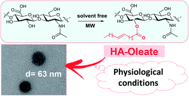
New self-assembled nano-particles were developed by chemical conjugation of natural fatty acids to the backbone of hyaluronan (HA). The chemical structure and the self-association behavior of them were studied by FT-IR, NMR, fluorescence and dynamic light scattering. The HA derivatives form stable spherical shape aggregates, as assessed by transmission electron microscopy.


During the past decade a broad range of HA (hyaluronan)-based materials have been developed by chemical modifications of the polysaccharide.1–3 The aim was the enhancement and modulation of the therapeutic properties of HA, to reach excellent and unique biological characteristics.

HA, a negatively charged naturally occurring polysaccharide, composed of repeating disaccharide units of D-glucuronic acid and N-acetyl glucosamine linked by β(1,4) and β(1,3) glucosidic bonds, has been extensively investigated for several biomedical applications,4–7 due to its unique advantages of non-toxicity, non-immunogenicity, biocompatibility and biodegradability.

More recently, it has also been studied as self-assembled nano-particles for drugs delivery applications.8–16 In this regard, it is noteworthy to underlay that native hyaluronan has limited use due to its short turnover rate and poor mechanical stability. Therefore, chemically modified HA derivatives have been developed to obtain biomaterials with improved and tailored characteristics.17 The prolonged in vivo stability and improved mechanical properties can be obtained by functionalizing the HA with groups that lower its aqueous solubility.18–20 A relatively simple coupling chemistry, due to the presence of the available carboxylic and the hydroxylic functional groups, allows the modification of the sugar units in order to develop a variety of functionalized HA derivatives, that can facilitate the self-assembly process of the polysaccharide and also a consequent encapsulation of drugs.

Several nano-systems were studied as nano-sized drug delivery system for cancer treatment.1 Among them, we cite the nano-system obtained by conjugating the HA backbone with polyamines12,13 and those obtained by functionalization with 5β-cholanic acid.9

The aim of our research was to develop self-assembled nano-systems, which could exhibit safe circulation, so that could be implemented as biocompatible drug carriers. Since HA-based nano-materials have received great attention, we prepared HA amphiphilic conjugates and investigated their ability to form nano-particles in an aqueous environment. Naturally occurring fatty acids were chosen as hydrophobic moieties to be conjugated to the hydrophilic HA biopolymer, whereby allowing the resulting polymeric nano-particles to imbibe only poorly water-soluble drugs in their hydrophobic inner cores. Moreover, it is expected that, after having released the drugs at the specific site, the biopolymer can degrade to nontoxic fragments before renal excretion, since all the starting materials, which were employed to prepare it, are provided by natural resources.

The chemical modification of the HA backbone was performed under solvent-free conditions by acylation of the polysaccharide alcoholic functions with several fatty acid anhydrides. In major details, oleic, linoleic and palmitic acids were activated as anhydride and then different amounts of them were reacted, by mechanical milling, with the polysaccharide in the presence of catalytic amount of K2CO3. The obtained mixture was irradiated with microwaves for two cycles of 3 min and for each sample the reaction was repeated in order to tune the technical conditions (see Table S1 in ESI). Then, after cooling, the solid was dissolved in water (Scheme 1) and the unreacted fatty acids were removed by extraction of the water layer with ethyl acetate. Subsequently, the pH of the aqueous solution was adjusted to the neutrality by adding several drops of 0.5 N HCl and the resultant salt KCl was removed by dialysis in Milli-Q water.21–24


image file: c5ra03107a-s1.tif
Scheme 1 Synthetic strategy for HA functionalization.

The final products were fully characterized by FT-IR and NMR spectroscopy.

The FT-IR spectra of HA–oleate (1a), HA–linoleate (2a) and HA–palmitate (3a) are shown in Fig. 1. For comparison purpose a spectrum of the native HA is also included. The chemical modifications of the polysaccharide produced the main changes of the infrared spectrum: a decrease of the O–H stretching band (in the 3450–3200 cm−1 range), related to the decrease of the hydroxyl HA groups upon reaction with the fatty acid anhydrides, is not clearly detectable, while an increase in the C–H stretching region (2850–2930 cm−1) and the appearance of well-defined band at 2853 cm−1 for each HA derivative was observed and is related to the introduction of the alkylic fatty acid chains on the polysaccharide.25–27 The region featuring the carboxylic groups (1740–1350 cm−1) is the more interesting. For each sample, the appearance of a new band in the C[double bond, length as m-dash]O ester stretching region (1740 cm−1) clearly accounted for the esterification of the employed fatty acid (Fig. 1). HA–oleate (1a) and linoleate (2a) derivatives were analyzed by NMR spectroscopy. The 1D [1H] spectra of the final HA derivatives are characterized by chemical shifts changes with respect to the free starting components in water (i.e. hyaluronan, oleic and linoleic acids); changes affect mainly the HA protons and to a minor degree the aliphatic protons from the lipid moieties (Fig. S1 and S2). The quality of NMR spectra is likely reduced by extensive aggregation phenomena that take place at the concentrations used to run the experiments. In detail, large line-broadening lowers signal intensities, especially those arising from the fatty acid chains (Fig. S1 and S2). In particular, this phenomenon forbids the NMR characterization for the HA–palmitate samples. The degree of substitution (DS) of HA conjugates can be determined, as mean value, by analysis of 1D [1H] NMR spectra (Table 1). In detail, previous works28,29 evaluated DS values from the ratio of relative peak integrations of protons belonging to the CH3 acetyl group of HA (around 2.0 ppm) and ad hoc chosen signals in the substituents.


image file: c5ra03107a-f1.tif
Fig. 1 Transmittance FT-IR spectra of HA conjugates (upper panel); expansion of a region of the transmittance FT-IR spectra (lower panel).
Table 1 Characteristic parameters of HA–fatty acid conjugates
Compound Mean diameter [nm] Poly-dispersity ζ [mV] CAC [mg mL−1] DS (%)
HA–oleate 1a 63.12 ± 0.77 0.050 −34.6 0.3 2.5
HA–oleate 1b 80.9 ± 0.27 0.154 −31.3 0.12 2.1
HA–linoleate 2a 107.1 ± 3.84 0.125 −21.7 0.30 1.9
HA–linoleate 2b 180.9 ± 4.39 0.263 −24.2 0.25 1.4
HA–palmitate 3a 146.8 ± 4.36 0.124 −31.0 0.12 n. d.
HA–palmitate 3b 266.2 ± 6.96 0.276 −29.8 0.16 n. d.


However for HA–oleate and linoleate, the HA's CH3 peak is overlapped with those from the fatty acid aliphatic chains thus, we evaluated integrals from sugar anomeric protons instead (4.50–4.54 ppm). DS values are rather similar in between compounds with identical substituents. Interestingly by increasing the number of unsaturated bonds in the acyl fatty acid chains i.e. passing from HA–oleate derivatives (1a and 1b, see Table 1) to HA–linoleate compounds (2a and 2b, see Table 1) we observe a slightly decrease in the DS values.

Moreover, because of the aggregation phenomena that decrease the NMR signal intensity, it is likely that the DS values could have been underestimated.

The self-assembling behavior of the synthesized amphiphilic polymers was investigated by using the pyrene fluorescence method.30 The solution was excited at 337 nm and the intensity ratio I373/I384 of the emission spectrum was evaluated versus the concentration of the polysaccharide derivative. The ratio I373/I384 decreases with the increase of HA–fatty acid concentration, thus indicating the encapsulation of the fluorophore into the hydrophobic interior of the aggregate. The critical aggregation concentration (CAC) values was determined as inflection point of the obtained curve and are in the range 0.12–0.30 mg mL−1 (Fig. S7).

The nano-particles of HA–oleate and HA–linoleate were prepared by using the so called emulsion evaporation method which involved the dissolution of the HA derivatives in chloroform, an equal volume of NaCl 0.9% aqueous solution was added.31 A fast shaking movement allowed the breaking of the emulsion and also the evaporation of the organic layer. As a result the aggregates were dissolved into the aqueous phase. On the other hand, HA–palmitate could easily form nano-particles by dissolving the conjugate in HEPES buffer (pH = 7.4, 10 mM) and sonicating the final solution. Subsequently, the size distribution of the prepared nano-systems was evaluated with the dynamic light scattering technique (DLS).

The mean diameters of the HA derivatives were in the range of 63–266 nm, depending on the fatty acid used to functionalize the hyaluronan. The particle size of the HA–oleate (1a) and HA–linoleate (2a) resulted smaller than HA–palmitate (3a) aggregates (Fig. S8). Moreover, a narrow size distribution was found for the HA–oleate derivatives, as indicated by the polydispersity factor which was always below 0.20 (Table 1) and also by the intensity correlation function (Fig. 2).


image file: c5ra03107a-f2.tif
Fig. 2 Intensity correlation functions for HA–oleate, HA–linoleate and HA–palmitate.

The ζ potential for each HA derivative was also evaluated and the calculated values ranged from −35 to −22 mV, thus indicating that, as expected, the nano-particle surface was covered by the negative charge of the HA carboxylic groups. It is worth noting that only the alcoholic group of the polysaccharide was involved into the reaction with the fatty acid, therefore the whole HA charge is not expected to change upon conjugation. The unchanged distribution of negative charge on the surface of the nano-systems provides electrostatic repulsion between the aggregates, and is responsible for their long term stability in aqueous conditions.

In fact, it was found that, under the used physiological conditions, the diameter size of each HA–fatty acid particle was maintained over the course of 7 days at room temperature (Fig. S12). Moreover, the particle size did not result affected by the different concentration used to prepare the samples (0.5–1 mg mL−1) for the DLS analysis, thus suggesting that interaction/aggregation among nano-particles did not occur. The morphology of the obtained aggregates was analyzed by transmission electron microscopy technique (TEM), which provided images of spherical shapes (Fig. 3). The diameter sizes observed from TEM images (Fig. 3) followed the same trend of those obtained by the DLS analysis, i.e. the smallest resulted the HA–oleate aggregates, while the biggest are confirmed to be the HA–palmitate system.


image file: c5ra03107a-f3.tif
Fig. 3 Morphology of HA–fatty acid nano-particles measured using transmission electron microscopy (TEM).

However, from TEM images, nano-system size is somewhat unreliable, since the aggregates flatten and spread on the grid, during the sample preparation, in dry conditions. A clear correlation among the degree of unsaturation of the lipid chains in the obtained conjugates and some investigated properties, like the particle sizes and their stability, was not established. However, it is likely that unsaturation modulates the cohesion of the particle hydrophobic core.32

In our case, its influence could not be investigated as, in this preliminary work, a DS value scale was not accurately investigate for each compound.

In conclusion, amphiphilic biopolymers composed of HA and natural fatty acids were synthesized and characterized. The reported synthetic procedure uses microwaves for activation and solvent-free conditions and offers the advantages of being efficient, clean and fast. The obtained HA-based materials were able to self-assemble, forming stable spherical nano-particles under physiological conditions. These preliminary results promise a potential employment of the HA-based nano-sized material as a stable and effective drug delivery system for biomedical applications. As activity in progress, we are tuning a microwave-assisted synthetic protocol which shall allow us to obtain, for each HA–fatty acid conjugate, samples characterized by different degree of substitution (DS). The final scope is to deeply investigate the effect of DS on the particle size, zeta potential and, as most important aspect, on physiological stability.

Notes and references

  1. C. E. Schanté, G. Zuber, C. Herlin and T. F. Vandamme, Carbohydr. Polym., 2011, 85, 469 CrossRef PubMed.
  2. G. Kogan, L. Soltés, R. Stern and P. Gemeiner, Biotechnol. Lett., 2007, 29, 17 CrossRef CAS PubMed.
  3. E. A. Balazs, in Chemistry and biology of hyaluronan, ed. H. G. Garg and C. A. Hales, Elsevier, Amsterdam, 2004, p. 415 Search PubMed.
  4. V. Mironov, V. Kasyanov, X. Zheng Shu, C. Eisenberg, L. Eisenberg, S. Gonda, T. Trusk, R. R. Markwald and G. D. Prestwich, Biomaterials, 2005, 26, 7628 CrossRef CAS PubMed.
  5. S. Manju and K. Sreenivasan, Langmuir, 2011, 27, 14489 CrossRef CAS PubMed.
  6. D. Peer and R. Margalit, Int. J. Cancer, 2004, 108, 780 CrossRef CAS PubMed.
  7. H. Lee, H. Mok, S. Lee, Y. K. Oh and T. G. Park, J. Controlled Release, 2007, 119, 245 CrossRef CAS PubMed.
  8. J. Huang, H. Zhang, Y. Yu, Y. Chen, D. Wang, G. Zhang, G. Zhou, J. Liu, Z. Sun, D. Sun, Y. Lu and Y. Zhong, Biomaterials, 2014, 35, 550 CrossRef CAS PubMed.
  9. K. Y. Choi, H. Chung, K. H. Min, H. Y. Yoon, K. Kim, J. H. Park, I. C. Kwon and S. Y. Jeong, Biomaterials, 2010, 31, 106 CrossRef CAS PubMed.
  10. H. J. Cho, H. Y. Toon, H. Koo, S. H. Ko, J. S. Shim, J. H. Lee, K. Kim, I. C. Kwon and D. D. Kim, Biomaterials, 2011, 32, 7181 CrossRef CAS PubMed.
  11. K. Y. Choi, K. H. Min, J. H. Na, K. Choi, K. Kim, J. H. Park, I. C. Kwon and S. Y. Jeong, J. Mater. Chem., 2009, 19, 4102 RSC.
  12. K. Y. Choi, S. Lee, K. Park, K. Kim, J. H. Park, I. C. Kwon and S. Y. Jeong, J. Phys. Chem. Solids, 2008, 69, 1591 CrossRef CAS PubMed.
  13. S. Ganesh, A. K. Iyer, D. V. Morrisey and M. M. Amiji, Biomaterials, 2013, 34, 3489 CrossRef CAS PubMed.
  14. H. J. Cho, I. S. Yoon, H. Y. Yoon, H. Koo, Y. J. Jin, S. H. Ko, J. S. Shim, K. Kim, I. C. Kwon and D. D. Kim, Biomaterials, 2012, 33, 1190 CrossRef CAS PubMed.
  15. G. Pitarresi, F. S. Palumbo, A. Albanese, C. Fiorica, P. Picone and G. Giammona, J. Drug Targeting, 2010, 18, 264 CrossRef CAS PubMed.
  16. Y. Liu, J. Sun, P. Zhang and Z. He, Curr. Med. Chem., 2011, 18, 2638 CrossRef CAS.
  17. E. J. Oh, K. Park, K. S. Kim, J. Kim, J.-A. Yang, J.-H. Kong, M. Y. Lee, A. S. Hoffman and S. K. Hahn, J. Controlled Release, 2010, 141, 2 CrossRef CAS PubMed.
  18. G. Huerta-Angeles, M. Bobek, E. Príkopová, D. Smejkalová and V. Velebny, Carbohydr. Polym., 2014, 111, 883 CrossRef CAS PubMed.
  19. P. Mlcochová, S. Bystrický, B. Steiner, E. Machová, M. Koós, V. Velebný and M. Krcmár, Biopolymers, 2006, 82, 74 CrossRef PubMed.
  20. M. Zhang and S. P. James, J. Mater. Sci.: Mater. Med., 2005, 16, 587 CrossRef CAS PubMed.
  21. L. Monfregola, M. Leone, V. Vittoria, P. Amodeo and S. De Luca, Polym. Chem., 2011, 2, 800 RSC.
  22. L. Monfregola, V. Bugatti, P. Amodeo, S. De Luca and V. Vittoria, Biomacromolecules, 2011, 12, 2311 CrossRef CAS PubMed.
  23. E. Calce, V. Bugatti, V. Vittoria and S. De Luca, Molecules, 2012, 17, 12234 CrossRef CAS PubMed.
  24. E. Calce, E. Mignogna, V. Bugatti, M. Galdiero, V. Vittoria and S. De Luca, Int. J. Biol. Macromol., 2014, 68, 28 CrossRef CAS PubMed.
  25. F. Donghui, W. Beibei, X. Zheng and G. Qisheng, J. Wuhan Univ. Technol., Mater. Sci. Ed., 2006, 21, 32 CrossRef.
  26. S. Manju and K. Sreenivasan, J. Colloid Interface Sci., 2011, 359, 318 CrossRef CAS PubMed.
  27. T. D. Lopes, I. C. Riegel-Vidotti, A. Grein, C. A. Tischer and P. C. Faria-Tischer, Int. J. Biol. Macromol., 2014, 67, 401 CrossRef CAS PubMed.
  28. S. A. Bencherif, A. Srinivasan, F. Horkay, J. O. Hollinger, K. Matyjaszewski and N. R. Washburn, Biomaterials, 2008, 29, 1739 CrossRef CAS PubMed.
  29. D. A. Ossipov, X. Yang, O. Varghese, S. Kootala and J. Hilborn, Chem. Commun., 2010, 46, 8368 RSC.
  30. K. Dan, N. Bose and S. Ghosh, Chem. Commun., 2011, 47, 12491 RSC.
  31. E. J. Cho, H. Holback, K. C. Liu, S. A. Abouelmagd, J. Park and Y. Yeo, Mol. Pharmaceutics, 2013, 10, 2093 CrossRef CAS PubMed.
  32. M. Monette and M. Lafleur, Biophys. J., 1996, 70, 2195 CrossRef CAS.

Footnote

Electronic supplementary information (ESI) available: Experimental section, NMR spectra, FT-IR spectra, critical aggregation concentration, DLS measurements, stability of fatty acid conjugates, TEM images. See DOI: 10.1039/c5ra03107a

This journal is © The Royal Society of Chemistry 2015
Click here to see how this site uses Cookies. View our privacy policy here.