DOI:
10.1039/C5RA02991K
(Communication)
RSC Adv., 2015,
5, 33767-33771
Folded three-dimensional graphene with uniformly distributed mesopores for high-performance supercapacitors†
Received
16th February 2015
, Accepted 7th April 2015
First published on 7th April 2015
Abstract
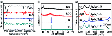
Folded three-dimensional graphene (FTG) is prepared through self-assembly of graphite oxide (GO) and liquid-phase exfoliation graphene (LG), followed by sonication and reduction processes. The obtained FTG consists of few-layer graphene. And it possesses a high degree of crystallinity, three-dimensional architecture and uniformly distributed mesopores. A supercapacitor based on an FTG electrode exhibits enhanced electrochemical performance. The FTG electrode exhibits high specific capacitance of 195.4 F g−1 at a scan rate of 1 mV s−1 and excellent cycling stability with 93.9% of its initial capacitance at a large scan rate of 500 mV s−1 after 8000 cycles. The supercapacitor fabricated with the FTG electrode delivers a high energy density of 27.1 W h kg−1 at a power density of 97.7 W kg−1. These results suggest that FTG is a promising material for high-performance supercapacitor applications.
1 Introduction
The investigation of novel, low-cost, environmentally friendly, and high-performance energy storage systems has been in ever increasing demand as a result of the needs of modern society and ecological concerns.1 Supercapacitors, known as electrochemical capacitors, play a very important role in energy storage for electric devices.2 Supercapacitors have many advantages such as high power density, long cycle life, ultrafast charge and discharge.3,4 Many carbon-based materials have been investigated as electrode materials for supercapacitors due to their intriguing properties including low cost, non-toxic, environmental friendliness and stability.5,6 In the family of carbon materials, graphene is considered as a promising material for the next-generation supercapacitors7 because of its high specific surface area, excellent electrical conductivity, and high theoretical specific capacitance.8 Ruoff and co-workers9 first explored graphene as electrode materials, and specific capacitance value of 135 F g−1 was achieved using aqueous electrolyte. However, this value is much lower than the theoretical maximum of 550 F g−1,9 which calculated by single-layer graphene.
Researchers have developed many strategies to improve the specific capacitance of graphene, such as treating graphene with KOH under high temperature,10 functionalization of graphene with metal oxide nanoparticles,11 assembly of graphene and carbon nanotubes.12–15 These methods effectively improve the capacitance of graphene materials, but the demand of high temperature, the toxicity of heavy metal and high cost of carbon nanotubes result in environmental destruction and economic pressures. To overcome these disadvantages, three-dimensional graphene,16,17 which alleviates the agglomeration and restacking of graphene nanosheets, shows a promising result.
In this work, we propose a simple liquid-phase exfoliation and chemical reduction method to fabricate folden three-dimensional graphene (FTG) with high degree of crystallinity, three-dimensional architecture and uniformly distributed mesopores. FTG shows excellent performance as an electrode material for supercapacitors. The specific capacitance of FTG electrode is 195.4 F g−1 at a scan rate of 1 mV s−1. The specific capacitance of FTG electrode decreases by only 6.1% after 8000 cycles at a large scan rate of 500 mV s−1. The energy density and specific power density of present supercapacitor are 27.1 W h kg−1 and 97.7 W kg−1, respectively. These results suggest that FTG is a promising material for high-performance supercapacitor applications.
2 Experimental section
Preparation of GO and LG
Graphite oxide (GO) was synthesized by oxidation of microcrystalline graphite (∼120 μm) with modified Hummers' method.17,18 In a typical reaction, 3.0 g of microcrystalline graphite was mixed with a mixture of concentrated H2SO (180 mL) and H3PO4 (20 mL) in a 500 mL flask. The mixture was stirred for 0.5 h in an ice-water bath. While maintaining stirring, 18.0 g of KMnO4 was added to the mixture slowly. The suspension was stirred for another 0.5 h and kept the reaction temperature below 5 °C. Then, removed the ice-water bath, and the mixture was kept stirring at 50 °C for 12 h. After that, 200 mL of deionized water was slowly added to the mixture. Following, 200 mL of hydrogen peroxide and deionized water at a volume ratio of 1
:
9 was slowly added into the mixture and the color of the mixture turned into yellow. Subsequently, GO was washed with deionized water several times and collected by centrifugation. Finally, GO powder was obtained by vacuum drying at 60 °C for 24 h.
LG sample was synthesized based on some reported literatures.19,20 Typically, 3 g of microcrystalline graphite was added to the mixed solvents of N-methyl-2-pyrrolidone (NMP) (90 mL) and toluene (60 mL), and the liquid was sonicated for 3 h. Then, the dark slurry was filtered through a 0.22 μm PTFE (Teflon) membrane and the filter cake was redispersed in 500 mL of NMP, which was sonicated for 7 h. The final liquid-phase exfoliation graphene (LG) dispersion was obtained. The dispersion was filtered through a PTFE membrane (0.22 μm). Finally, LG powder was obtained by vacuum drying at 60 °C for 24 h.
Preparation of FTG
40 mg of LG and 160 mg of GO were added to 200 mL of N,N-dimethylformamide (DMF), and the mixture was sonicated for 4 h. Then, 3 g of (NH4)2CO3 and 3 mL of hydrazine hydrate (80%) were added. The reaction was warmed to 100 °C and stirred for 2 h. Once the reduction process was completed, the mixture was filtered through a PTFE membrane and washed with deionized water and ethanol several times. Finally, FTG powder was obtained by vacuum drying at 60 °C for 24 h.
Characterization
Fourier transform infrared spectroscopy (FTIR) was tested by IR Affinity-1 (Japan) (KBr pellet). X-ray diffraction (XRD) analysis was carried out on a BRUKER D8-ADVANCE X-ray diffractometer using Cu (40 kV, 40 mA) radiation. Raman spectroscopy was tested by Raman microspectroscopic setup (RamLab-010) (LabRam, Horiba-Jobin-Yvon, Bensheim, Germany). The morphology of the products were investigated by scanning electron microscopy (SEM) (Hitachi S-4800, Japan) at an accelerating voltage of 5.0 kV. Transmission electron microscopy (TEM) images were obtained with a JEM-3010 (JEOL-3010, Japan). Nitrogen adsorption–desorption measurements were carried out on Autusorb-1C-TCD (Quantachrome, USA).
Electrochemical measurement
Initially the Ni foam (current collector) was repeatedly washed with acetone/deionized water to ensure that the surface is clean. The working electrode was prepared by mixing FTG (active material), acetylene black, and polyvinylidene fluoride (PVDF) (a mass ratio of 80
:
10
:
10) in NMP to form a slurry. The slurry was pasted on Ni foam and dried. Then, the foam was pressed under a pressure of 10 MPa to completely adhere with the electrode material. The mass loading of the working electrode is about 2 mg (weighing on AUW120D, which could reach a precision of 0.01 mg). The electrochemical properties of the sample was investigated under a three-electrode system with FTG electrode as working electrode, platinum wire as counter electrode, Hg/HgO electrode as reference electrode in 6 M KOH. Cyclic voltammetry (CV), galvanostatic charge–discharge (GCD) and electrochemical impedance spectroscopy (EIS) measurements (including equivalent circuit fitting) were performed with a CHI 660E electrochemical workstation (Chenhua, Shanghai). CV and GCD tests were recorded in the potential range of −0.8 V to 0.2 V. Impedance studies on electrode material were conducted between 0.1 Hz to 100 kHz at amplitude of 5 mV. Specific capacitance was calculated from CV plot (Cs, c) and GCD plot (Cs, g), respectively. Specific capacitance (Cs), energy density and power density are calculated by following formulas. |
Cs, c = (∫idV)/(2 × m × ΔV × S)
| (1) |
where, S is the scan rate, ΔV is the electrochemical window, m is the mass of active material, ΔI is the discharge current, td is the discharge time, respectively.
3 Results and discussion
Analysis of FTG
FTIR was carried out to characterize the chemical structures of GO, RGO, LG, FTG. Fig. 1(a) shows the FTIR spectra of GO, RGO, LG, FTG. In the spectrum of GO, following functional groups are identified: –OH groups center at 3450 cm−1, C
O groups center at 1720 cm−1, C
C groups center at 1635 cm−1,21,22 and the stretching vibration bands C–O of epoxy and alkoxy are observed at 1240 cm−1 and 1050 cm−1, respectively. It demonstrates that GO has abundant oxygen-containing functional groups. In the spectrum of RGO, however, it is markedly different from that of GO, where the intensities of all the absorption bands correlated to the oxygen-containing functional groups decreased markedly. The FTIR spectra of LG and FTG are similar to that of RGO, which due to there are few functional groups on them.
 |
| Fig. 1 FTIR spectra of GO, RGO, LG, FTG (a); XRD patterns of GO, RGO, LG, FTG (b); Raman spectra of GO, RGO, LG, FTG (c). | |
Fig. 1(b) shows the XRD patterns of GO, RGO, LG and FTG. The XRD patterns of these graphite carbons can be indexed to the graphite (JCPDS no. 75-2078). After oxidation, the peak at 10.2° is observed, which is corresponding to (001) diffraction peak of GO. According to Braggs equation (2d
sin
θ = nλ), the d-spacing of GO increases to 0.8663 nm from 0.3347 nm, which is ascribed to the oxide-induced O-containing functional groups that can be confirmed by FTIR. After reduced, there is only a small peak at 24.8° can be found in RGO, which indicating that RGO has been reduced and owns a low degree of crystallinity. The d-spacing of RGO decreased to 0.3582 nm from 0.8663 nm, but it is larger than the d-spacing of LG (0.3347 nm). The peak at 26.6° corresponding to (002) reflection of LG. For FTG, there is a strong peak at 26.4°. According to Braggs equation (2d
sin
θ = nλ), FTG had larger d-spacing (0.3368 nm) than LG (0.3347 nm) and had higher degree of crystallinity than RGO, which contributed to promoting of the capacitance of FTG, as shown in Fig. 4.
Typical Raman spectra of GO, RGO, LG and FTG are presented in Fig. 1(c). In carbon materials, G peak corresponds to a first-order scattering of the E2g mode,23 D peak is associated with defects or lattice distortion,24 so the degree of disorder and edge affects25 can be measured by the relative intensities ratio of D peak and G peak (ID/IG). 2D peak centers at 2703 cm−1 is typically used to indicate the quality of graphene.19
For GO and RGO, the peaks appear at 1340 cm−1 and 1600 cm−1 correspond to D and G peaks, respectively. For LG and FTG, the D and G peaks appear at 1340 cm−1 and 1590 cm−1. The downshifted G peak position in GO and RGO matches with many reported literatures.26,27 Compared with GO, the ID/IG of RGO increases from 1.09 to 1.37, which due to increase of sp2 domains and edge defects. However, LG displays a small number of defects (ID/IG = 0.21). The ID/IG of FTG is 1.11, which is smaller than RGO. It indicates that FTG owns fewer defects than RGO. The shape of 2D peak is indicative of the number of layers per flake, and for the flakes, the Raman spectrum is considerably different to that of graphene, which is thinner than 5 layers.28 As shown in Fig. 1(c), the shoulder of 2D peak of graphene disappears and the band becomes sharp and asymmetrical, suggesting that FTG flakes have less than five layers.
Fig. 2(a) shows the SEM images of RGO, in which the graphene nanosheet appears a plicated shape. Fig. 2(b) shows the SEM images of LG, it clearly shows that the LG nanosheet appeared as a thin film. RGO and LG assemble to form a folded three-dimensional architecture, which preventing the neighboring nanosheets from restacking with one another, as shown in Fig. 2(c). Some crimp can be obvious seen at the edge of graphene nanosheet. The folded three-dimensional structure of FTG is further confirmed by TEM analysis. Fig. 2(d) shows a typical TEM image of the three-dimensional graphene. This observation is similar to graphene scroll,29,30 which has an excellent performance in supercapacitors. Fig. 2(e) shows the selected area electron diffraction pattern (SAED) of FTG, it indicates that the thickness of FTG is very thin (<5 layers).19,31 This result agrees well with the Raman studies (see in Fig. 1(c)). Fig. 2(f) shows the HRTEM image of FTG. The inset in Fig. 2(f) shows the fold axes in the accordion structure.
 |
| Fig. 2 (a)–(c) SEM images of RGO, LG, FTG, (d) TEM image of FTG, (e) TEM and SAED of FTG, (f) HRTEM image of FTG. | |
The surface area and pore-size characterization of FTG was verified by measuring the nitrogen adsorption–desorption isotherms as shown in Fig. 3. According to the IUPAC classification, FTG exhibites the combined characteristics of type-II and type-IV isotherms.22,32,33 This reveals that FTG has a typical mesoporous structure, which is further verified from the Brunauer–Emmett–Teller (BET) pore size distribution data shown in Fig. 3(a). BET analysis indicates that the surface area is 93.2 m2 g−1 with a pore volume of 0.392 cm3 g−1 and average pore diameter of 3.8 nm. The existence of a pore size distribution in the open structure is also supported by the SEM and TEM data (see in Fig. 2(c), (e) and (f)). It is observed that FTG has a narrow pore distribution that concentrates on 3.8 nm.
 |
| Fig. 3 (a) Nitrogen adsorption–desorption isotherms and (b) pore-size distribution of the FTG. | |
The surface area of FTG is lower than many reported graphene, but this results is also similar to some reported literatures, graphene with a small surface area also provide a large capacitance.17 The traditional understanding of electrical double-layer capacitance (EDLC) believe that only a big surface area can achieve a high capacitance. However, many researchers find that there is no linear relationship between the area and capacitance.34–36 Taking a high specific surface area of 1000 m2 g−1 for carbon as an example, its ideal capacitance would be over 200 F g−1. However, the practically obtained values are of only a few tens of F g−1.37 Many latest research find that only the surface that is accessible to electrolyte ions can contribute to charge storage; therefore, optimization of pore size, pore structure, surface properties and conductivity of the electrode materials is required,38 a well distribute pore size and a high effective surface areas should be considered.39
The moderate specific surface area and porous structure of FTG provides the possibility of efficient transport of electrons and ions in the electrode,40,41 which can be further illustrated by the electrochemical impedance spectroscopic (EIS) results of FTG (see in Fig. 4(d)). The high degree of crystallinity bring a good conductivity, the three-dimensional architecture and uniformly distributed mesopores benefit the ion transport in electrode. Hence leading to enhanced electrochemical property.
 |
| Fig. 4 (a) CV curves of FTG, (b) galvanostatic charge–discharge of FTG, (c) cycle performance of FTG. (vs. Hg/HgO electrode.), (d) EIS studies of GO, RGO, LG, FTG. | |
Electrochemical performance of FTG electrode
Cyclic voltammograms for FTG and RGO samples are shown in Fig. S1 (see in ESI†) in the potential range of −0.8–0.2 vs. (Hg/HgO) electrode in a 6 M aqueous KOH electrolyte at the scan rate of 1 mV s−1. The specific capacitance of 195.4 F g−1 is obtained for FTG, in comparison with 130.6 F g−1 for RGO. The capacitance of FTG is higher than other graphene-based carbon materials (graphene, 135 F g−1;13 activated graphene-based carbons, 174 F g−1 (ref. 42)). Fig. 4(a) shows the cyclic voltammetry (CV) curves of FTG nanostructures electrode in 6 M KOH electrolyte at scan rates ranging from 1 to 500 mV s−1 in the potential window of −0.8–0.2 V (vs. Hg/HgO electrode). The well-defined rectangular CV shapes are observed for FTG supercapacitor, indicativing of a typical EDLC behavior and a fast charge propagation within the electrode of the supercapacitor.43 To further examine the electrochemical performances of FTG electrode, we performance galvanostatic constant current charge–discharge curves at various current densities (Fig. 4(b)). The presence of the triangular symmetry and linear slopes with respect to the charge–discharge curves confirm again a good electrochemical performance. The specific capacitance (CS) obtains from the discharging curves is calculated to be 138.6 F g−1 at the current density of 0.5 A g−1.
The long cycle stability is a very vital requirement for supercapacitors. Fig. 4(c) shows the cycling stability of FTG electrode by CV test at a scan rate of 500 mV s−1 for 8000 cycles. It can be seen that the specific capacitance of FTG electrode maintains 93.9% of its initial value even after 8000 cycles, indicating the excellent cycling stability. The CV curves of the first and last cycles of the electrode are shown in the inset of Fig. 4(c). It can be seen that all the CV curves are almost overlapping with each other, suggesting a high reversibility and excellent electrochemical stability that will be beneficial for the practical applications.
The EIS technique was used to study the electron and ion transport phenomena in GO, RGO, LG and FTG electrodes in Fig. S2.† The EIS curves are analyzed using simple method based on the basis of the equivalent circuit, which is given in the inset of Fig. 4(d). Rs is the resistance between the electrode and electrolyte, Rp is the charge transfer resistance in the supercapacitor electrode. The EIS studies results are shown in Fig. 4(d). The moderate specific surface area and porous structure of FTG provides the possibility of efficient transport of electrons and ions in the electrode, hence leading to enhanced electrochemical property. Fig. S3† shows the GCD plots of RGO and FTG at different current density. Compared to RGO, FTG has a good rate performance, which is important in supercapacitors. These results highlight the capability of FTG electrode to meet the requirements of both high specific capacitance and excellent cycling stability, which are important merits for high-performance supercapacitor.
4 Conclusion
In summary, FTG has been prepared by liquid-phase exfoliation and reducing method. With high degree of crystallinity, three-dimensional architecture and uniformly distributed mesopores, FTG shows excellent performance as an electrode material for supercapacitors. The specific capacitance of the FTG electrode is 195.4 F g−1 at a scan rate of 1 mV s−1. The specific capacitance of the FTG electrode decreases by only 6.1% after 8000 cycles at a large scan rate of 500 mV s−1. The energy density and specific power density of the present supercapacitor are 27.1 W h kg−1 and 97.7 W kg−1, respectively. The results suggest that FTG is a promising material for high-performance supercapacitor applications.
References
- A. S. Arico, P. Bruce, B. Scrosati, J. M. Tarascon and W. V. Schalkwijk, Nat. Mater., 2005, 4, 366 CrossRef CAS PubMed.
- Z. H. Liu, H. H. Zhou, Z. Y. Huang, W. Y. Wang, F. Y. Zeng and Y. F. Kuang, J. Mater. Chem. A, 2013, 1, 3454 CAS.
- M. Winter and R. J. Brodd, Chem. Rev., 2004, 104, 4245 CrossRef CAS.
- L. T. Le, M. H. Ervin, H. W. Qiu, B. E. Fuchs and W. Y. Lee, Electrochem. Commun., 2011, 13, 355 CrossRef CAS PubMed.
- H. Jiang, P. S. Lee and C. Z. Li, Energy Environ. Sci., 2013, 6, 41 CAS.
- G. Wang, Z. Ling, C. P. Li, Q. Dong, B. Q. Qian and J. S. Qiu, Electrochem. Commun., 2013, 31, 31 CrossRef CAS PubMed.
- Z. W. Xu, Z. Li, C. M. B. Holt, X. H. Tan, H. L. Wang, B. S. Amirkhiz, T. Stephenson, D. Mitlin and J. Phys, Chem. Lett., 2012, 3, 2928 CAS.
- M. F. El-Kady, V. Strong, S. Dubin and R. B. Kaner, Science, 2012, 335, 1326 CrossRef CAS PubMed.
- M. D. Stoller, S. J. Park, Y. W. Zhu, J. H. An and R. S. Ruoff, Nano Lett., 2008, 8, 3498 CrossRef CAS PubMed.
- Y. Zhu, S. Murali, M. D. Stoller, A. Velamakanni, R. D. Piner and R. S. Ruoff, Science, 2011, 332, 1537 CrossRef CAS PubMed.
- X. Z. Xu, B. G. Lu and Z. Xu, Adv. Mater., 2014, 26, 1044 CrossRef PubMed.
- N. Jia, P. Ramesh, E. Bekyarova, M. E. Itkis and E. C. Haddon, Adv. Energy Mater., 2012, 2, 438 CrossRef PubMed.
- M. Q. Zhao, Q. Zhang, J. Q. Huang, G. L. Tian, T. C. Chen, W. Z. Qian and F. Wei, Carbon, 2013, 54, 403 CrossRef CAS PubMed.
- J. L. Liu, C. K. Poh, D. Zhan, L. F. Lai, S. H. Lim, L. Wang, X. X. Liu, N. G. Sahoo, C. M. Li, Z. X. Shen and J. Y. Lin, Nano Energy, 2013, 2, 377 CrossRef CAS PubMed.
- J. L. Liu, L. L. Zhang, H. B. Wu, J. Y. Lin, Z. X. Shen and X. W. Lou, Energy Environ. Sci., 2014, 7, 3709 CAS.
- Y. X. Xu, Z. Y. Lin, X. Q. Huang, Y. Liu, Y. Huang and X. F. Duan, ACS Nano, 2013, 7, 4042 CrossRef CAS PubMed.
- J. Hu, Z. Kang, F. Li and X. Huang, Carbon, 2014, 67, 221 CrossRef CAS PubMed.
- D. C. Marcano, D. V. Kosynkin, J. M. Berlin, A. Sinitskii, Z. Z. Sun, S. Alexander, L. B. Alemany, W. Lu and J. M. Tour, ACS Nano, 2010, 8, 4806 CrossRef PubMed.
- E. C. Ou, Y. Y. Xie, C. Peng, Y. W. Song, H. Peng, Y. Q. Xiong and W. J. Xu, RSC Adv., 2013, 3, 9490 RSC.
- X. Y. Zhang, M. S. Han, S. Chen, L. Bao, L. Li and W. J. Xu, RSC Adv., 2013, 3, 17689 RSC.
- J. Oh, J. H. Lee, J. C. Koo, H. R. Choi, Y. Lee, T. Kim, N. D. Luong and J. D. Nam, J. Mater. Chem., 2010, 20, 9200 RSC.
- M. M. Sk and C. Y. Yue, RSC Adv., 2014, 4, 19910 Search PubMed.
- X. Zhou, J. Zhang, H. Wu, H. Yang, J. Zhang and S. Guo, J. Phys. Chem. C, 2011, 115, 11957 CAS.
- S. Reich and C. Thomsen, Philos. Trans. R. Soc., A, 2004, 362, 2271 CrossRef CAS PubMed.
- M. Q. Sun, G. C. Wang, X. W. Li and C. Z. Li, J. Power Sources, 2014, 245, 436 CrossRef CAS PubMed.
- H. Wang, J. T. Robinson, X. L. Li and H. J. Dai, J. Am. Chem. Soc., 2009, 131, 9910 CrossRef CAS PubMed.
- U. N. Maiti, J. Lim, K. E. Lee, W. J. Lee and S. O. Kim, Adv. Mater., 2014, 26, 615 CrossRef CAS PubMed.
- F. Mauri, S. Piscanec, D. Jiang, K. S. Novoselov, S. Roth and A. K. Geim, Phys. Rev. Lett., 2006, 97, 166802 CrossRef.
- F. Y. Zeng, Y. F. Kuang, Y. Wang, Z. Y. Huang, C. P. Fu and H. H. Zhou, Adv. Mater., 2011, 23, 4929 CrossRef CAS PubMed.
- F. Y. Zeng, Y. F. Kuang, G. Q. Liu, R. Liu, Z. Y. Huang, C. P. Fu and H. H. Zhou, Nanoscale, 2012, 4, 3997 RSC.
- M. Lotya, Y. Hernandez, P. J. King, R. J. Smith, V. Nicolosi, L. S. Karlsson, F. M. Blighe, S. De, Z. Wang, I. T. McGovern, G. S. Duesberg and J. N. Coleman, J. Am. Chem. Soc., 2009, 131, 3611 CrossRef CAS PubMed.
- Z. Ryu, J. Zheng, M. Wang and B. Zhang, Carbon, 1999, 37, 1257 CrossRef CAS.
- Y. Li, Z. Li and P. K. Shen, Adv. Mater., 2013, 25, 2474 CrossRef CAS PubMed.
- J. Gamby, P. L. Taberna, P. Simon, J. F. Fauvarque and M. Chesneau, J. Power Sources, 2001, 101, 109 CrossRef CAS.
- D. Y. Qu and H. Shi, J. Power Sources, 1998, 74, 99 CrossRef CAS.
- Y. P. Zhai, Y. Q. Dou, D. Y. Zhao, P. F. Fulvio, R. T. Mayes and S. Dai, Adv. Mater., 2011, 23, 4838 Search PubMed.
- L. M. Dai, D. W. Chang, J. B. Baek and W. Lu, Small, 2012, 8, 1151 Search PubMed.
- Y. Yoon, K. Lee, S. Kwon, S. Seo, H. Yoo, S. Kim, Y. Shin, Y. Park, D. Kim, J. Choi and H. Lee, ACS Nano, 2014, 8, 4580 CrossRef CAS PubMed.
- L. Zhang, X. Yang, F. Zhang, G. K. Long, T. F. Zhang, K. Leng, Y. W. Zhang, Y. Huang, Y. F. Ma, M. T. Zhang and Y. S. Chen, J. Am. Chem. Soc., 2013, 135, 5922 Search PubMed.
- A. Ghosh and Y. H. Lee, ChemSusChem, 2012, 5, 480 CrossRef CAS PubMed.
- M. Huang, R. Mi, H. Liu, F. Li, X. L. Zhao, W. Zhang, S. X. He and Y. X. Zhang, J. Power Sources, 2014, 269, 760 CrossRef CAS PubMed.
- T. Y. Kim, G. Jung, S. Yoo, K. S. Suh and R. S. Ruoff, ACS Nano, 2013, 7, 6899 CrossRef CAS PubMed.
- Z. Wen, X. Wang, S. Mao, Z. Bo, H. Kim, S. Cui, G. Lu, X. Feng and J. Chen, Adv. Mater., 2012, 24, 5610 CrossRef CAS PubMed.
Footnote |
† Electronic supplementary information (ESI) available. See DOI: 10.1039/c5ra02991k |
|
This journal is © The Royal Society of Chemistry 2015 |
Click here to see how this site uses Cookies. View our privacy policy here.