Unusual crystallographic existence of a hydrated zinc(II) bisulphate complex: experimental and theoretical observations

Dhananjay Deya, Sukanta Pala, Hare Ram Yadavb, Partha Sarathi Senguptac, Angshuman Roy Choudhuryb, Niranjan Kolea and Bhaskar Biswas*a
aDepartment of Chemistry, Raghunathpur College, Purulia-723133, India. E-mail: icbbiswas@gmail.com; Fax: +91 3251 255235; Tel: +91 3251 255235
bDepartment of Chemical Sciences, Indian Institute of Science Education and Research Mohali, Sector 81, S. A. S. Nagar, Manauli PO, Mohali 140 306, India. E-mail: angshurc@iisermohali.ac.in
cDepartment of Chemistry, Vivekananda Mahavidyalaya, Burdwan-713104, India. E-mail: anapspsmo@gmail.com

Received 12th February 2015 , Accepted 5th May 2015

First published on 5th May 2015


Abstract

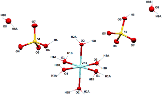
A unprecedented hydrated zinc(II) bisulphate complex, [Zn(H2O)6](HSO4·H2O)2 (1) has been synthesized along with a molecular ion salt, [4,4′-bipy-H+][NCS] (2) (4,4′-bipy = 4,4′-bipyridine) and characterized by different spectroscopic tools including a single crystal X-ray diffraction study. From the X-ray crystal structure of 1, it is revealed that the Zn(II) ion is in a perfect octahedral geometry with a ZnO6 core and the molecule crystallizes in the P21/c space group. Self-assembly of the bisulphate anions with lattice water molecules pack to form a 12-membered cyclic cluster through strong intermolecular H-bonding (O–H⋯O–S). Again four cyclic tetramers interact with one another via H⋯O bonding to form a 20-membered macrocyclic bisulphate–water cluster and extends as a 2D network along the a axis. It presents a new mode of association of water molecules with bisulphate molecules. Density Function Theory (DFT) studies agree well with the experimental findings.


Introduction

Apart from being fascinating subjects for scientific study in their own right, molecular clusters play an important role in many different areas of scientific and common interest. For instance, molecular clusters play a significant role in the atmosphere by acting as precursors for secondary particle formation. Uncertainties associated with the abundance and properties of clusters contribute to the uncertainties connected with atmospheric particles in general. The aerosol effect is a primary uncertainty in the prediction of changes in the greenhouse effect and global temperature.1 Molecular clusters are also of interest from the perspective of air quality and human health, because the clusters, being of nanometre size, can penetrate into the deepest part of the human respiratory tract. A molecular-level understanding of the solvation of acids in size-selected clusters can lend insight into their behaviour in bulk solution. Some experimental2–4 and theoretical5,6 studies have explored the extent of acid dissociation in clusters and the minimum number of water molecules needed for this process to occur. Bisulfate, a weak acid in aqueous solution with a pKa of 2.0 is also among the most prevalent negative ions in the troposphere and the stratosphere due to its high stability with respect to electron detachment.7–9 Therefore, studies of the hydrated anionic species, that is, anion–water clusters, are of special importance in understanding of the hydration phenomena of inorganic anions in nature, biochemistry and atmosphere. However, only a few examples are involved in discrete inorganic anion–water clusters, and they usually contain only one or two inorganic anions.10–12 On the other hand, the development of the anionic receptor has become a challenging area of research because of its importance for molecular recognition in biological systems as well as in supramolecular chemistry.13–15 A series of neutral amine, amide, urea, pyrrole, indole and imidazolium receptors have been reported in the literature.16–18 The design and synthesis of new materials with desired chemical and physical properties have been of interest, and this involves the generation and study of structural motifs in crystals, which is essentially guided by precise topological control through the manipulation of weak intermolecular interactions.19 Zinc is an essential metal and one of the most bio-relevant transition-metal ions for human beings. Zinc(II) cations, owing to their d10 electronic configuration, form complexes with a flexible coordination environment, and the geometries of these complexes can vary from tetrahedral to octahedral. Many features of zinc, such as its ability in assisting Lewis activation, nucleophile generation, fast ligand exchange, and leaving-group stabilization, make ZnII ideal for the preparation of different functional materials.20–22

In this present work, we focus on syntheses, crystallographic and spectroscopic characterization of hydrated zinc(II)bisulphate (1) and hybrid molecular salt (2). Also, we investigate the supramolecular aspects of macrocyclic water–bisulfate solvent clusters enforced by hexaaqua zinc(II) ion backbone which presents a new mode of association of water molecules with bisulphate anions. TGA data show its enhanced stability of the primary hydration sphere. We proposed a mechanistic pathway as proton transfer phenomenon behind the formation of 1. Investigation of molecular simulation studies using time dependent density functional theory (TD-DFT) of 1 and 2 compounds supports well in favour of experimental observation.

Experimental

Chemicals, solvents and starting materials

High purity 4,4′-bipyridine (Lancaster, UK), zinc(II) sulphate heptahydrate (E. Merck, India) and ammonium thiocyanate (E. Merck, India) were purchased from respective concerns and used as received. All the other reagents and solvents are of Analytical grade (A.R. grade) and were purchased from commercial sources and used as received.

Physical measurements

Infrared spectrum (KBr) was recorded with a FTIR-8400S SHIMADZU spectrophotometer in the range 400–3600 cm−1. 1H NMR spectrum in DMSO-d6 was obtained on a Bruker Avance 300 MHz spectrometer at 25 °C and was recorded at 299.948 MHz. Chemical shifts are reported with reference to SiMe4. Ground state absorption was measured with a JASCO V-530 UV-Vis spectrophotometer. Electrospray ionization (ESI) mass spectrum was recorded using a Q-tof-micro quadruple mass spectrometer. The pH value of the solutions was measured by Systronics pH meter at room temperature. Thermal analysis was carried out on a Perkin-Elmer Diamond TG/DTA system up to 700 °C in a static nitrogen atmosphere with a heating rate of 10 °C min−1. Elemental analyses were performed on a Perkin-Elmer 2400 CHN microanalyser.

Syntheses of the compounds

A methanolic solution (10 cm3) of 4,4′-bipyridine (0.312 g, 2 mmol) was added dropwise to a solution of ZnSO4·7H2O (0.287 g, 1 mmol) in the aqueous medium (10 cm3) and mixed slowly on a magnetic stirrer with slow stirring for 10 minutes. After that solid NH4NCS (0.152 g, 2 mmol) was added into the above solution and kept the mixture on a magnetic stirrer for another 10 minutes. The colourless clear solution was filtered and the supernatant liquid was kept in air for slow evaporation. After 5–7 days, the fine microcrystalline compound (1) was separated out from mother liquor. After complete collection of compounds (1), the mother liquor produced molecular salt (2) as colourless solid. Both the compounds were washed with hexane and dried in vacuo over silica gel indicator.

Yield of 1: 0.208 g (72.4% based on metal salt) anal. calc. for H18O16S2Zn (1): H, 4.49; found: H, 4.52. IR (KBr pellet, cm−1): 3417 (νOH); UV-Vis (λmax, nm): 197, 235.

Yield of 2: 0.0766 g (26.5% based on metal salt) anal. calc. for C11H9N3S (2): C, 61.37; H, 4.21; N, 19.52; found: C, 61.26; H, 4.39; N, 19.61. IR (KBr pellet, cm−1): 2072 (νNCS), 1608–1675 (νC[double bond, length as m-dash]N); UV-Vis (λmax, nm): 243, 277.

X-ray diffraction study

Single crystal X-ray diffraction data were collected using a Rigaku XtaLAB mini diffractometer equipped with Mercury CCD detector. The data were collected with graphite monochromated Mo-Kα radiation (λ = 0.71073 Å) at 298(2) K using ω scans. The data were reduced using Crystal Clear suite 2.0.23 and the space group determination was done using Olex2. The structure was resolved by direct method and refined by full-matrix least-squares procedures using the SHELXL24-2014/7 software package using OLEX2 suite.25 The crystallographic data and bond distance, bond angle have been calculated using PARST26 and given in Table 1 and S1.
Table 1 Crystallographic data and structure refinement parameters for 1
Parameters 1
Empirical formula H18O16S2Zn
Formula weight 403.63
Temperature (K) 100
Crystal system Monoclinic
Space group P21/c
a (Å) 6.265(7)
b (Å) 12.524(16)
c (Å) 9.236(12)
Volume (Å3) 694.5(15)
Z 2
ρ (g cm−3) 1.930
μ (mm−1) 2.147
F (000) 416
θ ranges (°) 3.0–25.0
Rint 0.056
R (reflections) 3806
wR2 (reflections) 1191
Final R indices 0.0583, 0.2469
Largest peak and hole (e Å−3) 1.61, −1.42


Thermogravimetric analysis of 1

The thermal behaviour of 1 was followed up to 700 °C in a static nitrogen atmosphere with a heating rate of 10 °C per minute.

Computational details

Full unconstrained geometry optimization and frequency calculation have been carried out at the two density Functional methods. These are three parameter fit non-local correlation provided by widely used gradient corrected correlated functional of Lee, Yang and Parr (LYP) expression and exchange functional suggested by Becke.27,28 The other DFT method is PBEPBE protocol as proposed by Perdew, Burke and Ernzerhof.29,30 All minimized geometries were checked by frequency calculations to be true minima. The basis set used in the present work is 6-31g(d).31,32 To incorporate solvent effect in the structure, Conductor-like Polarizable Continuum Model (CPCM) has been used.33 All the electronic structures calculations were carried out using the Gaussian 09 programme.34 Thermal contribution of the energetic properties was calculated at 298 K and 1 atmosphere.

Results and discussion

Syntheses of the compounds

The hydrated zinc(II) bisulphate complex and molecular ion salt were prepared by mixing hydrated zinc(II) sulphate, 4,4′-bipyridine and ammonium thiocyanate in aqueous methanolic medium. The coordination geometry of 1 was determined by mainly single crystal X-ray diffraction study along with different spectroscopic and analytical techniques. The colourless compound (2) was obtained from mother liquor by slow evaporation. The schematic presentation of syntheses is given below (Scheme 1):
image file: c5ra02640g-s1.tif
Scheme 1 Synthetic procedure of the compounds.

Proposed mechanistic pathway for the formation of 1 and 2

With an aim to synthesis a coordination polymer of zinc(II) ion we designed the course of reaction but unprecedentedly we found different products. To investigate the mechanistic aspects behind the formation of different products, we monitored pH of the solution at every stage of the reaction. Initially pH of the hydrated zinc(II) sulphate in aqueous-methanolic solution was 4.6 but after addition of 4,4′-bipyridine and NH4NCS into the solution pH increased to 5.4. Herein, we proposed a mechanistic aspect of proton carrier by 4,4′-bipyridine in presence of NH4NCS. Initially one or both of the end of 4,4′-bipy gets coordinated axially with Zn2+ ion and displaces axial water molecules from the primary zone of coordination. After that, N-atom of the other end of coordinated 4,4′-bipy is protonated by H+ ion from NH4+ or aqueous pool. Sulphate ion, existed nearby hydrated zinc(II) ion now plays its role. One of the anionic oxygen of tetrahedral SO42− ion in close proximity with coordinated 4,4′-bipy forms very strong H-bond with the proton of uncoordinated protonated N-atom and ultimately transformed from SO42− to HSO4. At this stage the proton transfer is occurred via a transition state as 4,4′-bipy-N+–H⋯O–SO3 and resultant Zn–N coordination becomes weaken which is again facilitated by the coordination of water molecule at Zn centre. Ultimately Zn–N bond dissociates and N-end of the uncoordinated 4,4′-bipy is again protonated which is strongly attracted by thiocyanate ion in solution to form an organic salt as [4,4′-bipy-H+][NCS] (2). The hybrid pyridinium–thiocyate molecular salt which we collected from mother liquor was purified and characterized by elemental analysis, IR spectroscopy, UV-Vis spectroscopy, 1H NMR and mass spectrometry. We tried different solvents to produce suitable crystals for X-ray data collection but failed to produce. Controlled experiments under the same conditions in the absence of either 4,4-bipy or NH4NCS revealed that for successful production of the hydrated zinc-bisulphate both 4,4-bipy or NH4NCS are essential (Scheme 2).
image file: c5ra02640g-s2.tif
Scheme 2 Proposed mechanistic pathway for the synthesis of 1 and 2.

Spectroscopic characterization

IR characterization

The FT-IR spectrum of 1 exhibit a moderately strong, flat peak centered at 3417 cm−1, which is ascribed to the O–H stretching frequency, therefore confirming the presence of OH groups mainly in hydrated and lattice water molecules along with bisulphate molecules in 1. The experimental IR spectrum (Fig. S1) also provides important information regarding bisulphate–water clusters. A number of characteristic bisulphate vibrational frequencies have been identified. The water bending mode is present in bisulphate–water cluster with a frequency ranging from 1652 to 1697 cm−1. This assignment is consistent with previous results on liquid water (1645 cm−1)35 and microhydrated sulfate dianion clusters (1674–1735 cm−1).36 In the present case the Td symmetry of sulphate anionic center is broken by the presence of the hydrogen in HSO4. The resulting vibrations for a C3v centre (the assumed averaged symmetry of bulk aqueous HSO4) are then the symmetric and anti symmetric SO3 stretches (singly and doubly degenerate, respectively).37

In C1 or Cs symmetry, the antisymmetric SO3 stretch further splits into an antisymmetric SO2 stretch and a stretching mode of the remaining S[double bond, length as m-dash]O bond (with some symmetric SO2 stretching). The peak at 1259 cm−1 has been assigned to S–O–H plane bending of HSO4 group. The S–O–H bend also appears in this region and is coupled to the S[double bond, length as m-dash]O stretching vibration. The character of the two resulting normal modes and the assignment of the corresponding experimental peaks depend on the degree of hydration. The peak appeared at 1097 cm−1 is assigned to asymmetric stretching of SO4 groups. Symmetric stretching of SO4 groups has appeared at 1062 cm−1. The peaks appeared below 700 cm−1 is assigned to the symmetric and asymmetric bending of SO4 groups.38,39 Molecular dynamics40 calculations predict a stronger interaction of the solvating water at the bisulfate hydrogen site than elsewhere on the ion. The peak at 372 cm−1 is also found for 1 which has been assigned to Zn–O stretching in theoretical IR spectra.41

In compound 2, the presence of the –C[double bond, length as m-dash]N– moiety is corroborated by the appearance of the typical strong band due to the imine vibration, which appears in the region 1608–1675 cm−1 (Fig. S2). Absorption band is also observed at 2072 cm−1 which characterize νC[triple bond, length as m-dash]N vibrations of thiocyanate anions.42 Theoretical spectroscopic investigation using TD-DFT also supports very well and produces highly similar IR spectra (Fig. S1 and S2) for 1 and 2 with closely matching frequencies.

UV-Visible spectroscopy

The electronic absorption spectrum of 1 and 2 were recorded in 10−4 M aqueous solution in the range 190–1100 nm at room temperature. The UV-Vis spectrum for 1 shows high intensity characteristic absorption bands at 198 and 235 nm which are assignable to a charge transfer transitions (Fig. S3).43 For the molecular salt (2), electronic spectrum in aqueous medium displays intense absorption bands at 243 and 277 nm which are assignable to n–π*and π–π* transition of –C[double bond, length as m-dash]N-chromophore of pyridyl rings of protonated 4,4′-bipyridine (Fig. S4).

Time dependent density functional theory (TD-DFT) is widely used to compute the electronic spectra of molecules. In order to have a better comprehension of the nature of UV-Vis spectra of 1 and 2 TD-DFT studies are carried out. The electrons in a molecule residing in frontier molecular orbital (FMO) are least bound to the molecule and are related to the charge transition properties of the compound. HOMO–LUMO energy gap is an indicator of stability in a molecule, the larger the gap, the greater is the stability of the compound for further reaction. The calculated absorption bands in gas phase are found at ∼230 and 198 nm (Fig. S3) for 1 and ∼241 and 262 nm for 2 (Fig. S4). The oscillator strengths of the transitions for 1 correspond to 230 and 198 nm are 0.0007 and 0.0019, respectively. The calculation of orbital contribution in the ground state for these absorptions shows that 230 nm peak is due to HOMO to LUMO transition, while peak at 198 nm arises from HOMO to LUMO+1 and HOMO to LUMO+2 transitions (Fig. 1). From the orbital contribution it is clear that the absorption bands of 1 are interpreted in terms of π → σ* and π → π* transitions. The major contribution of atomic orbital to molecular orbitals is presented in Table S2. The intensity of transition from HOMO to LUMO at ∼230 nm is small compared to other transition that signifies symmetry forbidden π → σ* transitions.


image file: c5ra02640g-f1.tif
Fig. 1 Frontier molecular orbitals involved in UV-Vis absorption spectrum of 1.

The orbital contribution in the ground state of 2 (Table S2) shows that both HOMO (−0.2276 a.u.) and HOMO−1 (−0.2304 a.u.) are from thiocyanate moiety while LUMO and LUMO+1 represents protonated 4,4′-bipyridine and protonated pyridine ring respectively (Fig. S5). From the contribution it is clear that HOMO's are mainly π type and LUMO's are π* type except the LUMO. The absorption bands of 2 are interpreted in terms of π → π* transitions. The experimental and calculated wavelength of bands observed for 1 and 2 is illustrated in Table 2.

Table 2 Comparison of electronic spectra for 1 and 2
  Expt wavelength (λ, nm) Theoretical wavelength (λ, nm) Molecular orbital Oscillator strength (f)
1 235 229.6 HOMO → LUMO 0.0007
198 197.2 HOMO → LUMO+1 0.0019
HOMO → LUMO+2
2 277 261.5 HOMO-4 → LUMO 0.502
243 241 HOMO-8 → LUMO+1 0.059
HOMO-5 → LUMO


Mass spectrometric characterization

The mass spectral study of the hydrated Zn(II) bisulphate compound (1) and hybrid salt (2) is in good agreement with the theoretical molecular mass of the compounds. The ESI mass spectrum of 1 in MeOH–H2O exhibited characteristic molecular ion peak (Fig. S6) at m/z 404.93 (calc. 404.65) for 1 and base peak for 2 at m/z 157.11 (calc. 157.08) respectively. The intensity of molecular ion peak for 1 indicates the stability of the molecular ion species including water–bisulphate molecular cluster in water–methanol medium. But, FAB mass spectral study (Fig. S7) of [(4,4′-bipy-H+)(NCS)] (2) consolidates its instability. Compound 2 with formula weight 215.27 melts at 93.4 °C in solid state produces base peak at m/z 156 for [4,4′-bipy] along with characteristic peak for thiocyanate ion at m/z 58 (Fig. S7).

1H NMR characterization

The NMR spectrum of the hybrid molecular salt (2) is recorded in dimethylsulphoxide (DMSO-d6) solution using tetramethylsilane (TMS) as an internal standard. Upon examinations it is found that for the molecular salt 2, all the aromatic-CH proton signals were found in the range 8.3 to ∼6.9 ppm (Fig. S8). The characteristic signals at ∼11.68 ppm indicate the proton attached to any of the nitrogen atom of the pyridine ring in 4,4′-bipyridine. Signals at ∼8.3 and ∼8.0 reflect the protons closest to nitrogen atoms of the pyridine ring in 4,4′-bipyridine.

Description of crystal structure

Single crystal X-ray diffraction analysis reveals that 1 crystallizes in the monoclinic unit cell with space group P21/c. The compound contains one zinc(II) ion surrounded by six water molecules in a perfect octahedral geometry and two bisulphate ions with two aqua molecules form a bisulphate–water dimer outside of the primary zone of coordination (Fig. 2). The primary zone of coordination links with bisulphate and water molecules by strong O–H⋯O/O–H⋯S intermolecular hydrogen bonds (Table 3).
image file: c5ra02640g-f2.tif
Fig. 2 ORTEP diagram of [Zn(H2O)6](HSO4·H2O)2 (1).
Table 3 Geometrical parameters of O–H⋯O hydrogen bonds (Å, °) involved in the supramolecular construction in 1. D = donor, A = acceptor (Å, °)a
D-H⋯A D-H H⋯A D⋯A D-H⋯A
a Translation of symmetry code to equivalent position: a = −1 + x, y, z; b = x, y, 1 + z; c = x, 3/2 − y, 1/2 + z; d = 1 − x, −1/2 + y, 3/2 − z; e = 1 − x, 1 − y, 2 − z; f = 2 − x, 1 − y, 1 − z; g = 2 − x, 1 − y, 1 − z; h = 2 − x, −1/2 + y, 3/2 − z.
O1–H1A⋯O5 0.88 1.95 2.820 166
O1–H1B⋯O6a 0.88 2.03 2.806 145
O2–H2A⋯O7b 0.89 1.84 2.710 165
O2–H2B⋯O5c 0.89 1.91 2.765 162
O3–H3A⋯O4d 0.89 1.86 2.70 156
O3–H3B⋯O6e 0.62 2.23 2.753 143
O6–H6⋯O8 0.84 2.14 2.836 141
O8–H8A⋯O4f 0.87 2.22 3.020 156
O8–H8A⋯O7g 0.87 2.44 3.164 141
O8–H8B⋯O4h 0.87 2.11 2.972 170


In comparison with the crystal structure and self assembly between this bisulphate molecule with reported zinc(II)sulphate heptahydrate, it is found that zinc ion in both compounds is octahedrally surrounded by aqua molecules in the primary zone of coordination while bisulphate/sulphate with solvate aqua molecules exist in outside the primary zone of coordination, and H-bonding pattern between the two is different. In case of the reported structure of ZnSO4·7H2O by Anderson et al.,44 the structure has been described as the O atoms of the Zn(H2O)6 octahedra to be connected to 9SO4 anions and 3 water molecules through hydrogen bonding (Fig. 5 of ref. 44). The hydrated zinc(II) sulphate structure contains linear arrays of sulphate tetrahedra linked via H bonds to linear arrays of Zn octahedra in a corrugated layer parallel to the perfect cleavage (010). The interstitial water molecule, Ow7 bridges these layers. The H-bond network of the zinc sulphate heptahydrate structure produces a void space in the form of a channel parallel to the c axis. In hydrated zinc(II) bisulphate structure 1, Zn occupies special position (0.5 0.5 0) and hence it has three unique O atoms (O1, O2 and O3) linked to it in the asymmetric unit. The atom O1 is hydrogen bonded to O5 and O6 of two different symmetry related HSO4 anions. Similarly O2 is hydrogen bonded to O5 and O7 of other two HSO4 anions. Interestingly the O3 atom is simultaneously hydrogen bonded to another O3 of the adjacent Zn(H2O)6 octahedra and to two more HSO4 anions through O4 and O6. The water molecule (O8) is hydrogen bonded to four different HSO4 anions through O4, O5 and O6.

Supramolecular aspects of water–bisulphate cluster

Investigations on clusters of water with ionic acids (or bases) are also of considerable interest8,38 since using clusters as model systems, it is possible to investigate solvation mechanisms of ions and electrons, and to extract information on thermodynamic properties and dynamics. Taking this fact into consideration, we have examined the supramolecular assembly of the cationic hydrated Zn(II) ions and the solvent molecules within. The crystal packing diagram shows that cationic [Zn(H2O)6]2+ (Fig. 3) are associated with neighbouring hydrated zinc(II) ion through O–H⋯O hydrogen-bonding interactions along the crystallographic b axis. Three of the four oxygen atoms (O5, O6, O7) in bisulphate molecules interacts with H atoms (H1A, H1B, H3B, H2A) of the coordinated water molecules via S–O–H⋯O hydrogen bonds (H1A⋯O5 = 1.95 Å, H1B⋯O6 = 2.03 Å, H3B⋯O6 = 2.22 Å, H2A⋯O7 = 1.84 Å) and grows through bisulphate mediation which also leads to the formation of macrocyclic rings (Fig. 3). The observed average O⋯H distances (2.01 Å) in the said supramolecular architectures indicates the existence of strong additional weak forces in this molecular structure. Close inspection on the assembly of the solvate molecules indicate that water molecules strongly interacts with two bisulphate molecules (H1SA⋯O10 = 2.19 Å, H1SA⋯O9 = 2.47 Å, H1SA⋯S1 = 2.77 Å, H2SA⋯O12 = 2.13 Å, H2SB⋯O8 = 2.18 Å, H7⋯O2S = 2.32 Å, H1SA⋯O11 = 2.04 Å, H1SA⋯S2 = 2.94 Å) to form a water–bisulphate tetramers as (HSO4·H2O)2 (Table 3).
image file: c5ra02640g-f3.tif
Fig. 3 Crystal packing diagram of 1; cationic hexaaqua zinc(II) ions are interconnected to each other via O⋯H interactions (violet dotted interactions).

Again four cyclic tetramers interact with one another via H⋯O bonding to form a chair shaped 20-membered macrocyclic bisulphate–water cluster and extends as 2D network along a axis. The individual tetrameric water–anion clusters are bridged by intermolecular H-bond involving O8 atom of bisulphate and H2SB atom of lattice water molecules along a axis, leading to 2D zigzag chain of anion–solvent cluster (Fig. 4). This water–bisulphate cluster is further stabilized by additional O⋯H/S⋯H hydrogen-bonding interaction (O1w⋯O4 = 2.955 Å). The average O⋯H and O⋯O hydrogen-bonding distance is measured to be 2.82 and 3.08 Å respectively in the water–bisulphate tetramers. The O⋯O (O⋯Oav = 3.08 Å) distances in the water–bisulphate clusters are larger than the O⋯O distances in reported liquid water (2.85 Å). The O⋯H distances and O–H–O angles between water–bisulphate clusters and hydrated Zn(II) ion range from 1.84 to 2.44 Å and from 141° to 170°, respectively. These values indicate that the configuration of the 2D water–bisulphate clusters is strongly enforced by the shape of the supporting backbone (Scheme 3).


image file: c5ra02640g-f4.tif
Fig. 4 The individual tetrameric water–anion clusters grows via intermolecular H-bonding interactions along a axis, leading to 2D zigzag chain of anion–solvent cluster.

image file: c5ra02640g-s3.tif
Scheme 3 Water–bisulphate tetrameric solvent cluster.

Thermogravimetric analysis

The thermal behaviour of 1 was followed up to 600 °C in a static nitrogen atmosphere with a heating rate of 10 °C per minute. Thermal analysis of 1 show that it decomposes in three steps (Fig. 5). In the first step, release of lattice aqua molecules is occurred with a mass loss 8.10% (calc. 8.91%). The release of aqua molecules requires higher temperature, probably due to presence of intrinsic H-bonding interactions with bisulphate molecules in solid state. In the second step, the mass corresponding to coordinated aqua molecules is lost. The experimental mass loss (28.14%) agrees well with calculated mass loss (26.75%).
image file: c5ra02640g-f5.tif
Fig. 5 TGA analysis spectrum of 1.

Typically, aquo ions in transition metal complexes are lost at temperatures below 150 °C.45–47 This correlates very well with the first loss of lattice water observed in 1. The temperature of the second mass loss then shows that the aquo ligands of the encapsulated metal center have been stabilized by >100 °C through the secondary coordination sphere, a very significant value given that only weak interactions are involved.

Density functional theory analysis

Geometry optimization for 1 and 2 are performed using B3LYP and PBEPBE density functional theory with 6-31G(d) basis sets as incorporated in the Gaussian 09W programme in gas phase. Both the theoretical structures are very similar to experimental findings. The optimized structure of 1 as [Zn(H2O)6](HSO4·H2O)2 has nearly a perfect octahedral geometry with slight distortion (Fig. 6). The two axial positions and four equatorial positions are being occupied by oxygen of aqua molecules. The second coordination sphere consists of two bisulphate and two lattice aqua molecules. The primary and secondary zone of coordination is strongly interacted via strong intermolecular H-bonds (Fig. S9). Optimized molecular geometry of 1 in gas and solvent phases, and single crystal structure produces very similar bond distances (Table S3) and angles which favours the structural existence in experimental and theoretical conditions.
image file: c5ra02640g-f6.tif
Fig. 6 Molecular modeling diagram of [Zn(H2O)6](HSO4·H2O)2 (1).

DFT study on 2 also produced identical structure of molecular salt [4,4′-bipy-H+][NCS] (2) (Fig. 7) consisting a protonated 4,4′-bipy as 4,4′-bipy-H+ and a NCS ion. Theoretical and experimental resembles on structural similarity reflects the formation of 2 via thermodynamically favourable process.


image file: c5ra02640g-f7.tif
Fig. 7 Optimized diagram of (2).

Conclusions

We have observed a well-resolved macrocyclic water–bisulphate cluster consisting of a novel chair-shaped cyclic tetrameric and water–bisulphate solvent cluster in a hexaaqua zinc(II) backbone. This demonstrates that the formation of water–bisulphate solvent cluster is highly associated with their coordination to metal ions and surrounding environments and it presents a new mode of association of water molecules with bisulphate molecules not found experimentally till date. 4,4′-bipyridine in presence of ammonium thiocyanate in aqueous methanolic solution act as a proton carrier in the synthesis of the compounds. Compound 2 demonstrates that the formation of hydrogen bond between protonated pyridinium and anions provides a sufficient driving force for the directed assembly of 2. Investigation of TD-DFT studies on the compounds in gas phase also consolidates the experimental findings concerning spectral characterization and generation of the structures.

Acknowledgements

DD acknowledges the Department of Science and Technology (DST), New Delhi for his fellowship. The work is supported financially by DST India under the FAST TRACK SCHEME for YOUNG SCIENTIST (no. SB/FT/CS-088/2013dtd. 21/05/2014). HRY gratefully acknowledges the research fellowship received from UGC, India through UGC-NET JRF programme. ARC thanks the X-ray facility of the Department of Chemical Sciences, IISER Mohali for single crystal X-ray diffraction data collection. PSS thanks UGC for granting him a minor research project (no. PSW-024/11-12, dated 3.8.2011).

Notes and references

  1. S. H. Lee, J. M. Reeves, J. C. Wilson, D. E. Hunton, A. A. Viggiano, T. M. Miller, J. O. Ballenthin and L. R. Lait, Science, 2003, 301, 1886 CrossRef CAS PubMed.
  2. M. Oncak, P. Slavicek, V. Poterya, M. Farnik and U. Buck, J. Phys. Chem. A, 2008, 112, 5344 CrossRef CAS PubMed.
  3. G. Sedo, J. L. Doran and K. R. Leopold, J. Phys. Chem. A, 2009, 113, 11301 CrossRef CAS PubMed.
  4. S. D. Flynn, D. Skvortsov, A. M. Morrison, T. Liang, M. Y. Choi, G. E. Douberly and A. F. Vilesov, J. Phys. Chem. Lett., 2010, 1, 2233 CrossRef CAS.
  5. S. Re, Y. Osamura and K. Morokuma, J. Phys. Chem. A, 1999, 103, 3535 CrossRef CAS.
  6. C. Conley and F.-M. Tao, Chem. Phys. Lett., 1999, 301, 29 CrossRef CAS.
  7. W. H. Robertson and M. A. Johnson, Annu. Rev. Phys. Chem., 2003, 54, 173 CrossRef CAS PubMed.
  8. K. R. Asmis and D. M. Neumark, Acc. Chem. Res., 2011, 13, 13287 Search PubMed.
  9. F. L. Eisele, E. R. Lovejoy, E. Kosciuch, K. F. Moore, R. L. Mauldin, J. N. Smith, P. H. McMurry and K. Iida, J. Geophys. Res.: Atmos., 2006, 111, 4305 CrossRef.
  10. T. Hu, X. Zhao, X. Hu, Y. Xu and D. S. D. Sun, RSC Adv., 2011, 1, 1682 RSC.
  11. Y. Li, L. Jiang, X.-L. Feng and T.-B. Lu, Cryst. Growth Des., 2008, 8, 3689 CAS.
  12. D. Liu, H.-X. Li, Z.-G. Ren, Y. Chen, Y. Zhang and J.-P. Lang, Cryst. Growth Des., 2009, 9, 4562 CAS.
  13. Y. Xia, B. Wu, Y. Liu, Z. Yang, X. Huang, L. He and X. J. Yang, CrystEngComm, 2009, 11, 1849 RSC.
  14. B. Wu, X. Huang, Y. Xia, X. J. Yang and C. Janiak, CrystEngComm, 2007, 9, 676 RSC.
  15. P. S. Lakshminarayanan, I. Ravikumar, E. Suresh and P. Ghosh, Chem. Commun., 2007, 5214 RSC.
  16. C. Caltagirone and P. A. Gale, Chem. Soc. Rev., 2009, 38, 520 RSC.
  17. P. A. Gale, Acc. Chem. Res., 2006, 39, 465 CrossRef CAS PubMed.
  18. B. Biswas, S.-P. Hung, H.-L. Tsai, R. Ghosh and N. Kole, J. Coord. Chem., 2012, 65, 2280 CrossRef CAS PubMed.
  19. D. Dey, H. R. Yadav, A. De, S. Chatterjee, M. Maji, A. R. Choudhury, N. Kole and B. Biswas, J. Coord. Chem., 2015, 68, 169 CrossRef CAS PubMed.
  20. D. Dey, G. Kaur, A. Ranjani, L. Gyathri, P. Chakraborty, J. Adhikary, J. Pasan, D. Dhanasekaran, A. R. Choudhury, M. A. Akbarsha, N. Kole and B. Biswas, Eur. J. Inorg. Chem., 2014, 3350 CrossRef CAS PubMed.
  21. D. Dey, G. Kaur, M. Patra, A. R. Choudhury, N. Kole and B. Biswas, Inorg. Chim. Acta, 2014, 421, 335 CrossRef CAS PubMed.
  22. B. Biswas, M. Patra, S. Dutta, M. Ganguly and N. Kole, J. Chem. Sci., 2013, 125, 1445 CrossRef CAS.
  23. CrystalClear 2.0, Rigaku Corporation, Tokyo, Japan.
  24. G. M. Sheldrick, Acta Crystallogr., Sect. A: Found. Crystallogr., 2008, 64, 112 CrossRef CAS PubMed.
  25. O. V. Dolomanov, L. J. Bourhis, R. J. Gildea, J. A. K. Howard and H. Puschmann, J. Appl. Crystallogr., 2009, 42, 339 CrossRef CAS.
  26. L. J. Farrugia, J. Appl. Crystallogr., 2012, 45, 849 CrossRef CAS.
  27. A. D. Becke, J. Chem. Phys., 1993, 98, 5648 CrossRef CAS PubMed.
  28. C. Lee, W. Yang and R. G. Parr, Phys. Rev. B: Condens. Matter Mater. Phys., 1988, 37, 785 CrossRef CAS.
  29. J. P. Perdew, K. Burke and M. Ernzerhof, Phys. Rev. Lett., 1996, 77, 3865 CrossRef CAS.
  30. J. P. Perdew, K. Burke and M. Ernzerhof, Phys. Rev. Lett., 1997, 78, 1396 CrossRef CAS.
  31. R. Ditchfield, W. J. Hehre and J. A. Popple, J. Chem. Phys., 1971, 54, 724 CrossRef CAS PubMed.
  32. W. J. Hehre, R. Ditchfield and J. A. Pople, J. Chem. Phys., 1972, 56, 2257 CrossRef CAS PubMed.
  33. V. Barone, M. Cossi and J. Tomasi, J. Phys. Chem. A, 1998, 102, 1995 CrossRef CAS.
  34. M. J. Frisch, G. W. Trucks, H. B. Schlegel, G. E. Scuseria, M. A. Robb, J. R. Cheeseman, G. Scalmani, V. Barone, B. Mennucci, G. A. Petersson, H. Nakatsuji, M. Caricato, X. Li, H. P. Hratchian, A. F. Izmaylov, J. Bloino, G. Zheng, J. L. Sonnenberg, M. Hada, M. Ehara, K. Toyota, R. Fukuda, J. Hasegawa, M. Ishida, T. Nakajima, Y. Honda, O. Kitao, H. Nakai, T. Vreven, J. A. Montgomery Jr, J. E. Peralta, F. Ogliaro, M. Bearpark, J. J. Heyd, E. Brothers, K. N. Kudin, V. N. Staroverov, T. Keith, R. Kobayashi, J. Normand, K. Raghavachari, A. Rendell, J. C. Burant, S. S. Iyengar, J. Tomasi, M. Cossi, N. Rega, J. M. Millam, M. Klene, J. E. Knox, J. B. Cross, V. Bakken, C. Adamo, J. Jaramillo, R. Gomperts, R. E. Stratmann, O. Yazyev, A. J. Austin, R. Cammi, C. Pomelli, J. W. Ochterski, R. L. Martin, K. Morokuma, V. G. Zakrzewski, G. A. Voth, P. Salvador, J. J. Dannenberg, S. Dapprich, A. D. Daniels, O. Farkas, J. B. Foresman, J. V. Ortiz, J. Cioslowski and D. J. Fox, Gaussian, Inc., Wallingford CT, 2010.
  35. A. N. Rusk, D. Williams and M. R. Querry, J. Opt. Soc. Am., 1971, 61, 895 CrossRef CAS.
  36. J. Zhou, G. Santambrogio, M. Brummer, D. T. Moore, G. Meijer, D. M. Neumark and K. R. Asmis, J. Chem. Phys., 2006, 125, 111102 CrossRef PubMed.
  37. K. L. Nash, K. J. Sully and A. B. Horn, Phys. Chem. Chem. Phys., 2000, 2, 4933 RSC.
  38. M. F. Bush, R. J. Saykally and E. R. Williams, J. Am. Chem. Soc., 2007, 129, 2220 CrossRef CAS PubMed.
  39. J. T. O'Brien, J. S. Prell, M. F. Bush and E. R. Williams, J. Am. Chem. Soc., 2010, 132, 8248 CrossRef PubMed.
  40. V. Vchirawongkwin, C. Kritayakornupong and B. M. Rode, J. Phys. Chem. B, 2010, 114, 11561 CrossRef CAS PubMed.
  41. M. H. Jamroz, Spectrochim. Acta, Part A, 2013, 114, 220 CrossRef CAS PubMed.
  42. K. Nakamoto, Infrared and Raman Spectra of Inorganic and Coordination Compounds part B: Applications in Coordination, Organometallic and Bioinorganic Chemistry, John Wiley & Sons Inc, New York, 1997, p. 116 Search PubMed.
  43. J. G. Sole, L. E. Bausa and D. Jaque, An Introduction to Optical Spectroscopy of Inorganic solids, John Wiley & Sons Ltd, 2005 Search PubMed.
  44. J. L. Anderson, R. C. Peterson and I. P. Swainson, Mineral. Mag., 2005, 69, 259 CrossRef CAS PubMed.
  45. A. W. Apblett, L. A. Cubano, G. D. Georgieva and J. T. Mague, Chem. Mater., 1996, 8, 650 CrossRef CAS.
  46. A. Maleki, R. Gajerski, S. Labus, B. Prochowska-Klisch and K. T. Wojciechowski, J. Therm. Anal. Calorim., 2000, 60, 17 CrossRef.
  47. S. B. Kanungo and S. K. Mishra, J. Therm. Anal., 1997, 48, 385 CrossRef CAS.

Footnote

Electronic supplementary information (ESI) available: Experimental and theoretical spectroscopic investigation such as IR spectra, UV-Vis spectra, ESI and FAB mass spectra, 1H NMR spectrum of 2, frontier molecular orbital contribution, shapes of HOMOs and LUMOs, involvement of H-bond in optimized structure 1, and bond length and angle parameters are given. CCDC 1012510. For ESI and crystallographic data in CIF or other electronic format see DOI: 10.1039/c5ra02640g

This journal is © The Royal Society of Chemistry 2015
Click here to see how this site uses Cookies. View our privacy policy here.