DOI:
10.1039/C4RA16957C
(Paper)
RSC Adv., 2015,
5, 42666-42680
Hierarchical coated metal hydroxide nanoconstructs as potential controlled release carriers of photosensitizer for skin melanoma†
Received
24th December 2014
, Accepted 20th April 2015
First published on 20th April 2015
Abstract
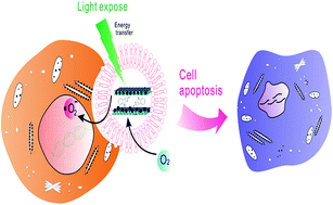
Inorganic nanostructured ensembles containing an anionic clay matrix with layered double hydroxide (LDH) were designed in nanooncology for photosensitizer delivery. A core–shell strategy was combined with host–guest chemistry by the intercalation of indole-3-acetic acid (IAA). This single formulation, with good drug loading percentage and a compact liposome coating (LDH–IAA–Lipo) over and around the positively charged layered surface establishes a controlled release property for the treatment of skin melanoma. All the data regarding synthesis, physical characterization, chemical stability by thermogravimetric analysis and coat stability by leaching test in solvent mixture containing triton X-100 and biological buffer was obtained. IAA was estimated using high-performance liquid chromatography (HPLC) under optimized conditions with an admirable outcome. An improvement in cytotoxic properties under visible light exposure has been confirmed by MTT, ROS levels, DNA fragmentation using comet assay and apoptosis by analysis of mitochondrial membrane potential (MMP) using the MitoProbe JC-1 assay. In contrast, this formulation depicted cytocompatibility in a normal fibroblast (3T3) cell line. Photodynamic therapy (PDT) can be suggestible for long term therapy since the combinatorial efficiency of drug molecules in addition to light irradiation was dramatically evidenced to treat melanoma effectively.
1. Introduction
Skin melanoma is a severe infirmity in the melanocytes, which metastasize and lead to malignancy and eventually death. Nevertheless the freckles that lead to skin cancer are in contrast with the moles possibly caused by UV irradiation from sunlight.1,2 Recently, photodynamic therapy (PDT) has fascinated researchers as an effective therapy using topically applied drugs (photosensitizers).3–5 In light irradiation these photosensitizers are activated and output in cells as porphyrin derivatives6,7 and reactive oxygen species,8,9 which play a key role in PDT for treating various malignancies.10 PDT is now applied to benign skin disorders such as warts, acne and various melanomas.11 It is advantageous over conventional treatments like surgery chemotherapy and radiation therapy in most of the cancers since it is very simple, non-invasive and most convenient to the patient12 and selectively targeted towards localized infections. Drugs like cisplatin13 and docetaxol14 are preferred for skin melanoma; these cause very serious adverse effects and can be bypassed by means of PDT. These days most of the research is being directed towards the development of typical photosensitizers for topical and systemic delivery to treat various kinds of infections besides cancer.15 A suitable formulation is desired to deliver a photosensitizer for its accretion in effective therapy.
Layered double hydroxide (LDH) nanoparticles have been explored widely owing to their biocompatibility16 as well as biodegradability in the biomedical field for cancer treatment, termed nanooncology.17 These inorganic layered solids are stable18 and have the ability to encapsulate or immobilize various bio- and organic molecules in the interlayer space.19–25 Many varieties of metal nanocomposites can also be synthesized using different metals with varying compositions, synthetic preparation routes at different hydrodynamic diameter range (30–100 nm) and encompassing various applications.17,20,26–40 Among all the classes of anionic clays, nanocomposites with magnesium and aluminum containing nanohybrids have potential drug delivery applications.41 These clay materials could be useful to tackle diversified cell lines and helpful in targeted therapy for various tumor environments.42–50 In general the tumor environment contains leaky vasculature and high permeability due to its inbuilt angiogenesis properties. These nanomaterials can effectively target the tumor due to the enhanced permeability and retention (EPR) effect,51 while the higher surface area of LDH plays a crucial role in developing an effective therapeutic strategy in the controlled delivery of photosensitizer.52–55 LDH nanocontainers are versatile carriers, which are more effective in terms of drug delivery compared with other inorganic nanoparticles like mesoporous silica nanoparticles (MSNs), which are poor in biodegradability and encapsulation efficiency.56 Most of these reports indeed suggest LDH as a major carrier system in nanooncology as far as drug delivery using nanovehicles is concerned.20,46,47,57–68
Liposome is selected as a supportive coat, since it is a well-known attractive system for the controlled release of various drugs used to treat different infections.69,70 In addition, liposomes are biocompatible, versatile and remarkable delivery systems with an enormous number of applications71 on account of their variability in composition. Biodegradability, as well as the structural and biological properties of a delivery system, can significantly decrease drug toxicity72 and accelerate the dissolution capability of drugs. Although a liposome coating is not that familiar in the delivery of photosensitizers, this coating could represent an advance in encapsulating both hydrophilic as well as lipophilic drugs. Eventually the coated nanohybrid is necessarily required for the controlled delivery73 of photosensitizer74,75 at selective sites on the LDH to improve its therapeutic activity as well as to prop up cell uptake.
Indole-3-acetic acid (IAA), a plant hormone, has gained the attention of researchers since it was first activated to produce free radicals when exposed to light at a particular wavelength, via the photo activation process.76 IAA itself is not toxic; indeed an agent for PDT resulted in the death of cancer cells by inducing apoptosis in prostate cancer and antimicrobial activity77 under Ultraviolet-B (UV-B) irradiation.78 Although previous reports suggest that IAA’s anti-neoplastic nature is a good example for combinational therapy with the oxidative decarboxylated product of horseradish peroxidase (HRP)79 , IAA alone can generate free radicals,80 since the treatment is so effective even at the low oxygen levels common in tumors. It can also be effective at lower light doses than conventional photodynamic therapy,81 and could thus act effectively on various cancers.82–84 We intended to synthesize and formulate a delivery system by encapsulating IAA intercalated LDH (LDH–IAA) within the coat of the liposome layer (LDH–IAA–Lipo) for the controlled delivery of a photosensitizer (Fig. 1). However, this is the first time to report using IAA as a photosensitizer and prove the apoptosis of the skin melanoma cell line under visible light irradiation and its potential for photosensitizing capability measured by optimizing all the conditions.
 |
| Fig. 1 Schematic representation of the uptake of liposome coated LDH nanohybrids by cell and resultant apoptosis by photodynamic therapy (PDT). | |
2. Experimental section
2.1 Materials
All the reagents, chemicals and organic solvents were of analytical grade at the highest purity and commercially available, and were used without further purification. Indole-3-acetic acid (IAA), magnesium nitrate hexahydrate [Mg(NO3)2·6H2O], lecithin (phosphatidylcholine), cholesterol, and ethylenediaminetetraacetic acid (EDTA) were purchased from Alfa Aesar (A Johnson Matthey company, Heysham, England). Aluminium nitrate nonahydrate [Al(NO3)3·9H2O], Triton X-100 was obtained from J. T. Baker chemicals Pvt. Ltd (Phillipsburg NJ, USA). Dicetyl phosphate, sodium phosphate dibasic (Na2HPO4), 3-(4,5-dimethylthiazol-2-yl)-2,5-diphenyltetrazolium bromide (MTT), potassium phosphate monobasic (KH2PO4), potassium phosphate dibasic (K2HPO4), citric acid, n-butanol, sodium hydroxide (NaOH), 2′,7′-dichlorodihydrofluorescein diacetate (H2DCFDA), ninhydrin (2,2-dihydroxyindane-1,3-dione), 4′,6-diamidino-2-phenylindole dihydrochloride (DAPI), formaldehyde (HCHO), and fluorescein isothiocyanate (FITC) were obtained from Sigma. Co. Ltd (St. Louis, MO, USA). Thiobarbituric acid (TBA) was obtained from Acros organics Ltd. Fetal bovine serum (FBS) and Dulbecco’s modified Eagle’s medium was purchased from GIBCO/BRL Life Technologies (Grand Island, NY, USA). Potassium bromide (KBr) (FT-IR grade) and (3-aminopropyl)trimethoxysilane (APTS) were obtained from Fisher scientific Ltd (Loughborough, UK) and Gelest (Morrisville, PA, USA), respectively. Rhodium phalloidin was purchased from Invitrogen Ltd (Eugene, Oregon, USA). The comet assay kit was obtained from Trevigen (Gaithersburg, MD, USA).
2.2 Instruments
Centrifugation during the cell culturing process and nanomaterial synthesis was performed at an appropriate temperature using Swing rotor Kubota KN-70 (Tokyo, Japan) and Hermle Z 36 HK (Wehingen, Germany) instruments, respectively. Ultraviolet-visible (UV-Vis) spectroscopic absorbance was recorded on a Genequant-300 series spectrophotometer while fluorescence imaging was captured on an Olympus microscope hybridized with Nikon CCD camera apparatus (Palo Alto, CA, USA) with BD pathway (BD biosciences, USA). 532 nm LED lights purchased locally. B16F10 cell lines were obtained from the Bioresource Collection and Research Center (Hsinchu, Taiwan). Fluorescence intensity and MTT absorbance were recorded using Perkin Elmer’s EnSpire Multi-label Plate Reader (Santa Clara California, USA). The flow cytometric quantification was achieved using a Beckmann Coulter (Cytomics FC-500) equipped with a FSC detection system and argon laser lamp (488 nm emission wavelength), while the date acquisition was in linear mode and the data was visualized in logarithmic mode.
2.3 Characterization
Fourier transform infrared (FT-IR) spectra were recorded on a Bruker Alpha spectrometer with a dried KBr pellet. Zeta (ζ)-potential as well as particle size distribution was measured by dynamic light scattering (DLS) (Malvern Nano-HT Zetasizer). The samples were prepared by diluting the nanoparticle suspension with de-ionized water until the counter rate was less than 1.5 Mcps (mega counts per second). The physical state and composition determination of IAA intercalated in the LDH and the strength of liposome coated LDH nanohybrids were investigated by thermogravimetric analysis-differential thermal analysis (TGA-DTA) curve on TGA Q50 V20, 13 Build 39 (Universal V4.5A TA Instruments). The temperature increased from ambient to 800 °C at a rate of 20 °C min−1 under a dry nitrogen purge at a flow rate of 20 mL min−1. Powder X-ray diffraction (PXRD) analysis of the samples was carried out using a powder X-ray diffractometer (XRD D8 Advanced, Bruker) to determine the existing nature of IAA in LDH. The diffraction angle (2-theta) was recorded from 2° to 70° with a scanning speed of 3° min−1. Cu Kα radiation was used as an X-ray source at 40 kV and 40 mA. Nitrogen adsorption–desorption isotherms were measured by Micrometric ASAP 2010 surface area analyzer at the temperature of liquid nitrogen (−196 °C) using ultra high purity nitrogen and helium as the adsorbate and carrier gas, respectively. For surface area and pore size distribution measurements, about 150 mg of each nanoparticle sample was degassed overnight at 80 °C under vacuum (10−3 Torr). TEM images were captured on a Hitachi H-7100 operating at 100 kV. Samples were prepared by dispersing LDH aqueous solution deposited on carbon coated copper (Cu) grids and dried at room temperature.
2.4 Synthesis of LDH nanoparticles
Inorganic LDH nanoparticles were synthesized by a co-precipitation method as reported previously.85–87 Mg(NO3)2·6H2O and Al(NO3)3·9H2O (3
:
1) were dissolved in 10 mL double-distilled water (dd-H2O) and added instantly to 40 mL of 0.2 M NaOH solution by stirring at room temperature under a nitrogen purge for 10 minutes. Atmospheric carbon dioxide (CO2) entrapment/contamination between the layers was prevented by maintaining an inert atmosphere and by using decarbonated dd-H2O, accomplished by simultaneous boiling and ultra-sonication. Eventually, the nanoparticles were collected and washed repeatedly with 40 mL of dd-H2O and centrifuged at 13
000 rpm for 15 minutes. The slurry was further suspended in 50 mL dd-H2O and the solution underwent hydrothermal treatment in a Teflon-lined autoclave at 100 °C for 16 hours. The nanoparticles were subsequently collected and washed repeatedly by centrifuging at 13
000 rpm for 15 minutes, and finally suspended in ethanol to prevent contamination.
2.5 Synthesis of LDH–IAA nanohybrids
Indole-3-acetic acid (IAA) intercalation was primed using the ion-exchange process as reported previously, with slight optimization.88 We accurately weighed 100 mg of IAA dissolved in 10 mL water, set to pH 9.0. The nanoparticles were then re-suspended in the prepared drug solution at the same ratio and the slurry was stirred at 70 °C for 3 days under a nitrogen atmosphere. Finally, the resultant product was centrifuged and washed repeatedly with dd-H2O and re-suspended in ethanol for further study. This sample was denoted as LDH–IAA (layered double hydroxide–indole acetic acid).
2.6 Loading efficiency of IAA in LDH
Quantification of intercalated IAA in LDH nanocontainers was estimated by UV-Vis spectroscopy. 5 mg of the nanohybrids, accurately weighed, was placed in a 10 mL volumetric flask, to which 0.5 mL of 0.1 M hydrochloric acid (HCl) solution was added to mortify LDH, and the rest was filled with ethanol. The exact concentration of the drug in solution was determined by monitoring the absorbance at 280 nm (λmax) and the concentration was calculated by regression analysis according to the standard curve obtained from a series of standard solutions of IAA. It was calculated as 19.5% (w/w) of LDH.
2.7 Synthesis of liposome coated LDH nanohybrids
Required amounts of lipid phase with three components lecithin, cholesterol and dicetyl phosphate were weighed and dissolved in chloroform in a molar ratio 7
:
2
:
1. Once the lipids were thoroughly mixed in the organic solvent, it was evaporated under reduced pressure at 40 °C to yield a thin lipid film; this film was dried to remove residual organic solvent by being placed under vacuum overnight. Finally the dried lipid film was hydrated by adding 60 mg nanoparticles dispersed in 20 mL of the aqueous phase followed by agitation for 1 hour at 45 °C. Eventually small unilamellar vesicles were prepared by bath sonication for 20 minutes at 45 °C. This sample was denoted as LDH–IAA–Lipo (layered double hydroxide–indole acetic acid–liposome).
2.8 Synthesis of surface functionalized LDH–FITC
Fluorescein isothiocyanate (FITC) on the surface of LDH was anchored using an amine linker and effectively surface functionalized using toluene as the reaction solvent, as reported previously.89 Since the surface activation of LDH attained at higher temperature is favored by toluene, 0.2 g of LDH nanoparticle was suspended in 30 mL toluene and stirred vigorously; 1 mL of 97% (3-aminopropyl)trimethoxysilane (APTS) was added after 30 min to the reaction mixture and stirred for 24 hours at 100 °C under a nitrogen purge. The resultant nanoparticles were collected and washed repeatedly with acetone and ethanol to remove the unconjugated silane. Conjugation was confirmed by the ninhydrin test, which is specific to primary amines. FITC was dissolved in dry methanol for successful immobilization of the respective amine on LDH surface. 10 mg of FITC was dissolved in 10 mL dry methanol and 100 mg nanoparticles (LDH-NH2) was re-suspended and stirred for 24 hours in the dark at room temperature. Liposome was coated on the FITC conjugated LDH as mentioned in the above section (Section 2.7).
2.9 Leaching/stability test
Liposome coat efficiency was measured by performing a leaching test of the liposome coated LDH–IAA (LDH–IAA–Lipo) samples in citrate buffer pH 5.0 with and without Triton X-100. This facilitates LDH degradation (only in citrate buffer) and liposome disruption (with Triton X-100). This test directly symbolizes the coating efficiency of liposome around LDH and indirectly represents the IAA intercalation in LDH when treated with the solvent mixture. 10 mg of the respective sample was placed in 200 μL Triton X-100 and the other with a citrate buffer for 30 min while a further 500 μL of citrate buffer was added to both samples and incubated for 4 hours in a rotary shaker at 150 rpm. Finally the supernatant was collected after centrifuging and analyzed using high-pressure/performance liquid chromatography (HPLC) under optimized conditions for the detection of IAA.
2.10 HPLC method for IAA determination
IAA detection using HPLC system containing a Hitachi L-2130 HPLC pump attached with a manual injection system and UV detector (Hitachi L-2400) at the specific absorption wavelength of 280 nm. IAA was separated using a Cosmosil C18 column (250 mm × 4.6 mm, 5 μm size). Separation was undertaken using the mobile phase with the gradient elution method, beginning with water
:
acetonitrile (50
:
50) for the first 8 minutes and continuing with 50
:
50 acetonitrile
:
methanol for 15 minutes. The flow speed was maintained at 1.0 mL min−1 and injection volume was fixed at 20 μL.
2.11 In vitro drug release study
IAA release study was performed by suspending the LDH–IAA and LDH–IAA–Lipo nanohybrids in phosphate buffer saline (PBS) solutions at various pHs (5.0 and 7.4). The intention is to simulate the passage and mimic the release behavior in different gastrointestinal environments for the purpose of establishing the in vitro–in vivo correlation. LDH–IAA–Lipo nanohybrids were additionally ensured in a pH 1.2 (0.1 M HCl) buffer at 37 °C while being stirred at 100 rpm and the respective simulated fluids were replaced at relevant time intervals. Aliquots were removed by centrifuging at 12
000 rpm for 10 minutes and analyzed at 280 nm, while the same volume of fresh buffer was replaced for a further time period. The released percentage of the drug was determined by measuring the concentration periodically using UV-Vis spectroscopy.
2.12 Cancer studies
To determine the photodynamic therapy using IAA, the B16F10 skin melanoma model cell line was used selectively towards topical delivery for PDT. B16F10 (mouse melanoma cell line) and normal fibroblast (3T3) cell line were cultured in a Dulbecco’s Modified Eagle Medium (DMEM) supplemented with 10% (v/v) fetal bovine serum and penicillin (100 units per mL)/streptomycin (100 μg mL−1). Cultures were maintained in a humidified incubator at 37 °C in 5% CO2.
2.13 Cell viability assay (MTT)
Cellular viability was measured using MTT assay, which was reported previously.90 Cells were inoculated into 96-well plates at a density of 1 × 10⁴ cells per well and incubated for cell attachment. After 24 hours they were subjected at various concentrations (B16F10 cell line 0–250 μg mL−1 and 3T3 cell line 0–500 μg mL−1) of LDH, LDH–IAA and LDH–IAA–Lipo samples in FBS free DMEM. They were further incubated for 4 hours to facilitate particle uptake and the delivery of photosensitizer; then the light was irradiated for the prescribed time with 532 nm LED green light (power per cm2 calculation, see ESI†) and incubated for another 20 hours. At the end of incubation, 50 μL MTT solution (1 mg mL−1 of MTT in PBS) was added and further incubated for 4 hours. Finally, the medium was pipetted out and the violet crystals (formazan) were dissolved with 150 μL dimethyl sulfoxide (DMSO) and the reduction of MTT by mitochondrial dehydrogenase was measured at 570 nm using ELISA reader. In addition, cells without nanoparticle suspension were taken as the control with the viability set as 100%. The percentage viable cells in each well was calculated from the absorbance of purple colored formazan crystals. The final report data was expressed as a percentage of the control (mean ± SD). The percentage inhibition of each compound was calculated using the following formula: % inhibition = (mean absorbance of treated cells/mean absorbance of control) × 100.
2.14 Free radical determination
In vitro free radical quenching by IAA loaded LDH nanohybrids was precisely quantified using 2′,7′-dichlorodihydrofluorescein-diacetate (H2DCF-DA) assay, with the fluorescence measurement taken after the H2DCF-DA is oxidized to fluorescent DCF underneath photo irradiation, as reported previously.78 Prior to the free radical determination, H2DCF-DA (1 mM) was activated using an ethanolic stock solution mixed with 0.01 M NaOH and the resulting reaction mixtures containing nanohybrid at various concentrations (250, 500 μg mL−1) placed in a 25 mM sodium phosphate buffer (pH 7.2) along with the control before 4 hours and incubated for 10 minutes in the dark. The whole reaction mixture (activated DCF-DA along with the LDH–IAA solution) was irradiated after 20 minutes of incubation at various pre-set times from 10–60 s at 532 nm laser light. The resultant absorbance was measured using an Elisa reader at Ex/Em 485/528 nm. Similarly, the free radical determination in the presence of the B16F10 cell line was performed as well as with a positive control hydrogen peroxide (H2O2). The assay was performed in triplicate for three independent experiments.
2.15 Cell uptake studies and fluorescence quantification using flow cytometric analysis
The drug delivery efficacy of liposome coated LDH nanoparticles (LDH–FITC–Lipo) was investigated by means of a cell uptake study using FITC conjugated LDH by visualization in BD pathway as reported previously with slight optimization.91 B16F10 skin melanoma cells after 80% confluence were harvested with a trypsin–EDTA solution and seeded into fluorescent plates (96-well) at a density of 1 × 10⁴ cells per well. LDH–FITC and liposome coated LDH samples (LDH–FITC–Lipo) were incubated for 4 hours with the cells then fixed with 3.7% paraformaldehyde and eventually incubated for 10 minutes with 0.5% Triton X-100 for cell wall dissociation; simultaneously 100 μL of 1% bovine serum albumin was added. The cytoskeleton and nucleus was further stained with rhodium phalloidin (100 μL, 0.05 mg mL−1) and DAPI (100 μL, 0.1 mg mL−1) respectively, intermittently washed thrice with PBS for each step. Eventually the cell images were captured using the BD pathway.
In addition, the harvested cells were seeded in a 6-well plate at a density of 3 × 10⁵ cells per well, and after 24 hours of incubation the cells were treated with liposome coated LDH–FITC (100 μg mL−1) in addition to the control i.e., nanoparticles devoid of FITC. Cells were collected in a tube and centrifuged at 400g for 5 minutes, washed once with PBS and re-suspended for fluorescence quantification using flow cytometry. The samples after treatment were immediately analyzed by measuring the fluorescence from the gated cell population using Beckmann coulter flow cytometry with the laser set at 530 nm in a 3 decade pulse area. The forward scattered (FS) and side scattered (SS) light profiles were adjusted for gating the cell population and the fluorescence parameters were recorded by collecting with logarithmic amplification.
2.16 Comet assay
DNA fragmentation was determined using the most reliable technique, alkaline comet single cell gel electrophoresis according to the manufacturer’s (Trevigen) instructions. B16F10 cell lines were treated with the LDH–IAA and LDH–IAA–Lipo nanohybrids. After 4 hours of incubation, the light was irradiated for 30 seconds and further incubated for 20 hours. Cells were harvested and pooled in a 1% low melting point agarose at a ratio of 1
:
10 (v/v) at 37 °C and immediately layered on custom frosted slides, which feature a clear centered window. The gel was run in the alkaline electrophoresis buffer composition (0.2 N NaOH, 0.5 mM EDTA, pH 12.5) for 20 minutes at 21 Volts and 350 mA. We washed the slides in water and ethanol for 5 minutes to reanneal the DNA and finally stained the dried smear with SYBR green (1 mg mL−1) for 30 minutes, with the micrographs captured using fluorescence image analysis.
2.17 Morphological analysis of mitochondria
Mitochondria membrane potential (MMP) was ratiometrically recognized by the indicator JC-1 stain (5,5′,6,6′-tetrachloro-1,1′,3,3′-tetraethyl benzimidazolyl carbocyanine iodide) observed in the cells by changes in coloration after treatment. The cultured B16F10 cell line at a density of 1 × 10⁴ cells per well was subjected to the treatments using LDH–IAA and LDH–IAA–Lipo in the dark and under light exposure. After 4 hours the light was irradiated for 30 seconds at a wavelength of 532 nm in the required wells and incubated for 20 hours. Subsequently, the indicator JC-1 stain was added at 5 μM and incubated for 30 minutes at 37 °C. Finally, the dye was cleaned off and washed twice with PBS; the cells were observed under the fluorescent microscope for dye aggregates showing different colors representing the mitochondrial potential.
2.18 Lipid peroxidation
Lipid peroxidation is an indicator of oxidative stress. The generated thiobarbituric acid reactive substances (TBARS) such as lipid hydroperoxides and aldehydes [e.g. malondialdehyde (MDA)] in the cell culture media combine with thiobarbituric acid (TBA) in a 1
:
2 ratio to form a fluorescent adduct. TBARS are expressed as MDA equivalents by following the procedure as reported previously.92,93 Cells were treated with LDH–IAA (100 μg mL−1) and LDH–IAA–Lipo (100 μg mL−1) and incubated for an hour, with diethyl maleate (DEM) at 10 mM used as a positive control. Cells from prepared flasks were harvested and seeded at a density of 5 × 10⁵ cells per mL in DMEM cell culture medium (2 mM L-glutamine, 10% FBS). After attaining 80% confluence after 24 hours, the cell culture medium was replaced with respective test nanomaterials suspended in PBS along with negative and positive controls and incubated at 37 °C in 5% CO2. After 2 hours of incubation the supernatant and cell lysate were extracted and the in vitro TBARS assay was performed. 200 μL supernatant was taken and a mixture containing 400 μL of 0.67% TBA/0.01% BHT in 2.5% TCA and 200 μL of 15% TCA was added. This mixture was heated at 95 °C for 30 minutes, allowed to cool down and a complex of MDA–TBA was extracted using n-butanol. Eventually, fluorescence was measured at Ex. 530 nm and Em. 550 nm, along with the reference recoded to a reagent blank.
3. Results and discussion
The current investigations are discussed in brief. Indole acetic acid (IAA) loaded within layered double hydroxide (LDH) was synthesized and eventually coated with the biocompatible liposome using negatively charged lipids for controlled release of photosensitizer. IAA acts as a prodrug involving energy exchange phenomena through a photoreaction process when exposed to visible light. This photosensitized IAA mainly generates free radicals on oxidation (indolyl radical cation, skatolyl radical), forming reactive toxins known as highly reactive species (peroxyl radical) responsible for phototoxicity.81 These harmful adducts seriously obstruct the biochemical process of the cancer cell and eventually lead to death. IAA as a photosensitizer is intercalated to deliver and prove the apoptosis of skin melanoma (B16F10 cell line from mouse) under photo activation and has potential for photosensitization in cancer therapy. In addition the cytocompatibility of IAA loaded nanoconstructs were tested in a normal fibroblast cell line.
Fourier transform infrared (FT-IR) spectra of pristine LDH and IAA loaded LDH and liposome coated LDH are characterized and represented in Fig. 2. It suggests that all the molecules were stabilized after intercalation of IAA in the interlayer space of LDH with preserved functional groups. A strong and extensive band centered around 3400 cm−1 is the result of the O–H group stretch of absorbed water molecules and surface hydroxyl groups of LDH layered structures. A sharp peak at 1382 cm−1 exemplifies the interlayer nitrate ion (Fig. 2a). The peaks at 553 and 683 cm−1 are attributed to the lattice vibrations of M–O and M–O–M, respectively,94 These are observed on the subsequent modification of LDH, which reveals the undissociative nature of LDH on modification. After the intercalation of IAA, two sharp peaks are seen at around 1560 and 1640 cm−1, corresponding to the N–H (amine) stretch and asymmetric carboxylate stretch of IAA, respectively, indicating the successful intercalation of IAA (Fig. 2b). However an increase in the intensities of the peak results in IAA loading, although the peak at 1382 cm−1 is reduced to some extent, which shows the presence of nitrate ions in the interlayer of LDH. The coating with the lipid blend on the surface of LDH displays the absorption bands at 1370 and 1742 cm−1 and embodies the CH2 bending and the C–O stretch of lecithin ester, respectively. A sharp peak at 1225 cm−1 confirms the PO4 stretch of lecithin and dicetyl phosphate (Fig. 2c). However the sharp and tapered bands at 2921, 2850 cm−1 confirm the C–H stretch of cholesterol and the aliphatic hydrocarbon chain of dicetyl phosphate. These spectra clearly reveal that the LDH has not degraded or dissociated during the intercalation of IAA and the liposome coating on the LDH surface.
 |
| Fig. 2 FT-IR spectra of (a) pristine LDH, (b) LDH–IAA and (c) LDH–IAA–Lipo. | |
Fig. 3 depicts the characteristic PXRD patterns of IAA intercalated LDH as well as liposome coated LDH. The pristine LDH has shown the characteristic diffraction peaks (2θ) at 11°, 24° and 35°, which correspond to the respective planes at (003), (006) and (009) of LDH (Fig. 3a). After the intercalation of IAA the spacing of d(003) planes shifts towards the left by an angle of 4.95 degrees and then d(006), d(009) shift at 10.1 and 11.4 degrees, respectively (Fig. 3b). This reveals the increase of d-spacing in d(003) to 1.79 nm after IAA intercalation from 0.82 nm (LDH) and is in clear agreement to what has been reported previously.95 This clearly supports the FT-IR data as determined for functional group analysis. Nevertheless the typical hexagonal structural arrangement of LDH is shown by the characteristic peaks at the planes d(110) and d(113) of the basal reflection at 60.8° and 62.2°. This arrangement facilitates the ease of the exchange process among interlayer anions without any destruction in the layered structure.96 Moreover after coating with the liposome the changes in the PXRD pattern were negligible in the arrangement. But the d-spacing was amplified at d(003) reflection to 1.98 nm because of the geometric arrangement from the influence of electrostatic interactions between the positively charged surface of the LDH and the counter charged liposome coat (Fig. 3c). These results could be evidence for LDH and its arrangement before and after the intercalation of IAA.
 |
| Fig. 3 Powder X-ray diffraction spectra of (a) pristine LDH, (b) LDH–IAA and (c) LDH–IAA–Lipo. | |
The thermal properties have robustly corroborated the loading efficiency and stable nature of the inorganic nanoparticle. Fig. S1† represents the TGA and DTG curves of the LDH nanoparticle and its consecutive modified samples. However the degradation temperature of LDH–IAA shifts towards the right (higher than the pristine LDH) and was reverted in the case of liposome coated LDH, whereas the termination temperatures are almost similar in all cases. The stages of degradation have varied in all the samples with different thermal behaviors; for instance, a well-known loss occurred because of the elimination of the absorbed interlayer water under 250 °C. Moreover, at higher temperatures it results in the dehydroxylation of layers and the formation of double oxide-hydroxide and carbon dioxide from the layers.97 Pristine LDH exhibited different stages of weight loss, of which the first is attributed to water loss from the surface due to physical absorption and the interlayer space of LDH nanoparticles (Fig. S1B-a†).98,99 The next weight loss event was followed by 28.3% weight loss in the final stage of decomposition around 350 °C, which represents the final combustion of inorganic fractions of the nanoparticles. In the LDH–IAA (Fig. S1B-b†) sample, the weight loss in the early stages is same as the pristine LDH sample. However the final stage of the combustion profile disappeared and shifted right to 413 °C with 45% weight loss signifying that the drug and LDH were packed together and shrouded with layers.95 Since drug loaded LDH nanovehicles are degraded at higher temperatures, the enhanced thermal stability supports the strength of interactions between the molecules in the interlayer gallery as well.46 The loading of IAA was confirmed and the percentage nearly matches the quantitative UV detection obtained, i.e., 19.5% (w/w) of LDH (Fig. S1A†).
Recently, Zhang and An and co-workers have designed innovative formulations such as photosensitizer-doped perylene nanoparticles,100 as well as carrier free stable nanocrystals101 with a very high loading percentage of the photosensitizer. These are highly effective and very impressive in treating cancer cells. In a similar fashion, using biodegradable LDH nanocarriers, which have their own advantages, such as surface functionalization and high surface area with controlled release properties, could also be supportive in preventing the instant degradation/decay of the photosensitizer. In addition, these nanoconstructs are not only stable but also enhance the solubility of poorly soluble photosensitizers. In comparison to other inorganic nanocontainers like MSN, with less than 5% drug loading, usage of biocompatible and biodegradable LDH with improved IAA loading efficiency (nearly 20% w/w of LDH) is beneficial. Eventually, liposome coated LDH–IAA has been subjected to TGA analysis and surprisingly the 1st event of weight loss was much less (5%), than the adsorbed water molecules up to 200 °C, while the final combustion stage started at 180 °C and lasted up to 500 °C with a weight loss of 62% (Fig. S1B-c†). The lipids are thermo sensitive organic compounds that are degraded in the earlier stages, and inorganic moieties also follow at their respective temperatures at a higher combustion rate with the loss of more weight. The reduction in the degradation temperature of LDH–IAA–Lipo was evidenced by the liposome bilayer, which formed a compact sheet around the positively charged layered surface of the pristine LDH. Furthermore, the degradation of LDH was extended up to 450 °C, which demonstrates the shrouded coat on the nanoparticles.
The particle size measurement and the ζ-potential of various samples of LDH after modification were measured using DLS and are depicted in Fig. 4 and Table S1,† respectively. The average hydrodynamic diameter of positive charged layers of pristine LDH is 167 nm (Fig. 4a), while the potential is +47.2 ± 0.97 mV (Table S1a†), due to magnesium and aluminium metals in the framework. The IAA loaded LDH size has been enhanced by 50 nm (Fig. 4b) because of aggregation, and the potential was drastically reduced and attained a nearly neutral state (Table S1-b†). The charge was balanced by the high loading capacity of IAA achieved by the anion exchange mechanism. This confirms the intercalation of IAA in LDH. The zeta potential measurements of all the respective samples were carried out by adjusting to physiological pH 7.4. After coating with negatively charged lipids, the surface potential decreased to a large extent and terminated at −42.5 mV (Table S1c†), with the final mean hydrodynamic diameter at 320 nm (Fig. 4c). Herewith, negatively charged lipid dicetyl phosphate was used to establish charge based interactions between positively charged layers and negatively charged lipids for a controlled release and to ensure the compactness of the coat. The thickness of the coat is not only helpful in the controlled release of IAA but also enhances the stability of the nanoparticles that assist in preventing the leaching of intercalated photosensitizer. Meanwhile, this strategy offers the advantage of charge balancing capacity with a negatively charged liposome at which the intercalated anions can remain stable in their respective positions from the start. Therefore, it is worth noting that formulations with such size distribution could be established as delivery systems for various routes of administration. A stability investigation for the liposome was carried out by a leach test using the surfactant Triton X-100 (polyethylene glycol octyl phenyl ether) in citrate buffer (2
:
5 ratio). This creates the leakage of IAA by LDH degradation in the citrate buffer after its exposure to the wear and tear of the coat using Triton X-100. Simultaneously, a comparison can be made with the control i.e. exposure of LDH–IAA–Lipo using the citrate buffer alone, in which it indirectly signifies the stability of the liposome coat. However, the IAA release was highly negligible. In the HPLC determination of IAA (see in Experimental section), the spectra reveal that IAA separation is achieved at a retention time of 9.45 minutes in a 15 minute run (Fig. 5a). Sharp peaks were observed in the respective samples for IAA detection, which clearly shows IAA intercalation in LDH (Fig. 5b). The liposome coated sample (Fig. 5d), when subjected to the Triton X-100–citrate buffer mixture, leads to the disorganization of the lipid layer and eventually the LDH framework is disintegrated after coming into contact with the citrate buffer. Similarly, when the sample is subjected to treatment in the citrate buffer alone (Fig. 5c), no peak was observed at the retention time of IAA and this demonstrates that the coat is stable, and denotes that the LDH was encapsulated in liposome.
 |
| Fig. 4 Particle size distribution of (a) pristine LDH, (b) LDH–IAA and (c) LDH–IAA–Lipo. | |
 |
| Fig. 5 Leaching/stability test and HPLC determination representing (a) IAA alone, (b) LDH–IAA, (c) LDH–IAA–Lipo subjected to citrate buffer alone and (d) LDH–IAA–Lipo subjected to Triton X-100–citrate buffer mixture. | |
Fig. S2† reveals supportive evidence for the stability study. The FT-IR spectrum was recorded for the sample after being subjected to the leach test and compared with the samples of LDH–IAA (Fig. S2a†), LDH–IAA–Lipo (before stability (Fig. S2b†); it is devoid of the N–H stretching peak of IAA at 1560 cm−1). This discloses the leak of IAA to a maximum extent in liposome coated LDH (Fig. S2c†). In addition, all the peaks that exemplify the LDH framework and the constituents of the liposome organization were disturbed. These concerns are evidence that the liposome coat and its integrity are stronger and well established on the positively charged metal layers, and helpful for the controlled delivery of the intercalated molecules. The surface area of the as-prepared LDH nanohybrids was recorded based on the nitrogen adsorption–desorption isotherms using Brunauer–Emmett–Teller (BET). The samples were degassed under a vacuum (at 80 °C for 16 hours) prior to analysis and the results revealed a higher surface area of 74.3 m2 g−1 for pristine LDH (Fig. 6a). The surface area of IAA loaded LDH shows a drastic reduction in its final surface area than the LDH alone and was recorded as 38 m2 g−1 (Fig. 6b). The adsorption data represents the type IV isotherm with the hysteresis loop in the curve. Similarly to the BET surface area, the pore volume was also reduced from 0.690 cm3 g−1 for pristine LDH to 0.175 cm3 g−1 for LDH–IAA. This reduction in surface area and pore volume accentuates the hybridization by intercalation of IAA in the layers of pristine LDH. Liposome coated on LDH has shown a drastic decrease in the surface area at 1 m2 g−1 (data not shown), which indicates that the lipid mixture enclosed the LDH layers. This means that liposome indeed has less scope to get for the adsorption of nitrogen that supports the liposome coating on the LDH.
 |
| Fig. 6 Nitrogen adsorption–desorption isotherms of (a) pristine LDH and (b) LDH–IAA. | |
Fig. 7 illustrate the transmission electron microscopy (TEM) observations of coated and uncoated samples. As far as the size related to these micrographs is concerned, it clearly reflects that the particles are of uniform size and in proportion with the hexagonal arrangement. DLS characterized the size around 150 nm in uncoated LDH (Fig. 4a), whereas it was double the size in the liposome coated sample (Fig. 4c). But in the TEM we found no variation in size as a consequence of agglomeration. The liposome coated sample (Fig. 7b) is different in comparison with the uncoated sample (Fig. 7a) in the sense that the appearance is clumpy or misty in nature, while the arrow represents the hexagonal shape of the LDH on the left (Fig. 7a) and the layer of coating on the surface of LDH in the right image (Fig. 7b). These hexagonal plate-like structures are clearly seen, and, after intercalation and coating, some of them turned to nanorods by agglomeration or were further modified due to crystal growth.89 TEM showed the individual morphologies of the nanoparticles before and after coating; they are partially clustered and in the size range ∼100 nm.
 |
| Fig. 7 TEM images (a) pristine LDH and (b) LDH–IAA–Lipo. | |
An IAA cumulative release study from LDH nanohybrids was performed in the simulated body fluids at pH 5.0 and 7.4 to mimic IVIVC (in vitro–in vivo correlation). It was not conducted in pH 1.2 (0.1 M HCl) because of the LDH instability in acidic environments. The release curves of the drug intercalated LDH nanohybrids in pH 5.0, 7.4 (PBS) indicate the burst release mechanism in both environments due to the high exchange capacity of IAA at around 90% in first 3 hours. Similar circumstances were provided for the samples coated with liposome. In addition, the test was also performed in a pH 1.2 buffer.
The release rates of all the samples are depicted in Fig. 8, with the plots of percentage drug release against time. However the controlled release behavior in the respective buffers was attained by the interlocking of lipids in the liposome coat around the LDH. The release is approximately 70 and 90% in pH 5.0 (nearly skin pH) and 7.4, respectively. The metal hydroxide layer in the lipid was more stable in these buffers and facilitated good ion-exchange capacity, at which the IAA release ended conveniently. The highest amount of drug release was achieved because of its ample solubility in alkaline pH. However, this surprising and controlled behavior even ensued at pH 5.0 because of the liposome coat, since the disintegration of the inorganic host was prevented, which is characterized by the arrest of the IAA intercalated LDH inside the liposome coat. Such a discrepancy is possible because of the release exchange mechanism of the interlayer ions and the integrity of the lipids in the liposome anticipated for sustained delivery was satisfactory. This is in contrast to pH 1.2 where the release was very low (nearly 40%) compared to other buffers, because the liposome is stable in acidic pH. This kind of optimized release behavior could help the delivery system to achieve controlled release of IAA for PDT.
 |
| Fig. 8 IAA release from LDH and LDH–IAA–Lipo samples at various time intervals in physiological simulated fluids (PBS) (a) LDH–IAA in pH 5.0 (b) LDH–IAA at 7.4 (c) LDH–IAA–Lipo at pH 7.4. (d) LDH–IAA–Lipo at pH 5.0. (e) LDH–IAA–Lipo at pH 1.2. | |
Cellular internalization of nanocontainers was visualized using fluorescent tags immobilized by amine linkage on the LDH surface. The surface amination was confirmed by the ninhydrin reagent, the most common method of detecting primary amines. The UV-Vis absorption spectrum of all the LDH samples on successive modification at 580 nm after being subjected with ninhydrin reagent is displayed in Fig. S3.† Among all the samples, LDH-NH2 alone displayed a strong band, which was missing in all the others. It is evident from the inset figure that the tubes display positive confirmation for ninhydrin.
Primary amines on LDH surfaces generate Ruhemann’s purple coloration when heated (Fig. S3c†). However, it is devoid of the color before modification (Fig. S3b†) and after conjugation with FITC (Fig. S3d†) as well as liposome coated samples (Fig. S3e†). The disappearance of purple coloration in FITC modified samples of LDH validates the complete conjugation of the FITC molecules, as ninhydrin is known to be inactive for the secondary amine of LDH-NH-FITC molecules. Recently, many groups have studied the cellular uptake of uncoated inorganic nanoparticles,91,102–106 which are size dependent, since the internalization of these nanovehicles is challenging due to their increasing size after aggregation. This cellular uptake (internalization) plays an important role in efficient delivery with respect to activity of photosensitizer from the nanoparticle.4,102 However, the advantage of the liposome coated nanoparticles (LDH-NH-FITC–Lipo) is that they are tuned to establish the ease of internalization in a comfortable way and thus prove that the liposome coated systems are size and charge independent. The uptake study of uncoated nanohybrid (LDH-NH-FITC) was observed concurrently, where smaller amounts of green fluorescence were seen inside the cell, which might correspond to the aggregation encountered, suggesting naked LDH. Additionally, flow cytometric analysis is performed for quantification of fluorescence inside the cell; Fig. 9c reveals the maximum internalization at 85% confluence of the cells gated in comparison with the control in the B16F10 cell line. Adherent fluorescence was not observed and successful cellular internalization was achieved because of the coating of lipids to the nanoparticle surface, which does not have any influence on charge interactions on the cell line surface even after 24 hours of incubation.
 |
| Fig. 9 Cell uptake study, i.e. images captured using the BD pathway represent nuclei stained with DAPI (blue) and cytoskeletons in rhodium phalloidin (red) of (a) LDH-NH-FITC (b) LDH-NH-FITC–Lipo in green, respectively; (c) flow cytometric quantification of fluorescence using liposome coated LDH-NH-FITC (100 μg mL−1) compared with the control (devoid of nanoparticles) in the B16F10 cell line. | |
Free radicals are a highly reactive species measured by using non-specific chemiluminescent dye, called 2′,7′-dichlorodihydrofluorescein-diacetate (H2DCF-DA) assay. The scavenging capacity was determined by measuring the DCF (dichlorofluorescein), the oxidized form of DCFDA, which correlates the amount of free radicals generated on light irradiation. Furthermore, the higher levels of free radicals generated can damage the cellular constituents and are responsible for cytotoxicity. To demonstrate that IAA can produce free radicals under light irradiation, we treated DCFDA with LDH–IAA in PBS buffer. On successively increasing the time of light exposure, a simultaneous increase in the amount of free radical generation was observed (Fig. S4a†). However, after 30 seconds, the plateau was attained by increasing the exposure time and finally this optimized unit was fixed as the effective time period for PDT and further cytotoxic studies were continued. Anti-cancerous activity is completely dependent on the amount of light energy available in cell oriented studies. For the liposome-coated LDH–IAA sample, the lipid membranes may affect the addition reaction of free radicals and DCFDA and therefore a quantitative result did not show in our data. Previously, a study has revealed anti-microbial activity with activated IAA by using UV-light and visible light; however it was the first time using photosensitizer IAA against a skin melanoma cell line.78 The in vitro generation (devoid of cell line) of free radicals by the released IAA from the LDH was higher on exposure to the visible light for a span of 30 seconds (Fig. S4b†). It is clear from the graph that the free radical scavenging/fluorescence is achieved neither by light alone, nor by DCFDA alone, but by a blend of the two.78 The influence of the nanoparticle in delivering the photosensitizer can be understood from Fig. S4b,† where the free radicals generated by an equivalent amount of IAA are less than the IAA intercalated nanovehicle. Because LDH enhances the drug solubility107 with high carrying capacity and eventual photodynamic therapy, free radical generation and in vitro anti-cancerous activity was continued with the same light irradiation time.
IAA acts as a prodrug by generating reactive oxygen species (ROS) involving energy exchange through a photoreaction process when exposed to visible light harmless to the human body. Eventually the ability of generated ROS to damage cellular components leads to cell death. This is possibly due to intrinsically increased oxidative stress and susceptibility to free radical assaults. This is evident from the intracellular ROS measured by DCFDA assay when compared with the positive control H2O2. IAA in the presence of light has generated more ampoule fluorescence than in the dark, which is almost comparable to the H2O2 in both the samples of LDH–IAA and LDH–IAA–Lipo (Fig. S4c†). Light irradiation time and area of exposure should necessarily be taken into account while treating the patient with skin cancer; however laser light in the visible region around 532 nm is not that harmful. In contrast to the other photosensitizers, it could be established as a potential drug with no adverse effects when activated using light in the visible range. These results have proved that IAA itself acts as a free radical generator and gets accumulated in cells, which provide a strong substantiation for anti-cancer activity with photodynamic therapy on the melanoma cell line.
Cell proliferation studies were performed using a MTT assay (Fig. 10a) in the mouse melanoma (B16F10) cell line. The rationale behind choosing the B16F10 cell line is because the activated IAA compound on delivery acts as a photosensitizer, which is efficient in proving surface photodynamic therapy. These experiments were designed using various conditions such as the PDT effect of IAA, determined using bare as well as liposome-coated LDH nanoconstructs in the presence and absence of light. The activity of the delivered photosensitizer against the B16F10 cell line decreased in accordance with the concentration and the difference was significant across the range 250 μg mL−1. However, in contrast, the LDH alone, as well as other nanoparticles (both LDH–IAA and LDH–IAA–Lipo) in the dark seems to be inactive even at higher concentrations. On augmentation of the time for irradiation of light (Fig. S4d†), it was apparent that activation, as well as anti-cancer activity of IAA, was proportionate in both liposome-coated and uncoated LDH nanoconstructs.
 |
| Fig. 10 Cell viability (MTT assay) of LDH, LDH–IAA LDH–IAA–Lipo at increasing concentrations, (a) in mouse melanoma (B16F10) cell line (0–250 μg mL−1) and (b) in normal fibroblast (3T3) cell line (0–500 μg mL−1), in the presence and absence of light irradiation (532 nm). * represents p < 0.001 (one way ANOVA using Tukey test). | |
In a normal fibroblast cell line, surprisingly liposome coated and uncoated LDH–IAA (PDT) at 500 μg mL−1 (Fig. 10b) resulted in more viable cells than in a melanoma cell line (LDH–IAA (PDT) and LDH–IAA–Lipo (PDT) at 250 μg mL−1 (Fig. 10a)). This confirms the cytocompatibility of layered nanoconstructs under irradiation in the normal fibroblast cell line. The remaining samples (LDH, LDH–IAA and LDH–IAA–Lipo in the dark) showed similar results to the B16F10 cell line. The reason behind this surprise is the greater abridged uptake of nanoparticles by normal fibroblast compared to cancer cells.108
Intracellular free radicals generated on irradiation are likely one of the possible causes of cell death, by destabilizing cell functions and cell metabolism with morphological and biochemical changes typical in apoptosis. Therefore we infer that the IAA is delivered from the LDH nanovehicle after its successful uptake by endocytosis, and on activation with light it tends to act inside the cell. A delivery system with a liposome coat around the LDH layers facilitates the delayed release of photosensitizer, which also favors the LDH nanoparticle’s cell uptake mechanism. These results demonstrate that the activated IAA in the presence of green light (532 nm) could act as a treatment option for the delivery of a potent anticancer agent specifically designed for skin related cancers when PDT has been suggested. However, programmed cell death was confirmed by the DNA fragmentation study using the comet assay. The DNA damage/fragmentation seen after oxidative stress smash up was visualized using an alkaline comet assay by staining the nucleic acid using SYBR green (Fig. 11). There was a significant amount of DNA damage in cells treated with IAA immobilized LDH (Fig. 11d) as well as liposome coated LDH nanocontainers (Fig. 11f) on irradiation of light for 30 seconds. When compared with the control experiments (Fig. 11a), we have verified that the comet with light treatment (Fig. 11b) and drug loaded nanoparticles alone (Fig. 11c) inflicts no significant damage on the DNA. This exhibited a supercoiled compact DNA without any transformation, which demonstrates that light alone cannot affect the DNA too. This typically reveals that the free radicals generated from the activated form of IAA on exposure to light are eventually responsible for their fragmentation with elongated tails. However, it was a characteristic move that the uncoiled and damaged DNA was broken into fragments representing the photodynamic ability of free radicals generated by IAA in the B16F10 cell line.
 |
| Fig. 11 DNA fragmentation was detected by single cell gel electrophoresis (comet assay); (a) control in dark (b) control in presence of light for 30 seconds (c) LDH–IAA in dark (250 μg mL−1), (d) LDH–IAA in presence of light (250 μg mL−1), (e) LDH–IAA–Lipo in dark (250 μg mL−1) and (f) LDH–IAA–Lipo in presence of light (250 μg mL−1). Nucleic acid was stained by SYBR green. Scale bar 20 μm. | |
Apoptosis was virtually visualized by means of mitochondrial membrane potential (MMP) loss performed using the vital cationic carbocyanine dye, the sensitive marker JC-1. It is one of the tools for detecting cell death by emitted fluorescence. This is exhibited as a monomer at low concentrations and yields a green fluorescence, which looks similar to fluorescein in a depolarized state i.e. dead cells.109 In the hyperpolarization state, the dye aggregates preferentially in the mitochondria by a Δψ-dependent mechanism and fluoresces red as seen in the healthy cells i.e. the control in the dark (Fig. S5a†), control in the light (Fig. S5b†), LDH–IAA in the dark (Fig. S5c†) and LDH–IAA–Lipo in the dark (Fig. S5e†). In contrast, the treated cells underwent depolarization because of cell apoptosis, due to the free radical attack of IAA in the cytosol on light irradiation. Since it is an execution of apoptosis as pro-apoptotic signals generated by the caspase family of proteins,110 the alterations in the mitochondria are due to the permeabilization of the outer mitochondrial membrane and the consequent release of apoptotic proteins.
In addition, the loss of the electrochemical gradient, which is regulated by the respective mechanism inside the cell, is central to the apoptotic pathway.111 Eventually, it was clear that the IAA from LDH (Fig. S5d†) as well as LDH–Lipo (Fig. S5f†) in the presence of light gets activated and the green fluorescence is dominant with depolarization levels. This represents the damage of the mitochondrial membrane that maintains the electrochemical gradient (Fig. 12). In comparison, it was not that active when applied with light and the drug individually. This kind of approach reflects the combinatorial therapy.
 |
| Fig. 12 Graphical representation for quantification of the J aggregates (mitochondria membrane potential using JC-1 stain) analyzed at respective emission wavelength regions of red and green fluorescence using different conditions of LDH–IAA at 250 μg mL−1 and LDH–IAA–Lipo at 250 μg mL−1. The values are represented as the mean ± SD of three individual experiments. Statistical significance between treated and control samples is denoted by * (p < 0.001) using a one way ANOVA (Tukey test). | |
Oxidative deterioration of the cell membrane lipids was measured using an MDA
:
TBA adduct formed by lipid peroxidation, which occurred during free radical generation, which causes subsequent cell cycle alteration and cell death.112 This can be correlative data for fatty peroxide formation and the bursting out of the cell wall. The underlying mechanism involved in the detection of lipid peroxidation was the formation of malondialdehyde on deterioration of the conjugated dienes in the cell membrane, which acts as an index. The generation of the malondialdehyde levels of LDH–IAA is shown in Fig. 13. At the respective concentrations of LDH–IAA and in the absence of light irradiation, the MDA levels are almost equal to the control, while light alone did not have any effect on the cell line. Liposome coated samples of LDH–IAA have generated higher MDA levels during detection since the lecithin (one of the substituents of the liposome coat) is unsaturated. This interferes in the absorbance of the MDA–TBA adduct during lipid peroxidation because the formulation surprisingly shows higher MDA levels of LDH–IAA–Lipo in the dark. Eventually the result of LDH–IAA correlates to the subsequent generation of free radicals in the presence of light, which was the most likely cause of cancer cell death.
 |
| Fig. 13 Lipid peroxidation determined using a TBARS assay of LDH–IAA and LDH–IAA–Lipo (both treated at 100 μg mL−1). Diethyl maleate (DEM) is the positive control at 10 mM concentration compared to in the absence of the B16F10 cell line. The values are given as the mean ± SD of three individual experiments. Statistical significance between treated and control is denoted by * (p < 0.001) using a one way ANOVA (Tukey test). | |
4. Conclusions
In summary, we have formulated a delivery system suitable for the controlled delivery of non-cytotoxic photosensitizer (IAA) loaded nanocontainer (LDH) coated with liposome for therapy against skin melanoma. These biodegradable and biocompatible LDH nanoconstructs are active in the presence of laser light in the visible region (532 nm). Liposome coated nanohybrids are advantageous over conventional dosage systems in their controlled release behavior, with significant improvement in surface area and the highest carrying capacity. The mechanism underlying the IAA cytotoxicity in treating skin melanoma was the photo activation phenomenon. In order to prove this, we performed cell activity based assays and cell uptake studies as well as mitochondria membrane potential, which is responsive to PDT. These findings could suggest that photodynamic therapy is one of the most useful techniques in drug therapy, and the liposome coated LDH nanocontainers could serve as the best systems for sustained delivery of various drugs including photosensitizers. This system using a nano-photosensitizer shows great promise in melanoma phototherapy.
Acknowledgements
We are thankful to the Ministry of Science and Technology (MOST), Taiwan for research grants (NSC 101-2113-M-259-003-MY2, MOST 103-2113-M-259-005-MY2).
References
- V. Saranga-Perry, C. Ambe, J. S. Zager and R. R. Kudchadkar, Ca-Cancer J. Clin., 2014, 64, 171–185 CrossRef PubMed.
- V. Gray-Schopfer, C. Wellbrock and R. Marais, Nature, 2007, 445, 851–857 CrossRef CAS PubMed.
- D. Kessel, Photodiagn. Photodyn. Ther., 2004, 1, 3–7 CrossRef CAS.
- R. R. Allison, G. H. Downie, R. Cuenca, X.-H. Hu, C. J. H. Childs and C. H. Sibata, Photodiagn. Photodyn. Ther., 2004, 1, 27–42 CrossRef CAS.
- D. E. Dolmans, D. Fukumura and R. K. Jain, Nat. Rev. Cancer, 2003, 3, 380–387 CrossRef CAS PubMed.
- S. K. Pushpan, S. Venkatraman, V. G. Anand, J. Sankar, D. Parmeswaran, S. Ganesan and T. K. Chandrashekar, Curr. Med. Chem.: Anti-Cancer Agents, 2002, 2, 187–207 CrossRef CAS.
- P. Juzenas, R. Sørensen, V. Lani and J. Moan, Photochem. Photobiol., 1999, 69, 478–481 CrossRef CAS PubMed.
- T. J. Dougherty, C. J. Gomer, B. W. Henderson, G. Jori, D. Kessel, M. Korbelik, J. Moan and Q. Peng, J. Natl. Cancer Inst., 1998, 90, 889–905 CrossRef CAS PubMed.
- K. R. Weishaupt, C. J. Gomer and T. J. Dougherty, Cancer Res., 1976, 36, 2326–2329 CAS.
- L. Minai, D. Yeheskely-Hayon and D. Yelin, Sci. Rep., 2013, 3, 2146 Search PubMed.
- S. H. Ibbotson, Br. J. Dermatol., 2002, 146, 178–188 CrossRef CAS.
- S. B. Brown, E. A. Brown and I. Walker, Lancet Oncol., 2004, 5, 497–508 CrossRef CAS.
- G. Serša, B. Štabuc, M. Čemažar, D. Miklavčič and Z. Rudolf, Clin. Cancer Res., 2000, 6, 863–867 Search PubMed.
- W. Sun and L. Schuchter, Curr. Treat. Options Oncol., 2001, 2, 193–202 CrossRef CAS PubMed.
- G. B. Kharkwal, S. K. Sharma, Y. Y. Huang, T. Dai and M. R. Hamblin, Lasers Surg. Med., 2011, 43, 755–767 Search PubMed.
- L. N. M. Ribeiro, A. C. S. Alcântara, M. Darder, P. Aranda, F. M. Araújo-Moreira and E. Ruiz-Hitzky, Int. J. Pharm., 2014, 463, 1–9 CrossRef CAS PubMed.
- S. Bhattacharyya, R. A. Kudgus, R. Bhattacharya and P. Mukherjee, Pharmaceut. Res., 2011, 28, 237–259 CrossRef CAS PubMed.
- Z. Hu and G. Chen, RSC Adv., 2013, 3, 12021–12025 RSC.
- A. I. Khan and D. O’Hare, J. Mater. Chem., 2002, 12, 3191–3198 RSC.
- X. Wu, H. Li, S. Song, R. Zhang and W. Hou, Int. J. Pharm., 2013, 454, 453–461 CrossRef CAS PubMed.
- X. Pang, X. Ma, D. Li and W. Hou, Solid State Sci., 2013, 16, 71–75 CrossRef CAS PubMed.
- L. Dong, L. Yan, W.-G. Hou and S.-J. Liu, J. Solid State Chem., 2010, 183, 1811–1816 CrossRef CAS PubMed.
- S. Mallakpour, M. Dinari and V. Behranvand, RSC Adv., 2013, 3, 23303–23308 RSC.
- V. R. L. Constantino and T. J. Pinnavaia, Inorg. Chem., 1995, 34, 883–892 CrossRef CAS.
- W.-G. Hou and Z.-L. Jin, Colloid Polym. Sci., 2007, 285, 1449–1454 CAS.
- J.-H. Choy, S.-J. Choi, J.-M. Oh and T. Park, Appl. Clay Sci., 2007, 36, 122–132 CrossRef CAS PubMed.
- V. Rives, M. del Arco and C. Martín, Appl. Clay Sci., 2014, 88–89, 239–269 CrossRef CAS PubMed.
- S. Anandan, C.-Y. Chen and J. J. Wu, RSC Adv., 2014, 4, 55519–55523 RSC.
- M. de Victoria Rodriguez, E. Brunet, M. Nocchetti, F. Presciutti and F. Costantino, RSC Adv., 2014, 4, 26912–26917 RSC.
- U. Costantino, M. Nocchetti, L. Tammaro and V. Vittoria, Recent Pat. Nanotechnol., 2012, 6, 218–230 CrossRef CAS.
- L. Perioli and C. Pagano, Expert Opin. Drug Delivery, 2012, 9, 1559–1572 CrossRef CAS PubMed.
- E. Conterosito, W. Van Beek, L. Palin, G. Croce, L. Perioli, D. Viterbo, G. Gatti and M. Milanesio, Cryst. Growth Des., 2013, 13, 1162–1169 CAS.
- T. Selvam, A. Inayat and W. Schwieger, Dalton Trans., 2014, 10365–10387 RSC.
- H. Li, J. Wen, R. Yu, J. Meng, C. Wang, C. Wang and S. Sun, RSC Adv., 2014, 13, 9341–9347 RSC.
- B. Yang and Z. Yang, RSC Adv., 2013, 3, 12589–12592 RSC.
- K. Xu, Z. Zhang, G. Chen and J. Shen, RSC Adv., 2014, 4, 19218–19220 RSC.
- Y. Li, W.-G. Hou and W.-Q. Zhu, Colloids Surf., A, 2007, 303, 166–172 CrossRef CAS PubMed.
- Z. Zhang, G. Chen and J. Liu, RSC Adv., 2014, 4, 7991–7997 RSC.
- M. Zhang, B. Gao, J. Fang, A. E. Creamer and J. L. Ullman, RSC Adv., 2014, 4, 28171–28175 RSC.
- Y. Wang, Y. Zhou, T. Zhang, M. He and X. Bu, RSC Adv., 2014, 4, 29968–29974 RSC.
- K. Ladewig, Z. P. Xu and G. Q. Lu, Expert Opin. Drug Delivery, 2009, 6, 907–922 CrossRef CAS PubMed.
- Z. P. Xu, M. Niebert, K. Porazik, T. L. Walker, H. M. Cooper, A. P. J. Middelberg, P. P. Gray, P. F. Bartlett and G. Q. Lu, J. Controlled Release, 2008, 130, 86–94 CrossRef CAS PubMed.
- J. M. Oh, C. B. Park and J. H. Choy, J. Nanosci. Nanotechnol., 2011, 11, 1632–1635 CrossRef CAS PubMed.
- U. Riaz and S. M. Ashraf, Mini-Rev. Med. Chem., 2013, 13, 522–529 CrossRef CAS.
- S. J. Choi, J. M. Oh, H. E. Chung, S. H. Hong, I. H. Kim and J. H. Choy, Curr. Pharm. Des., 2013, 19, 7196–7202 CrossRef CAS.
- J. H. Choy, J. S. Jung, J. M. Oh, M. Park, J. Jeong, Y. K. Kang and O. J. Han, Biomaterials, 2004, 25, 3059–3064 CrossRef CAS PubMed.
- S. Yan, B. E. Rolfe, B. Zhang, Y. H. Mohammed, W. Gu and Z. P. Xu, Biomaterials, 2014, 35, 9508–9516 CrossRef CAS PubMed.
- L. Li, W. Gu, J. Chen, W. Chen and Z. P. Xu, Biomaterials, 2014, 35, 3331–3339 CrossRef CAS PubMed.
- R. Liang, M. Wei, D. G. Evans and X. Duan, Chem. Commun., 2014, 50, 14071–14081 RSC.
- K. Ladewig, M. Niebert, Z. P. Xu, P. P. Gray and G. Q. M. Lu, Biomaterials, 2010, 31, 1821–1829 CrossRef CAS PubMed.
- A. S. Thakor and S. S. Gambhir, Ca-Cancer J. Clin., 2013, 63, 395–418 CrossRef PubMed.
- S. Nazir, T. Hussain, A. Ayub, U. Rashid and A. J. MacRobert, Nanomedicine, 2014, 10, 19–34 CrossRef CAS PubMed.
- K. H. Bae, H. J. Chung and T. G. Park, Mol. Cells, 2011, 31, 295–302 CrossRef CAS PubMed.
- D. Banerjee, R. Harfouche and S. Sengupta, Vasc. Cell, 2011, 3, 3 CrossRef CAS PubMed.
- Z. P. Xu and G. Q. Lu, Pure Appl. Chem., 2006, 78, 1771–1779 CrossRef CAS.
- D. Carriazo, M. del Arco, C. Martín, C. Ramos and V. Rives, Microporous Mesoporous Mater., 2010, 130, 229–238 CrossRef CAS PubMed.
- J. H. Choy, S. Y. Kwak, J. S. Park, Y. J. Jeong and J. Portier, J. Am. Chem. Soc., 1999, 121, 1399–1400 CrossRef CAS.
- L. Qin, M. Wang, R. Zhu, S. You, P. Zhou and S. Wang, Int. J. Nanomed., 2013, 8, 2053–2064 CrossRef PubMed.
- Z. Gu, A. C. Thomas, Z. P. Xu, J. H. Campbell and G. Q. Lu, Chem. Mater., 2008, 20, 3715–3722 CrossRef CAS.
- M. S. Gasser, Colloids Surf., B, 2009, 73, 103–109 CrossRef CAS PubMed.
- M. del Arco, A. Fernández, C. Martín and V. Rives, Appl. Clay Sci., 2009, 42, 538–544 CrossRef CAS PubMed.
- S.-J. Ryu, H. Jung, J.-M. Oh, J.-K. Lee and J.-H. Choy, J. Phys. Chem. Solids, 2010, 71, 685–688 CrossRef CAS PubMed.
- R. Rojas, A. F. Jimenez-Kairuz, R. H. Manzo and C. E. Giacomelli, Colloids Surf., A, 2014, 463, 37–43 CrossRef CAS PubMed.
- S. S. Yoo, R. Razzak, E. Bedard, L. Guo, A. R. Shaw, R. B. Moore and W. H. Roa, Nanotechnology, 2014, 25, 425102 CrossRef PubMed.
- Z. L. Liu, D. Y. Tian, S. P. Li, X. D. Li and T. H. Lu, Int. J. Pharm., 2014, 473, 414–425 CrossRef CAS PubMed.
- D. Dorniani, M. Z. Hussein, A. U. Kura, S. Fakurazi, A. H. Shaari and Z. Ahmad, Drug Des., Dev. Ther., 2013, 7, 1015–1026 CrossRef PubMed.
- Z. P. Xu, T. L. Walker, K. L. Liu, H. M. Cooper, G. Q. Lu and P. F. Bartlett, Int. J. Nanomed., 2007, 2, 163–174 CAS.
- B. Saifullah, M. Z. Hussein, S. H. Hussein-Al-Ali, P. Arulselvan and S. Fakurazi, Drug Des., Dev. Ther., 2013, 7, 1365–1375 Search PubMed.
- C. E. Ashley, E. C. Carnes, K. E. Epler, D. P. Padilla, G. K. Phillips, R. E. Castillo, D. C. Wilkinson, B. S. Wilkinson, C. A. Burgard, R. M. Kalinich, J. L. Townson, B. Chackerian, C. L. Willman, D. S. Peabody, W. Wharton and C. J. Brinker, ACS Nano, 2012, 6, 2174–2188 CrossRef CAS PubMed.
- J. Liu, X. Jiang, C. Ashley and C. J. Brinker, J. Am. Chem. Soc., 2009, 131, 7567–7569 CrossRef CAS PubMed.
- A. Laouini, C. Jaafar-Maalej, I. Limayem-Blouza, S. Sfar, C. Charcosset and H. Fessi, J. Colloid Sci. Biotechnol., 2012, 1, 147–168 CrossRef PubMed.
- R. Liu, L. Gan, X. Yang and H. Xu, J. Biosci. Bioeng., 2011, 111, 98–103 CrossRef CAS PubMed.
- W. Sun, N. Zhang, A. Li, W. Zou and W. Xu, Int. J. Pharm., 2008, 353, 243–250 CrossRef CAS PubMed.
- M. Merchan, T. S. Ouk, P. Kubat, K. Lang, C. Coelho, V. Verney, S. Commereuc, F. Leroux, V. Sol and C. Taviot-Gueho, J. Mater. Chem. B, 2013, 1, 2139–2146 RSC.
- K. Lang, P. Bezdička, J. L. Bourdelande, J. Hernando, I. Jirka, E. Káfuňková, F. Kovanda, P. Kubát, J. Mosinger and D. M. Wagnerová, Chem. Mater., 2007, 19, 3822–3829 CrossRef CAS.
- J. I. Na, S. Y. Kim, J. H. Kim, S. W. Youn, C. H. Huh and K. C. Park, Lasers Surg. Med., 2011, 43, 200–205 CrossRef PubMed.
- S. Y. Huh, J. I. Na, C. H. Huh and K. C. Park, Ann. Dermatol., 2012, 24, 56–60 CrossRef CAS PubMed.
- S. Y. Kim, J. S. Ryu, H. Li, W. J. Park, H. Y. Yun, K. J. Baek, N. S. Kwon, U. D. Sohn and D. S. Kim, Anticancer Res., 2010, 30, 4607–4612 CAS.
- P. Wardman, Curr. Pharm. Des., 2002, 8, 1363–1374 CrossRef CAS.
- L. P. Candeias, L. K. Folkes, M. F. Dennis, K. B. Patel, S. A. Everett, M. R. L. Stratford and P. Wardman, J. Phys. Chem., 1994, 98, 10131–10137 CrossRef CAS.
- L. K. Folkes and P. Wardman, Cancer Res., 2003, 63, 776–779 CAS.
- K. C. Park, S. Y. Kim and D. S. Kim, Biol. Pharm. Bull., 2009, 32, 1609–1613 CAS.
- D. S. Kim, S. Y. Kim, Y. M. Jeong, S. E. Jeon, M. K. Kim, S. B. Kwon, J. I. Na and K. C. Park, Biol. Pharm. Bull., 2006, 29, 2404–2409 CAS.
- S. Y. Kim, M. K. Kim, S. B. Kwon, J. I. Na, K. C. Park and D. S. Kim, Arch. Dermatol. Res., 2009, 301, 319–322 CrossRef CAS PubMed.
- Z. P. Xu, G. Stevenson, C. Q. Lu and G. Q. Lu, J. Mater. Chem. B, 2006, 110, 16923–16929 CAS.
- J.-M. Oh, S.-J. Choi, G.-E. Lee, S.-H. Han and J.-H. Choy, Adv. Funct. Mater., 2009, 19, 1617–1624 CrossRef CAS PubMed.
- Z. P. Xu, G. S. Stevenson, C.-Q. Lu, G. Q. Lu, P. F. Bartlett and P. P. Gray, J. Am. Chem. Soc., 2006, 128, 36–37 CrossRef CAS PubMed.
- A. M. Fogg, J. S. Dunn, S.-G. Shyu, D. R. Cary and D. O’Hare, Chem. Mater., 1998, 10, 351–355 CrossRef CAS.
- P.-R. Wei, S.-H. Cheng, W.-N. Liao, K.-C. Kao, C.-F. Weng and C.-H. Lee, J. Mater. Chem., 2012, 22, 5503–5513 RSC.
- T. Mosmann, J. Immunol. Methods, 1983, 65, 55–63 CrossRef CAS.
- F. Lu, S. H. Wu, Y. Hung and C. Y. Mou, Small, 2009, 5, 1408–1413 CrossRef CAS PubMed.
- W. Q. Ding, B. Liu, J. L. Vaught, R. D. Palmiter and S. E. Lind, Mol. Cancer Ther., 2006, 5, 1864–1872 CrossRef CAS PubMed.
- W. G. Niehaus Jr and B. Samuelsson, Eur. J. Biochem., 1968, 6, 126–130 CrossRef CAS PubMed.
- J. Theo Kloprogge and R. L. Frost, Appl. Catal., A, 1999, 184, 61–71 CrossRef.
- S. H. Hussein Al Ali, M. Al-Qubaisi, M. Z. Hussein, M. Ismail, Z. Zainal and M. N. Hakim, Int. J. Nanomed., 2012, 7, 4251–4262 CrossRef PubMed.
- H. Zhang, D. Pan, K. Zou, J. He and X. Duan, J. Mater. Chem., 2009, 19, 3069–3077 RSC.
- S. Velu, K. Suzuki, M. Okazaki, T. Osaki, S. Tomura and F. Ohashi, Chem. Mater., 1999, 11, 2163–2172 CrossRef CAS.
- J.-H. Yang, Y.-S. Han, M. Park, T. Park, S.-J. Hwang and J.-H. Choy, Chem. Mater., 2007, 19, 2679–2685 CrossRef CAS.
- Z. Wang, E. Wang, L. Gao and L. Xu, J. Solid State Chem., 2005, 178, 736–741 CrossRef CAS PubMed.
- J. Zhang, F. An, Y. Li, C. Zheng, Y. Yang, X. Zhang and X. Zhang, Chem. Commun., 2013, 49, 8072–8074 RSC.
- F.-F. An, Y. Li and J. Zhang, Mater. Lett., 2014, 122, 323–326 CrossRef CAS PubMed.
- Z. P. Xu, Q. H. Zeng, G. Q. Lu and A. B. Yu, Chem. Eng. Sci., 2006, 61, 1027–1040 CrossRef CAS PubMed.
- J. M. Oh, S. J. Choi, S. T. Kim and J. H. Choy, Bioconjugate Chem., 2006, 17, 1411–1417 CrossRef CAS PubMed.
- J. M. Oh, S. J. Choi, G. E. Lee, J. E. Kim and J. H. Choy, Chem.–Asian J., 2009, 4, 67–73 CrossRef CAS PubMed.
- S. J. Choi and J. H. Choy, Nanomedicine, 2011, 6, 803–814 CrossRef CAS PubMed.
- S.-J. Choi and J.-H. Choy, Nanomedicine, 2011, 6, 803–814 CrossRef CAS PubMed.
- T.-H. Kim, G. J. Lee, J.-H. Kang, H.-J. Kim, T.-i. Kim and J.-M. Oh, BioMed Res. Int., 2014, 2014, 11 Search PubMed.
- R. Ma, Z. Wang, L. Yan, X. Chen and G. Zhu, J. Mater. Chem. B, 2014, 2, 4868–4875 RSC.
- J. D. Ly, D. R. Grubb and A. Lawen, Apoptosis, 2003, 8, 115–128 CrossRef CAS.
- N. Zamzami and G. Kroemer, Curr. Biol., 2003, 13, R71–R73 CrossRef CAS.
- J. D. Ly, D. R. Grubb and A. Lawen, Apoptosis, 2003, 8, 115–128 CrossRef CAS.
- M. A. Tirmenstein, F. A. Nicholls-Grzemski, J. G. Zhang and M. W. Fariss, Chem.–Biol. Interact., 2000, 127, 201–217 CrossRef CAS.
Footnote |
† Electronic supplementary information (ESI) available: Additional figures including TGA curves, table representing particle size distribution and ζ-potential values, FT-IR spectrum (for leaching study), UV-Vis spectra and sample images and free radical determination (by dichlorofluorescein (DCF) fluorescence). Mitochondria membrane potential images, light power calculation. See DOI: 10.1039/c4ra16957c |
|
This journal is © The Royal Society of Chemistry 2015 |
Click here to see how this site uses Cookies. View our privacy policy here.