Facile synthesis of zeolite-encapsulated iron oxide nanoparticles as superior catalysts for phenol oxidation

Lei Luoa, Chengyi Daia, Anfeng Zhanga, Junhu Wangb, Min Liua, Chunshan Song*ac and Xinwen Guo*a
aState Key Laboratory of Fine Chemicals, PSU-DUT Joint Center for Energy Research, School of Chemical Engineering, Dalian University of Technology, Dalian 116024, P. R. China. E-mail: guoxw@dlut.edu.cn; Fax: +86-0411-84986134; Tel: +86-0411-84986133
bMossbauer Effect Data Center, Dalian Institute of Chemical Physics, Chinese Academy of Sciences, Dalian, 116023, P. R. China
cEMS Energy Institute, PSU-DUT Joint Center for Energy Research and Department of Energy & Mineral Engineering, Pennsylvania State University, University Park, Pennsylvania 16802, USA. E-mail: csong@psu.edu; Fax: +1-814-865-3573; Tel: +1-814-863-4466

Received 4th February 2015 , Accepted 19th March 2015

First published on 19th March 2015


Abstract

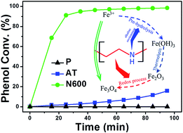
Meso-ZSM-5 modified by polyethyleneimine has been found to be an excellent support for iron oxide with improved physicochemical properties of iron oxide particles including size and chemical state. The resulting ZSM-5 encapsulated iron nanoparticles exhibit superior catalytic activity for phenol oxidation.


Advanced oxidation processes (AOPs) are effective methods for the destruction of the organic contaminants present in wastewater, and as such are drawing much attention in a wide range of investigations.1 Owing to their unique catalytic performance, iron-based materials together with hydrogen peroxide constitute important AOP systems.2 Development of a strategy to control the properties or features of iron-based catalysts including Fe3+/Fe2+ ratio and dispersion, is of great significance to optimize the catalytic efficiency.

Since the iron-based heterogeneous catalyst was first introduced in 1996,3 various molecular sieves including microporous zeolites, mesoporous material (SBA-15,4 MCM-41 (ref. 5)) and metal organic frameworks (MOFs),6 have been developed as iron supports. Compared with the latter two supports, zeolite-supported iron shows higher catalytic activity due to its approximate acidity which is beneficial for the adsorption of the organics.7 However, the supported iron in most cases remains on the outer surface of the zeolite as bulky materials8 which decrease the iron dispersion. Recently, mesoporous zeolite has been developed as a support to decrease the metal particle size.9 However, the chemical nature of iron species (e.g. Fe3+/Fe2+ ratio) is hardly controlled by conventional impregnation methods.

Herein, a facile synthesis strategy was designed for enhancing the dispersion as well as adjusting the chemical state of iron (Fig. 1a). By using mesoporous zeolite modified with polyethyleneimine (PEI) as a support, iron particle size was decreased through amine immobilization which prevents the iron precursor from migrating during thermal treatment. At the same time, the thermal decomposition of PEI is accompanied with a redox process leading to the transition from Fe3+ to Fe2+, which shows excellent performance in phenol degradation.10


image file: c5ra02194d-f1.tif
Fig. 1 (a) Process of N600 preparation, (b, c, d) STEM images of P, AT and N600, respectively, (e) TEM-EDS image of N600. Red color represents iron.

Besides PEI/meso-ZSM-5 composite, two other supports were used in this work as benchmarks, including micro-ZSM-5 and meso-ZSM-5, which were prepared through hydrothermal synthesis11 and alkaline treatment,12 respectively. Through incipient wetness impregnation using aqueous solutions of iron nitrates (Fe(NO3)3·9H2O), three catalysts with 5 wt% Fe loading were prepared. The as-prepared catalysts are referred to as P, AT, and N600. Each went through the same thermal treatment at 873 K for 3 h in N2. Fig. 1b shows the STEM image of sample P. Iron particles with ca. 40 nm are evident on the surface of ZSM-5. When using meso-ZSM-5 as a support, iron particle size clearly decreased (Fig. 1c), which is attributed to the increased external surface area (Table S1). Upon using PEI modified meso-ZSM-5 as a support; the iron particle size was further decreased, as can be seen from Fig. 1d and e.

The XRD patterns of P, AT and N600 are shown in Fig. S1. These clearly exhibit the MFI diffraction peaks of ZSM-5. The diffraction peaks at around 33.2° and 35.7° 2θ of sample P indicate the existence of α-Fe2O3, the particle size calculated from surface [104] and surface [110] by the Scherrer equation is 22.2 nm and 40.4 nm, respectively. While sample AT and N600 show the absence of those diffraction peaks suggesting highly dispersed iron, in line with the STEM images. Fig. S2 shows the Ar adsorption/desorption isotherms of those catalysts. Compared with sample P, the isotherms of AT and N600 exhibit a steep increase from P/P0 = 0.6 to P/P0 = 1, which is evidence of the presence of mesopores. The external surface area of AT and N600 are 240 and 222 m2 g−1, respectively, which is almost twice that of P (125 m2 g−1). Furthermore, AT and N600 possess mesopores of around 10 nm (inset of Fig. S2).

To investigate the reducibility of sample P, AT, and N600, H2-TPR experiments were conducted (shown in Fig. S3). For sample P, the H2 consumption peaks correspond to the combined reduction of Fe3+ and (or) α-Fe2O3 nanoparticles to Fe(3−δ)+ (α1) with intermediate valence as that in Fe3O4 and then to Fe2+ (α2). At higher temperature, the peaks represent partial reduction of Fe2+ to Fe0 (α3, α4).13 Sample N600 shows a decrease of the H2 consumption peak above 773 K and a shift to lower temperature, which is attributed to the strong interaction between iron oxide nanoparticles and the support. Compared with AT, the H2 consumption of N600 decreased, which can be attributed to a previous transition from Fe2O3 to Fe3O4 during the thermal treatment step during the preparation.

The Fe 2p XPS spectra of P, AT, and N600 are shown in Fig. S4. These clearly show that sample P has a higher intensity of the Fe 2p peak, while sample AT shows the weakest Fe 2p intensity. Considering that the detection depth of XPS is limited to about 2 nm, the content of iron located on the outer surface of sample AT is much lower than sample P and N600. From the deconvolution of the Fe 2p XPS spectra of Fe 2P3/2, peaks which centered at 710.8 eV and 708.5 eV are attributed to Fe3+ and Fe2+, respectively. For sample P, only one distinct peak centered at 710.8 eV was observed, indicating that iron in sample P is present only as Fe3+ and, therefore, the iron oxide species is Fe2O3.14 In the case of sample N600, two distinct peaks are present indicating the coexistence of Fe2+ and Fe3+, confirming the existence of Fe3O4 species.

To further investigate the metal oxide species of the three samples in our work, 57Fe Mossbauer spectra of sample P, AT and N600 are shown in Fig. 2, with the hyperfine interaction parameters summarized in Table S2. The peak type can reflect the particle size by determining whether the iron oxide particle is superparamagnetic or not. When the particle size of the iron oxide is below a critical point, super paramagnetic phenomenon converts the peak to a sextet. Contrarily, above the critical point the peak presents as a doublet. On the other hand, the 57Fe Mossbauer parameter can reflect the metal species according to the literature.16 In the case of sample P, the Mossbauer peak of IS = 0.38 mm s−1 and QS = −0.21 mm s−1 are in agreement with those values for α-Fe2O3 in the form of a sextet.15 For sample AT, peaks of α-Fe2O3 in the form of both a sextet and a doublet15 are observed simultaneously with the sextet relative intensity (RI) decreasing from 100% to 41.2%. The doublet, with 57Fe Mossbauer parameters of IS = 0.34 mm s−1 and QS = 0.82 mm s−1, accounted for about 58.8%. This change from sextet to doublet is due to the decrease in particle size, which has been confirmed by STEM and XRD. As for sample N600, the particle size is further minimized because the RI of the sextet further decreased to 22.6% and 57Fe Mossbauer parameters of IS = 0.31 mm s−1 and QS = −0.05 mm s−1 are attributed to Fe3O4.16 This indicates that PEI did have a positive impact on minimizing the particle size as well as reducing the iron oxide.


image file: c5ra02194d-f2.tif
Fig. 2 57Fe Mossbauer spectroscopy of (a) P, (b) AT, (c) N600.

The initial catalytic activity of the prepared samples was evaluated for the phenol degradation reaction. As shown in Fig. 3, the initial catalytic activity of P and AT was lower than that of N600, which is attributed to the increased content of Fe2+ as well as the minimized nanoparticle size. Sample AT exhibited a smaller iron oxide size compared with P. The reaction temperature has a great influence on the catalytic oxidation. As shown in the inset of Fig. 3, when the temperature was increased from 308 to 338 K, the advantages of the minimized particle size began to appear, where sample AT shows a much higher catalytic activity than sample P. It is the minimized particle size that made the catalytic difference of P and AT, and the reduction of iron by PEI led to the great catalytic difference of N600 at lower temperature. Meso-ZSM-5 has the effect on minimizing the particle size of iron oxide, meanwhile, the presence of PEI can enhance this effect by preventing Fe(NO3)3 from migrating as well as reducing Fe3+ during the thermal treatment preparation.


image file: c5ra02194d-f3.tif
Fig. 3 The phenol degradation catalyzed by samples P, AT, and N600. Reaction conditions: n(H2O2):n(phenol) = 14, C(phenol) = 1 g L−1, C(H2O2) = 0.69 M, pH = 6.2, temperature (a) 308 K, (b) 338 K.

In summary, PEI has been successfully used for the first time to fabricate a catalyst with superior activity in phenol oxidation. By using meso-ZSM-5 modified with PEI as a support, iron particle size has been decreased through amine immobilization which prevented the iron precursor from migrating during thermal treatment. At the same time, the thermal decomposition of PEI is accompanied with a redox process leading to the transition from Fe3+ to Fe2+, which is good for the enhancement of catalytic performance in phenol oxidation.

This work provides a guide for improved impregnation processes using low melting point metal precursor with minimized particle size and the facile preparation of transition metal oxides with adjustable physicochemical state.

Acknowledgements

This work was supported by the State Key Program of National Natural Science Foundation of China (grant no. 21236008).

Notes and references

  1. (a) F. C. Moreira, S. Garcia-Segura, R. A. R. Boaventura, E. Brillas and V. J. P. Vilar, Appl. Catal., B, 2014, 160–161, 492–505 CrossRef CAS PubMed; (b) M. Antonopoulou, E. Evgenidou, D. Lambropoulou and I. Konstantinou, Water Res., 2014, 53, 215–234 CrossRef CAS PubMed; (c) A. D. Bokare and W. Choi, J. Hazard. Mater., 2014, 275, 121–135 CrossRef CAS PubMed.
  2. (a) J. V. Coelho, M. S. Guedes, R. G. Prado, J. Tronto, J. D. Ardisson, M. C. Pereira and L. C. A. Oliveira, Appl. Catal., B, 2014, 144, 792–799 CrossRef CAS PubMed; (b) S. Fukuchi, R. Nishimoto, M. Fukushima and Q. Zhu, Appl. Catal., B, 2014, 147, 411–419 CrossRef CAS PubMed; (c) R. Prihod Ko, I. Stolyarova, G. Gündüz, O. Taran, S. Yashnik, V. Parmon and V. Goncharuk, Appl. Catal., B, 2011, 104, 201–210 CrossRef CAS PubMed.
  3. K. Fajerwerg and H. Debellefontaine, Appl. Catal., B, 1996, 10, L229–L235 CrossRef CAS.
  4. (a) C. H. Christensen, I. Schmidt, A. Carlsson, K. Johannsen and K. Herbst, J. Am. Chem. Soc., 2005, 127, 8098–8102 CrossRef CAS PubMed; (b) A. Y. Khodakov, A. Griboval-Constant, R. Bechara and V. L. Zholobenko, J. Catal., 2002, 206, 230–241 CrossRef CAS; (c) H. Song, R. M. Rioux, J. D. Hoefelmeyer, R. Komor, K. Niesz, M. Grass, P. Yang and G. A. Somorjai, J. Am. Chem. Soc., 2006, 128, 3027–3037 CrossRef CAS PubMed; (d) R. M. Rioux, H. Song, J. D. Hoefelmeyer, P. Yang and G. A. Somorjai, J. Phys. Chem. B, 2005, 109, 2192–2202 CrossRef CAS PubMed; (e) F. Su, J. Zeng, X. Bao, Y. Yu, J. Y. Lee and X. S. Zhao, Chem. Mater., 2005, 17, 3960–3967 CrossRef CAS; (f) X. Li, W. Ji, J. Zhao, S. Wang and C. Au, J. Catal., 2005, 236, 181–189 CrossRef CAS PubMed; (g) L. Chmielarz, P. Kuśtrowski, R. Dziembaj, P. Cool and E. F. Vansant, Appl. Catal., B, 2006, 62, 369–380 CrossRef CAS PubMed; (h) F. Martínez, G. Calleja, J. A. Melero and R. Molina, Appl. Catal., B, 2007, 70, 452–460 CrossRef PubMed; (i) M. Mureddu, I. Ferino, A. Musinu, A. Ardu, E. Rombi, M. G. Cutrufello, P. Deiana, M. Fantauzzi and C. Cannas, J. Mater. Chem. A, 2014, 2, 19396–19406 RSC.
  5. (a) J. F. Bengoa, M. V. Cagnoli, N. G. Gallegos, A. M. Alvarez, L. V. Mogni, M. S. Moreno and S. G. Marchetti, Microporous Mesoporous Mater., 2005, 84, 153–160 CrossRef CAS PubMed; (b) M. Xia, M. Long, Y. Yang, C. Chen, W. Cai and B. Zhou, Appl. Catal., B, 2011, 110, 118–125 CrossRef CAS PubMed; (c) A. I. Carrillo, E. Serrano, R. Luque and J. García-Martínez, Appl. Catal., A, 2013, 453, 383–390 CrossRef CAS PubMed.
  6. N. A. Khan and S. H. Jhung, J. Hazard. Mater., 2012, 237–238, 180–185 CrossRef CAS PubMed.
  7. (a) K. Na, C. Jo, J. Kim, K. Cho, J. Jung, Y. Seo, R. J. Messinger, B. F. Chmelka and R. Ryoo, Science, 2011, 333, 328–332 CrossRef CAS PubMed; (b) X. Zhang, W. Yu, Y. Qin, S. Dong, T. Pei, L. Wang and L. Song, Acta Biochim. Biophys. Sin., 2013, 29(06), 1273–1280 CAS.
  8. (a) C. Knapp, A. Obuchi, J. O. Uchisawa, S. Kushiyama and P. Avila, Microporous Mesoporous Mater., 1999, 31, 23–31 CrossRef CAS; (b) C. H. Christensen, I. Schmidt, A. Carlsson, K. Johannsen and K. Herbst, J. Am. Chem. Soc., 2005, 127, 8098–8102 CrossRef CAS PubMed.
  9. (a) C. Dai, A. Zhang, L. Li, K. Hou, F. Ding, J. Li, D. Mu, C. Song, M. Liu and X. Guo, Chem. Mater., 2013, 25, 4197–4205 CrossRef CAS; (b) D. Verboekend, R. Caicedo-Realpe, A. Bonilla, M. Santiago and J. Pérez-Ramírez, Chem. Mater., 2010, 22, 4679–4689 CrossRef CAS; (c) J. Mielby, J. O. Abildstrøm, F. Wang, T. Kasama, C. Weidenthaler and S. Kegnaes, Angew. Chem., Int. Ed., 2014, 53, 12513–12516 CAS.
  10. (a) R. Costa, M. Lelis, L. Oliveira, J. Fabris, J. Ardisson, R. Rios, C. Silva and R. Lago, J. Hazard. Mater., 2006, 129, 171–178 CrossRef CAS PubMed; (b) R. C. C. Costa, F. C. C. Moura, J. D. Ardisson, J. D. Fabris and R. M. Lago, Appl. Catal., B, 2008, 83, 131–139 CrossRef CAS PubMed.
  11. M. M. Forde, R. D. Armstrong, C. Hammond, Q. He, R. L. Jenkins, S. A. Kondrat, N. Dimitratos, J. A. Lopez-Sanchez, S. H. Taylor, D. Willock, C. J. Kiely and G. J. Hutchings, J. Am. Chem. Soc., 2013, 135, 11087–11099 CrossRef CAS PubMed.
  12. (a) D. Verboekend, G. Vilé and J. Pérez-Ramírez, Adv. Funct. Mater., 2012, 22, 916–928 CrossRef CAS; (b) K. Möller and T. Bein, Chem. Soc. Rev., 2013, 42, 3689 RSC; (c) M. Milina, S. Mitchell, P. Crivelli, D. Cooke and J. Pérez-Ramírez, Nat. Commun., 2014, 5, 3922 Search PubMed.
  13. F. Liu, H. He, C. Zhang, Z. Feng, L. Zheng, Y. Xie and T. Hu, Appl. Catal., B, 2010, 96, 408–420 CrossRef CAS PubMed.
  14. (a) J. Gao, X. Ran, C. Shi, H. Cheng, T. Cheng and Y. Su, Nanoscale, 2013, 5, 7026 RSC; (b) T. Yamashita and P. Hayes, Appl. Surf. Sci., 2008, 254, 2441–2449 CrossRef CAS PubMed.
  15. R. Zboril, M. Mashlan and D. Petridis, Chem. Mater., 2002, 14, 969–982 CrossRef CAS.
  16. K. Woo, J. Hong, S. Choi, H. Lee, J. Ahn, C. S. Kim and S. W. Lee, Chem. Mater., 2004, 16, 2814–2818 CrossRef CAS.

Footnote

Electronic supplementary information (ESI) available: Synthesis of P, AT and N600, catalyst performance, Ar adsorption and desorption isotherms, XRD, XPS. See DOI: 10.1039/c5ra02194d

This journal is © The Royal Society of Chemistry 2015
Click here to see how this site uses Cookies. View our privacy policy here.