Antibacterial hybrid cellulose–graphene oxide nanocomposite immobilized with silver nanoparticles

Soon Wei Chooka, Chin Hua Chia*a, Sarani Zakariaa, Mohd Khan Ayobb, Nay Ming Huangc, Hui Min Neohd and Rahman Jamald
aSchool of Applied Physics, Faculty of Science and Technology, Universiti Kebangsaan Malaysia, 43600 UKM Bangi, Selangor, Malaysia. E-mail: chiachinhua@yahoo.com; chia@ukm.edu.my
bSchool of Chemical Sciences and Food Technology, Faculty of Science and Technology, Universiti Kebangsaan Malaysia, 43600 UKM Bangi, Selangor, Malaysia
cPhysics Department, Faculty of Science, University of Malaya, 50603 Kuala Lumpur, Malaysia
dUKM Medical Molecular Biology Institute, Universiti Kebangsaan Malaysia, 56000 Cheras, Kuala Lumpur, Malaysia

Received 30th January 2015 , Accepted 2nd March 2015

First published on 2nd March 2015


Abstract

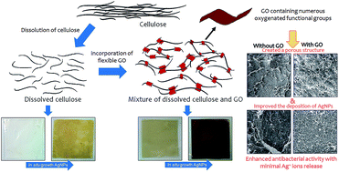
A hybrid nanocomposite cellulose membrane containing graphene oxide and silver nanoparticles was produced via a two step synthesis method. First, regenerated cellulose membranes containing different percentages of graphene oxide (GO) were produced by coagulating the mixture in an acid coagulating bath. Afterward, silver nanoparticles (AgNPs) were in situ synthesized onto the membranes using the modified Tollens' method. The presence of GO on the cellulose membranes significantly enhanced the deposition of AgNPs due to the electrostatic interaction between the positively charged silver ammonia complex and negatively charged oxygenated functional groups of GO before the reduction to AgNPs. The AgNPs content of the membrane with 1 wt% of GO was approximately 26 times greater than that of the neat cellulose membrane. More interestingly, the presence of GO significantly lowered the release of Ag ions and leaching of AgNPs into the aqueous solution. The produced composite membranes exhibited strong antibacterial activity against S. aureus and E. coli.
1. Introduction

For the past few decades, silver nanoparticles (AgNPs) have gained immense attention for their fascinating properties, especially their antibacterial properties against a wide range of bacteria.1 The high surface area to volume ratio of AgNPs allows more direct contact with bacteria or more efficient Ag+ ions release to inflict damage on bacterial components.2–4 Despite showing great potential in various applications such as water disinfection5 and wound dressings,6 the implementation of AgNPs into real life applications has been limited by issues, such as the leaching of Ag particles or ions,7,8 which has created safety and risk concerns in relation to the environment and human health.9 Thus, several endeavours to immobilize AgNPs on substrate or supported material such as textile,10,11 carbonaceous nanomaterials12–14 and polymers9,15,16 via in situ synthesis have been proposed to overcome these problems.

Cellulosic materials have often been used as a platform for nanoparticle immobilization17–21 because of their macromolecule structure consisting of abundant reactive hydroxyl groups (–OH) which can act as the nano nucleation site for particles formation. By nature, cellulose exhibits non-specific functional properties and could be benefited with an introduction of new functionality through functionalization of nanoparticles. Among that, several works has demonstrated that the utilization of regenerated cellulose as a template to produce functional nanocomposite by incorporating various kind of nanoparticles.22–27 Briefly, regenerated cellulose is obtained via dissolution of native cellulose, followed with regeneration process. Cellulose is non-soluble in common solutions, hence an alkaline solution composed of sodium hydroxide and urea was developed to dissolve cellulose at low temperature.28 Cellulose dissolution provides a versatile approach that allowed cellulose to be processed into various forms, such as hydrogel,29 membrane,30 fiber31 and aerogel.32

Graphene oxide (GO) has emerged as one of the most studied nanomaterials in recent years. Apart from graphene, GO exhibits rather different characteristics, such as good solvent dispersity, mainly due to the presence of oxygenated functional groups. Owing to these functional groups, GO has been widely used for immobilizing numerous metallic nanoparticles33,34 and pollutants removal studies.35–37 Besides, numerous studies has reported the excellent antibacterial properties of GO based materials. This including colloidal GO,38,39 GO paper,40 GO modified cotton fabrics,41 GO–bacterial cellulose,42 AgNPs–GO nanocomposite (AgGO)12,43 and AgGO–polymer composites.27,44 Recently, several studies have demonstrated that GO is excellently compatible within cellulosic matrix, where the physical properties of the composite can be improved significantly.45–47 Hereby, GO embedded regenerated cellulose nanocomposite membrane prepared via a feasible solution mixing and casting method, followed by the in situ synthesis of AgNPs. The role of GO on the formation of AgNPs on the cellulose–GO membranes and the antibacterial activity of the membranes against Staphylococcus aureus (S. aureus) and Escherichia coli (E. coli) were investigated. The release or leaching of the Ag+ ions of the membranes was also studied.

2. Experimental

2.1. Materials and chemicals

Cotton linter was supplied by Hubei Chemical Fiber Co. Ltd. (Xiangfan, China) with a weight-average molecular weight (Mw) of 9.0 × 10⁴, as determined by static laser light scattering (DAWN DSP, Wyatt Technology Co., USA). Silver nitrate (AgNO3), sodium hydroxide (NaOH), ammonium hydroxide (NH4OH, 25%), potassium permanganate (KMnO4, 99.9%), hydrogen peroxide (H2O2, 30%), sulphuric acid (H2SO4, 98%) and phosphoric acid (H3PO4, 85%) were purchased from Merck (Darmstadt, Germany). Glucose and urea were purchased from Sigma Aldrich. Graphite flake was purchased from Asbury Graphite Mill, Inc. (Asbury, NJ, USA). All the chemicals were analytical grade and used without further purification.

2.2. Preparation of GO

GO was prepared using the simplified Hummer's method.48 First, the graphite flake was oxidized to graphite oxide with H2SO4 and KMnO4 by stirring the mixture for 3 days. In order to stop the oxidation reaction, H2O2 was added into the graphite oxide solution, which was followed by centrifugation to remove the excess chemicals. The graphite oxide was washed repeatedly and then placed in an ultra-sonication bath to obtain exfoliated graphene oxide.

2.3. In situ preparation of AgNPs on GO-embedded regenerated cellulose membrane

Cotton linter fiber was added to a precooled (−12 °C) aqueous solution of NaOH/urea (7.0 wt%/12.0 wt%), and stirred vigorously at room temperature until a transparent cellulose solution (4.0 wt%) was obtained after centrifugation. A plain membrane without GO was prepared and labelled as CM. Three GO–cellulose solution mixtures containing 0.25, 0.50 and 1.00 wt% of GO were produced and regenerated in a 5 wt% acetic acid coagulation bath. These were labelled CMGO1, CMGO2, and CMGO3, respectively. The membranes were collected and washed repeatedly with DI water to remove excess chemicals. Each membranes with a same dimension of 5 × 5 cm was immersed into a solution containing silver ammonia complex, Ag(NH3)2+, separately. After 30 min, glucose was added to induce the formation of AgNPs under microwave irradiation for 1 min.

2.4. Characterizations

The X-ray diffraction pattern of each membrane was obtained using an X-ray diffractometer (XRD, Bruker Advance). The dried membranes were observed under a field emission scanning electron microscope (FESEM, LEO 1450VP) using SE2 and backscattering mode. FESEM sample preparation was carried out by froze the membrane using liquid nitrogen and freeze dried overnight. Furthermore, the membrane sample was immersed in a 10 wt% of nitric acid and DI water for 4 h and continuously shaken at 120 rpm. Both solutions were withdrawn and further diluted to measure the AgNPs content and Ag ions release using inductively coupled plasma-optical emission spectroscopy (ICP-OES, Perkin-Elmer Optima 4300 DV). X-ray photoelectron (XPS) spectra of the CM–Ag and CMGO3–Ag was obtained using a high resolution Auger electron spectroscopy with XPS (AES-XPS, Kratos Shidmazu).

2.5. Antibacterial test

CM–Ag and CMGO–Ag membranes were cut into five sheets with the dimensions of 2 cm × 1 cm and immersed into nutrient broth inoculated with 105 CFU mL−1 of both S. aureus and E. coli, respectively. Control containing bacterial inoculated broth was prepared without a membrane. The samples were incubated in an incubator shaker at 37 °C at 120 rpm for 4 h. Then, the OD600 values for the control (ODc) and samples (ODs) were obtained at 600 nm. The antibacterial performance was calculated based on the equation depicted in eqn (1).
 
Inhibition rate (%) = [1 − ODs/ODc] × 100% (1)

3. Results and discussion

Visual observations of the CM and CMGO before and after the in situ growth of AgNPs were captured digitally and are depicted in Fig. 1. The GO was observably well distributed within the regenerated cellulose without obvious aggregations. The membranes has further transformed into yellowish colour as an indication of the AgNPs formation on the membranes.
image file: c5ra01897h-f1.tif
Fig. 1 Digital images of membranes before and after in situ synthesis of AgNPs.

As shown in Fig. 2, a diffraction pattern of cellulose II was recorded for all the samples at the peak of 12°, 20° and 22°, proving the accomplishment of the dissolution and regeneration of cellulose. As depicted in Fig. 2a, the calculated degree of crystallinity (χc) for the samples has decreased from 50.1 to 44.0 as the GO content increased, which may have been due to the incorporation of GO interrupting the rearrangement of cellulose crystals during the regeneration process.46 On the other hand, the XRD diffraction pattern of the membrane after the immobilization of the AgNPs (Fig. 2b) revealed the successful formation of crystallite AgNPs, where a significant peak (indicated by the red arrow) corresponding to the Ag crystal (no. JCPDS: 04-0783) was found for each sample.


image file: c5ra01897h-f2.tif
Fig. 2 XRD diffraction pattern for CM and CMGO (a) before and (b) after the synthesis of AgNPs.

The membranes surface structure was observed under FESEM, and the images are shown in Fig. 3. Obvious physical structure changes with the incorporation of the GO can be observed, with the CMGO–Ag samples (Fig. 3b–d) exhibiting a more porous structure compared to the denser structure of CM–Ag (Fig. 3a). The formation of intermolecular hydrogen bonds between the GO and dissolved cellulose was accounted for the transformation, where the insertion of the flexible GO has disrupted the regeneration of the cellulose network,46 as depicted in Fig. 4. The formation of AgNPs was confirmed using the backscattered mode of SEM (Fig. 3e–h), where an increased distribution of AgNPs as white spots can be observed on the surface of membranes.


image file: c5ra01897h-f3.tif
Fig. 3 Surface morphologies of (a) CM–Ag and (b) CMGO1–Ag, (c) CMGO2–Ag and (d) CMGO3–Ag; distribution of AgNPs on (e) CM–Ag, (f) CMGO1–Ag, (g) CMGO2–Ag and (h) CMGO3–Ag.

image file: c5ra01897h-f4.tif
Fig. 4 Schematic illustration of interactions between dissolved cellulose and GO via intermolecular hydrogen bonding.

The AgNPs content of the membranes and Ag+ ions released, as measured using ICP-OES, are listed in Table 1. The AgNPs content of the membranes increased as the amount of embedded GO increased. First, Ag(NH3)2+ was allowed to diffuse through the porous structure of the CMGO with the immersion into Ag(NH3)2+ solution. The positively charged Ag(NH3)2+ was electrostatically attracted toward the negatively charged oxygen functional groups available on CMGO, in which GO introduced extra available oxygen functional groups. With the introduction of glucose under the condition of microwave irradiation, Ag(NH3)2+ was immediately reduced to zero valence Ag0 nuclei which deposited and continuously grew into crystalline AgNPs on the membrane surface.

Table 1 ICP-OES determination of AgNPs content and Ag+ release for CM–Ag and CMGO–Ag
Sample GO content (wt%) ICP-OES measurement
AgNPs content (μg cm−2) Ag+ ions release (μg mL−1)
CM–Ag 0.75 0.85
CMGO1–Ag 0.25 7.88 0.98
CMGO2–Ag 0.50 14.95 0.76
CMGO3–Ag 1.00 20.00 0.56


Interestingly, the amount of Ag+ ions released from the membranes decreased despite the increase in the AgNPs content from CMGO1 to CMGO3. The Ag+ ions release rate was calculated considering the total amount of Ag ions released to the total amount of AgNPs present on the membranes. The Ag+ ions release rates of CM–Ag, CMGO1–Ag, CMGO2–Ag and CMGO3–Ag were 34.1%, 3.7%, 1.5% and 0.84%, respectively. The significant reduction in the Ag+ ion release rate could be due to the increase in the GO–AgNPs interaction, which enhanced the stability of the AgNPs and rendered the oxidative dissolution of Ag+ ions. The XPS results shown in Fig. 5 revealed that both samples contained a characteristic Ag 3d spectrum with a spin orbit splitting value of 6.0 eV. In particular, CM–Ag (Fig. 5a) had a lower binding energy Ag 3d (367.9 eV and 373.9 eV) compared to the pure Ag standard (368.2 eV and 374.2 eV) and CMGO3–Ag (368.1 eV and 374.1 eV), as shown in Fig. 5b. This indicated that the AgNPs of CM–Ag underwent greater oxidation that led to the oxidative dissolution of Ag+ ions, which explained the higher release rate of CM–Ag compared to CMGO–Ag.3,49 Commonly, positive shift of binding energy for metal upon oxidation was observed due to the electronegativity difference. However, the binding energy of Ag 3d in this study underwent a negative shift upon oxidation is similar to several reported studies,50–52 which can be attributed to other factors such as lattice potential, work function changes and extra-atomic relaxation energy.53


image file: c5ra01897h-f5.tif
Fig. 5 High resolution XPS spectra of Ag 3d for (a) CM–Ag and (b) CMGO3–Ag.

The antibacterial activity of the CMGO–Ag is presented in Fig. 6. In general, the antibacterial activities of the nanocomposite membranes produced with higher amounts of AgNPs were more effective at inhibiting the growth of both S. aureus and E. coli. Albeit it had the highest release rate of Ag+ ions, CM–Ag exhibited the weakest antibacterial activity against both bacteria due to its low AgNPs content. In contrast, CMGO–Ag membranes with higher AgNPs contents and lower Ag ions released showed better antibacterial performance, especially against S. aureus. In addition, it is important to note that the porous structure of the CMGO–Ag membranes permitted the bacteria to diffuse through, which facilitated more direct interaction with the AgNPs deposited on the structure. In contrast, CM and CMGO exhibited non-effective antibacterial activity without the presence of AgNPs (Fig. S3). By combining the obtained results, we suggest that the strong inhibition effect of the CMGO–Ag membranes could mainly be attributed to the direct contact between the nanoparticles and bacteria, instead of the released Ag+ ions.


image file: c5ra01897h-f6.tif
Fig. 6 Antibacterial activities of CM–Ag and CMGO–Ag against S. aureus and E. coli.

4. Conclusions

In summary, this work demonstrated that the dissolution of cellulose has provided an advantage for cellulose to be processable and improved its functionality by incorporating GO during regeneration process. The surface functionality of the GO-embedded regenerated cellulose membranes was significantly improved as compared to the neat membrane, where the deposition of AgNPs can be significantly enhanced with the increase of GO content. In return, the GO-embedded membranes showed an effective bacterial inhibition effect with a minimal Ag+ ion release, in which the membrane's porous structure improved the interactions between bacteria and AgNPs. Given these advantages, the produced membrane is envisioned to be applicable for wound dressing and wastewater disinfection applications. The practical or realization of the membranes in those area of application should be further accessed to fully understand the performance of the membranes, which would involve complicated factors and conditions.

Acknowledgements

The authors acknowledge the financial support provided by the research project grants of DIP-2014-013 and LRGS/TD/2012/USM-UKM/PT/04. N. M. Huang acknowledges obtaining a High Impact Research Grant from the Ministry of Higher Education of Malaysia (UM.C/P/HIR/MOHE/SC/21). S. W. Chook appreciates Prof. Dr Lina Zhang for her kind support at Wuhan University.

References

  1. M. Rai, A. Yadav and A. Gade, Biotechnol. Adv., 2009, 27, 76–83 CrossRef CAS PubMed.
  2. S. Agnihotri, S. Mukherji and S. Mukherji, Nanoscale, 2013, 5, 7328–7340 RSC.
  3. Z.-M. Xiu, Q.-B. Zhang, H. L. Puppala, V. L. Colvin and P. J. J. Alvarez, Nano Lett., 2012, 12, 4271–4275 CrossRef CAS PubMed.
  4. G. A. Sotiriou and S. E. Pratsinis, Environ. Sci. Technol., 2010, 44, 5649–5654 CrossRef CAS PubMed.
  5. X. Wang, Y. Dai, J.-L. Zou, L.-Y. Meng, S. Ishikawa, S. Li, M. Abuobeidah and H.-G. Fu, RSC Adv., 2013, 3, 11751–11758 RSC.
  6. B. S. Atiyeh, M. Costagliola, S. N. Hayek and S. A. Dibo, Burns, 2007, 33, 139–148 CrossRef PubMed.
  7. X. Li and J. J. Lenhart, Environ. Sci. Technol., 2012, 46, 5378–5386 CrossRef CAS PubMed.
  8. C.-N. Lok, C.-M. Ho, R. Chen, Q.-Y. He, W.-Y. Yu, H. Sun, P.-H. Tam, J.-F. Chiu and C.-M. Che, JBIC, J. Biol. Inorg. Chem., 2007, 12, 527–534 CrossRef CAS PubMed.
  9. Y. Cheng, L. Yin, S. Lin, M. Wiesner, E. Bernhardt and J. Liu, J. Phys. Chem. C, 2011, 115, 4425–4432 CAS.
  10. B. H. Dong and J. P. Hinestroza, ACS Appl. Mater. Interfaces, 2009, 1, 797–803 Search PubMed.
  11. I. Osório, R. Igreja, R. Franco and J. Cortez, Mater. Lett., 2012, 75, 200–203 CrossRef PubMed.
  12. S. Chook, C. Chia, S. Zakaria, M. Ayob, K. Chee, N. Huang, H. Neoh, H. Lim, R. Jamal and R. Rahman, Nanoscale Res. Lett., 2012, 7, 541 CrossRef PubMed.
  13. C.-H. Xue, R.-J. Zhou, M.-M. Shi, Y. Gao, G. Wu, X.-B. Zhang, H.-Z. Chen and M. Wang, Nanotechnology, 2008, 19, 325605 CrossRef PubMed.
  14. C. Tang, W. Sun and W. Yan, RSC Adv., 2014, 4, 523–530 RSC.
  15. W. D. Cook, Q. D. Nghiem, Q. Chen, F. Chen and M. Sangermano, Macromolecules, 2011, 44, 4065–4071 CrossRef CAS.
  16. S. Gupta, P. Uhlmann, M. Agrawal, S. Chapuis, U. Oertel and M. Stamm, Macromolecules, 2008, 41, 2874–2879 CrossRef CAS.
  17. H. Dong, J. F. Snyder, D. T. Tran and J. L. Leadore, Carbohydr. Polym., 2013, 95, 760–767 CrossRef CAS PubMed.
  18. G. Yang, J. Xie, F. Hong, Z. Cao and X. Yang, Carbohydr. Polym., 2012, 87, 839–845 CrossRef CAS PubMed.
  19. E. Smiechowicz, P. Kulpinski, B. Niekraszewicz and A. Bacciarelli, Cellulose, 2011, 18, 975–985 CrossRef CAS.
  20. H. Liu, D. Wang, Z. Song and S. Shang, Cellulose, 2011, 18, 67–74 CrossRef CAS.
  21. J. You, M. Xiang, H. Hu, J. Cai, J. Zhou and Y. Zhang, RSC Adv., 2013, 3, 19319–19329 RSC.
  22. J. Cai, S. Kimura, M. Wada and S. Kuga, Biomacromolecules, 2008, 10, 87–94 CrossRef PubMed.
  23. S. Hribernik, M. S. Smole, K. S. Kleinschek, M. Bele, J. Jamnik and M. Gaberscek, Polym. Degrad. Stab., 2007, 92, 1957–1965 CrossRef CAS PubMed.
  24. A. John, H.-U. Ko, D.-G. Kim and J. Kim, Cellulose, 2011, 18, 675–680 CrossRef CAS.
  25. Z. Liu, M. Li, L. Turyanska, O. Makarovsky, A. Patanè, W. Wu and S. Mann, Chem. Mater., 2010, 22, 2675–2680 CrossRef CAS.
  26. J. Wu, N. Zhao, X. Zhang and J. Xu, Cellulose, 2012, 19, 1239–1249 CrossRef CAS.
  27. S. Chook, C. Chia, S. Zakaria, M. Ayob, N. Huang, H. Neoh, M. He, L. Zhang and R. Jamal, Cellulose, 2014, 21, 4261–4270 CrossRef CAS.
  28. X. Luo and L. Zhang, Food Res. Int., 2013, 52, 387–400 CrossRef CAS PubMed.
  29. C. Chang, S. Chen and L. Zhang, J. Mater. Chem., 2011, 21, 3865–3871 RSC.
  30. H. Kaco, S. Zakaria, C. H. Chia and L. Zhang, Bioresources, 2014, 9, 2167–2178 CrossRef.
  31. G. Jiang, W. Huang, L. Li, X. Wang, F. Pang, Y. Zhang and H. Wang, Carbohydr. Polym., 2012, 87, 2012–2018 CrossRef CAS PubMed.
  32. J. Cai, S. Kimura, M. Wada, S. Kuga and L. Zhang, ChemSusChem, 2008, 1, 149–154 CrossRef CAS PubMed.
  33. X. Wang, P. Huang, L. Feng, M. He, S. Guo, G. Shen and D. Cui, RSC Adv., 2012, 2, 3816–3822 RSC.
  34. A. M. Golsheikh, N. M. Huang, H. N. Lim and R. Zakaria, RSC Adv., 2014, 4, 36401–36411 RSC.
  35. C. H. Chia, N. F. Razali, M. S. Sajab, S. Zakaria, N. M. Huang and H. N. Lim, Sains Malays., 2013, 42, 819–826 CAS.
  36. S.-T. Yang, S. Chen, Y. Chang, A. Cao, Y. Liu and H. Wang, J. Colloid Interface Sci., 2011, 359, 24–29 CrossRef CAS PubMed.
  37. X. Yang, C. Chen, J. Li, G. Zhao, X. Ren and X. Wang, RSC Adv., 2012, 2, 8821–8826 RSC.
  38. L. Hui, J.-G. Piao, J. Auletta, K. Hu, Y. Zhu, T. Meyer, H. Liu and L. Yang, ACS Appl. Mater. Interfaces, 2014, 6, 13183–13190 CAS.
  39. S. Liu, T. H. Zeng, M. Hofmann, E. Burcombe, J. Wei, R. Jiang, J. Kong and Y. Chen, ACS Nano, 2011, 5, 6971–6980 CrossRef CAS PubMed.
  40. W. Hu, C. Peng, W. Luo, M. Lv, X. Li, D. Li, Q. Huang and C. Fan, ACS Nano, 2010, 4, 4317–4323 CrossRef CAS PubMed.
  41. J. Zhao, B. Deng, M. Lv, J. Li, Y. Zhang, H. Jiang, C. Peng, J. Li, J. Shi, Q. Huang and C. Fan, Adv. Healthcare Mater., 2013, 2, 1259–1266 CrossRef CAS PubMed.
  42. W. Shao, H. Liu, X. Liu, S. Wang and R. Zhang, RSC Adv., 2015, 5, 4795–4803 RSC.
  43. S. Barua, S. Thakur, L. Aidew, A. K. Buragohain, P. Chattopadhyay and N. Karak, RSC Adv., 2014, 4, 9777–9783 RSC.
  44. Z. Fan, B. Liu, J. Wang, S. Zhang, Q. Lin, P. Gong, L. Ma and S. Yang, Adv. Funct. Mater., 2014, 24, 3933–3943 CrossRef CAS.
  45. M. Tian, L. Qu, X. Zhang, K. Zhang, S. Zhu, X. Guo, G. Han, X. Tang and Y. Sun, Carbohydr. Polym., 2014, 111, 456–462 CrossRef CAS PubMed.
  46. W. Ouyang, J. Sun, J. Memon, C. Wang, J. Geng and Y. Huang, Carbon, 2013, 62, 501–509 CrossRef CAS PubMed.
  47. J. Zhang, Y. Cao, J. Feng and P. Wu, J. Phys. Chem. C, 2012, 116, 8063–8068 CAS.
  48. N. M. Huang, H. N. Lim, C. H. Chia, M. A. Yarmo and M. R. Muhamad, Int. J. Nanomed., 2011, 6, 3443–3448 CrossRef CAS PubMed.
  49. P. Prieto, V. Nistor, K. Nouneh, M. Oyama, M. Abd-Lefdil and R. Díaz, Appl. Surf. Sci., 2012, 258, 8807–8813 CrossRef CAS PubMed.
  50. C. Y. Dong, D. S. Shang, L. Shi, J. R. Sun, B. G. Shen, F. Zhuge, R. W. Li and W. Chen, Appl. Phys. Lett., 2011, 98, 072107 CrossRef PubMed.
  51. T. C. R. Rocha, A. Oestereich, D. V. Demidov, M. Havecker, S. Zafeiratos, G. Weinberg, V. I. Bukhtiyarov, A. Knop-Gericke and R. Schlogl, Phys. Chem. Chem. Phys., 2012, 14, 4554–4564 RSC.
  52. T. C. Kaspar, T. Droubay, S. A. Chambers and P. S. Bagus, J. Phys. Chem. C, 2010, 114, 21562–21571 CAS.
  53. J. F. Weaver and G. B. Hoflund, J. Phys. Chem., 1994, 98, 8519–8524 CrossRef CAS.

Footnote

Electronic supplementary information (ESI) available: The FTIR and Raman spectrum of the samples. See DOI: 10.1039/c5ra01897h

This journal is © The Royal Society of Chemistry 2015
Click here to see how this site uses Cookies. View our privacy policy here.