Tunable green to red ZrO2:Er nanophosphors

M. R. N. Soares*a, T. Holza, F. Oliveirab, F. M. Costaa and T. Monteiroa
aPhysics Department and I3N, Aveiro University, Aveiro, Portugal
bMaterials and Ceramics Engineering Department and CICECO, Aveiro University, Aveiro, Portugal. E-mail: rosasoares@ua.pt

Received 5th January 2015 , Accepted 12th February 2015

First published on 12th February 2015


Abstract

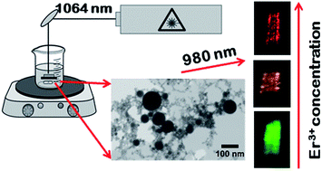
Pulsed laser ablation in water was validated as an effective method to produce highly crystalline erbium doped ZrO2 nanoparticles. Different concentrations of erbium doped ZrO2 ceramic precursor targets were used in the ablation, to study the efficiency of erbium incorporation in the zirconia lattice during nanoparticle synthesis by this method. The spherical nanoparticles produced, with a diameter of up to 200 nm, preserve the crystallinity and optical properties of the precursor target, even for the higher dopant amounts. The optical activation of Er3+ ions was achieved without the need for any additional thermal annealing, usually required for particles produced by several chemical routes. A tunable green to red color of the ZrO2:Er3+ nanoparticles is accomplished through the manipulation of the erbium ion concentration. Particularly, through the sequential absorption of two infrared photons, intense visible up conversion luminescence was observed at room temperature highlighting the doped nanoparticles as promising alternative imaging agents.


1. Introduction

In the last years an exponential interest in the field of optically active trivalent lanthanides (Ln3+) embedded in nanomaterials has been observed, as confirmed by the high number of scientific reports in this subject. Particularly, the demand for new routes of synthesis and the search for nanomaterials with improved properties that can fulfill the requisites for applications in biological sciences, such as luminescent nano-bioprobes, have become increasingly strong.1–8 This is mostly due to the need to overcome the limitations associated to the conventional luminescent bioprobes used nowadays (organic dyes, lanthanide chelates and II–VI semiconductor quantum dots).4–6 These conventionally bioprobes are based on prompt luminescence upon ultra-violet (UV) or visible light excitation. Such high excitation energy constitutes a drawback for the aforementioned biological applications since it results in a high auto-fluorescence background and photo-damage of the biological tissues. Moreover, the low photochemical stability, the short luminescence lifetimes, the large emission bandwidth and the toxicity associated to some of these materials, motivated an intense search for new alternatives.9,10 The Ln3+ ions embedded in inorganic nanomaterials, such as the oxides and fluorides, are considered as a new generation of optically active bioprobes. The luminescence of Ln3+ doped inorganic materials is characterized by atomic-like narrow emission lines with long lifetimes due to the parity forbidden electronic transitions. Moreover, inorganic materials possess high photo and chemical stability enhancing the ability to observe efficient intraionic emission up to room and higher temperatures. Particularly, Ln3+ doped nanoparticles able to produce up-conversion (UC) luminescence constitute a viable alternative as bioprobes.1,5,7,9,11–16 The most efficient mechanisms of up-conversion occur in solid state materials doped with lanthanide ions such as Er3+, Tm3+ and Ho3+.7 These ions have a ladder-like free-ion energy level scheme with similar gaps between the fundamental and the two first electronic excited states. Hence, and considering the long lifetime of the intermediate excited states, it is possible for an already excited ion to absorb a second photon, thus reaching a high energetic excited state from which the radiative de-excitation results in ultraviolet and/or visible emission, using low energy photons as excitation (normally near infrared light (NIR)). The use of NIR light as excitation reduce the problems associated with the auto-fluorescence background and the photo-damage being so favorable for the mentioned biological applications.4–6

The most promising UC materials are those based on Ln3+ doped fluorides, including Er, Yb:NaYF4.7,16,17 The low phonon energies of fluoride lattices reduce the probability of phonon assisted non-radiative transitions which, associated with the longer lifetimes of the intermediated Ln3+ electronic excited states, yields to very efficient up-conversion processes. However, fluoride materials have limited chemical and thermal stability that can hamper their practical applications.10 Moreover, the complex and expensive processes used in the synthesis of these materials associated with the toxicity of some of its precursors are also limitative.10 In other hand, wide band gap metal oxide hosts, including Y2O3,18–20 Y3Al5O12,21 Lu2O3,22 Lu3Ga5O12[thin space (1/6-em)]23,24 and ZrO2,25–28 despite less efficient than fluoride materials, can have high chemical, thermal and photo stability and can be produced by simple and low cost routes, being also interesting hosts for UC. In particular zirconia, ZrO2, a very hard and stable oxide, is also bio-inert. Its wide bandgap energy (from 4 to 6 eV depending on polymorph nature29) associated with a relative low phonon cut-off energy constitute relevant physical properties to consider zirconia as a promising material for the production of efficient lanthanides doped phosphors.30–32 The applications of this or other oxide hosts in bio-applications, including as bio-imaging agents, require the production of high quality doped nanoparticles which can be further functionalized into biological labels. This way, the search for new approaches to synthesize improved doped nanoparticles in an easy and low cost way is increasing. Several chemical and physical methods have been used to produce UC nanophosphors.3,7,33 Recently, pulsed laser ablation in liquids (PLAL) arises as a reliable technique to produce nanomaterials.34–38 In this technique a high power pulsed laser is used to irradiated a solid target, immersed in an appropriated liquid medium. PLAL allows the formation of chemical pure and stable nanoparticles in a liquid medium. Moreover, the simplicity and the low cost of the process associated with the absence of chemical reagents or ligands increases the interest of this method to produce metal, oxides, or even core–shell nanoparticles. The control of laser parameters and the nature of surrounding media allows manipulate the size, shape, chemical composition and functional properties of the nanoparticles (NPs). The synthesis of Ln3+ doped oxides nanoparticles, such as YAG and Y2O3 by PLAL were already reported in literature.39–45 However, it is noteworthy that although there are studies on zirconia nanoparticles produced by PLAL, all works are about undoped zirconia.46–49 Thus, the work here presented regarding erbium doped zirconia nanoparticles produced by pulsed laser ablation in water, constitutes a novelty, as far as we know. The spectroscopic characteristics of prompt and up conversion luminescence of the ZrO2:Er3+ nanoparticles and targets are analyzed in detail by means of energy and excitation dependent steady state photoluminescence (PL) and photoluminescence excitation (PLE). With a 4f11 electron configuration Er3+ ions introduce a wide range of energetic levels in the zirconia band gap allowing the observation of the parity forbidden intra-4f transitions from the visible to infrared. Besides the well-known infrared ∼1.54 μm 4I13/24I15/2 emission, visible light in the green and red spectral regions can be achieved via the 4S3/24I15/2 and 4F9/24I15/2 multiplet transitions, respectively, namely under infrared photon excitation, which is of interest for the aforementioned luminescent-based biosensors.

2. Experimental

Laser ablation was performed using a nanosecond Q-switched Nd:YAG laser with 1064 nm wavelength photons, 10 Hz pulse repetition rate and 7 ns pulse width. The maximum pulse energy is 685 mJ on a focus area of 0.38 cm2. The ceramic ZrO2:Er targets were placed in the bottom of a cell immersed in distilled water as shown in Fig. 1.
image file: c5ra00189g-f1.tif
Fig. 1 Experimental setup used in PLAL technique.

The height of water column above the target was kept constant at ∼10 mm. The ceramic targets were prepared through the uniaxial pressing and densification of Er3+ doped ZrO2 powders prepared by solution combustion synthesis (CS). In CS process, a solution of metal nitrates and an organic fuel (urea) was heated at 450 °C to promote auto-ignition of the solution and the production of the powders as described elsewhere.50 For the ZrO2:Er3+ samples the nominal Er3+ concentration was changed from 1 to 16 mol%. The produced powders were uniaxial pressed into pellets and heat treated at 1350 °C for 3 days to promote densification. The ceramic targets were taken as reference samples for the measured optical properties. The samples characteristics analyzed in this work are summarized in Table 1.

Table 1 Summary of synthesis conditions of the studied samples
Sample name Nominal dopant concentration Preparation Detected phases
Target ZrO2:1Er 1 mol% Er Targets produced by sintering of CS powders Monoclinic
Target ZrO2:2Er 2 mol% Er Monoclinic
Target ZrO2:5Er 5 mol% Er Monoclinic and tetragonal
Target ZrO2:10Er 10 mol% Er Tetragonal
Target ZrO2:16Er 16 mol% Er Cubic
NPs ZrO2:1Er 1 mol% Er NPs produced by pulsed laser ablation in liquid Monoclinic and tetragonal
NPs ZrO2:2Er 2 mol% Er Monoclinic and tetragonal
NPs ZrO2:5Er 5 mol% Er Monoclinic and tetragonal
NPs ZrO2:10Er 10 mol% Er Tetragonal
NPs ZrO2:16Er 16 mol% Er Cubic


For the crystalline phase identification, the targets and NPs were analyzed by RT Raman spectroscopy performed under backscattering geometry with the 325 nm line of a He–Cd laser as excitation, using a Jobin-Yvon HR800 system. The samples morphology was analyzed by scanning electron microscopy/scanning transmission electron microscopy (SEM/STEM) on Hitachi SU-70 equipment. Energy Dispersive X-ray Spectroscopy (EDS) analysis were performed in the SEM, for chemical elemental identification. The samples luminescence was assessed at RT using ultraviolet, visible and infrared light as excitation, enabling the analysis of prompt and up converted intraionic emission. Moreover, the identification of the preferential ion luminescence excitation pathways was measured by RT PLE. Dispersive systems were used in the PL and PLE measurements fitted with cooled Hamamatsu R928 photomultiplier tubes. In the first case a SPEX 1704 monochromator (1 m, 1200 g mm−1) was used whereas the PLE spectra were recorded in a Fluorolog-3 Horiba Scientific modular equipment with a double additive grating scanning monochromator (2 × 180 mm, 1200 g mm−1) in the excitation and a triple grating iHR550 spectrograph in the emission (550 mm, 1200 g mm−1). The measurements were realized using a front face acquisition mode, and were corrected to the optical components and to the Xe lamp spectral responses.

3. Results and discussion

3.1. Structural, morphological and compositional analysis

Fig. 2 shows the Raman spectra of the ceramic targets and the PLAL produced ZrO2 nanoparticles doped with different nominal erbium amounts. As expected, the number of Raman active modes decreases with the rise of dopant content due to increase of the zirconia crystal symmetry with the phase transformation from monoclinic to tetragonal and to cubic.51 For the monoclinic phase (C2h), the one stable at RT and ambient pressure for pure zirconia, 18 Raman active vibrational modes are predicted by group theory (9Ag + 9Bg) in the Γ point of the first Brillouin zone.51 For the high temperature tetragonal and cubic zirconia phases, stabilized with increasing dopant amount, group theory predicts 6 fundamental Raman active modes (1A1g + 2B1g + 3Eg) for the tetragonal phase (D4h), whereas in the case of cubic zirconia (Oh) only one Raman active vibrational mode (F2g) is expected.51
image file: c5ra00189g-f2.tif
Fig. 2 Raman spectra of the targets (left) and PLAL produced NPs (right) by using 325 nm wavelength excitation in backscattering geometry.

As evidenced in the Raman spectra shown in Fig. 2 – left, ceramic target doped with the lower Er3+ concentration (1 mol%) crystallizes in the monoclinic phase of zirconia. Increasing erbium concentration favors the stabilization of the high temperature phases at RT. A mixture of monoclinic and tetragonal phases were found for the intermediate contents (2–5 mol% of Er3+), and for nominal concentrations of 10 and 16 mol% Er3+ the Raman spectra evidence that zirconia doped targets crystallizes in tetragonal and cubic phases, respectively. These results, regarding the crystalline phase identification were corroborated by powders X-ray diffraction analysis (not shown). The main phases identified in the nanoparticles perfectly match those of the reference targets for the high dopant concentrations, where only the high temperature phases are present. In the case of lower dopant concentrations, additionally to the monoclinic phase identified in the targets, nanoparticles crystallized in the tetragonal phase were also present. This mismatch in the crystalline phases in the targets and NPs can be explained based in the existence of additional pathways to stabilize the high temperature metastable zirconia phases in the NPs. As well reported in literature, these phases can be stabilized in pure zirconia by reducing the particle size below a critical value.52–54 No secondary crystalline phases, such as Er2O3 or other Er based oxide, were detected, even for the nominal high dopant concentrations, meaning that the PLAL process allows an efficient incorporation of the ion in the zirconia lattice.

The morphology and grain size of the doped zirconia nanoparticles is shown in Fig. 3. The STEM images reveal that the PLAL produced ZrO2 nanoparticles have spherical shape with reduced dimensions as expected for a very fast process far from-equilibrium due to the narrow quenching times. These results agree well with the previous reported data by H. Zeng et al.37 where the particle dimensions were directly related with the plasma plume cooling rate. In fact, the size of the synthesized nanocrystals is at the nanometer scale, when the quenching time of the plasma plume is of the order of nanoseconds. However, as can be observed in the STEM images, the formed NPs have a large size distribution and a high degree of agglomeration, particularly in the smaller ones. This behavior is a typical characteristic in NPs produced by PLAL.55,56 This problem can be, apparently, overpassed through the control and optimization of growth parameters. In fact, in the last years worldwide researchers focus their attention on the size control of both metal, and oxide nanoparticles, produced by the PLAL as reported in the literature.57,58 Parameters such as laser wavelength, laser fluence, time of irradiation, height of liquid above the target, addition of surfactants, pH of the liquid medium and angle between target and laser beam, seems to influence, at different scales, both the average particle size and size distribution of produced NPs. Some examples are the works reported by the S. A. Al-Mamun et al.58,59 in which they studied almost all these parameters for the case Al2O3 NPs produced by PLAL in water, yielding to narrower size distribution. Although the effect of each of these parameters in the NPs size distribution cannot be directly generalized to all materials (since the interaction between the material and the laser beam strongly depends on the materials physical properties), it is expected to be possible to control the average size and size distribution in the doped ZrO2 NPs as achieved for other oxides NPs produced by PLAL. Concerning the morphology, no differences were observed with the increase of dopant amount. The EDS analysis revealed a uniform distribution of the erbium ions in the produced zirconia nanoparticles (Fig. 3(f–h)) meaning that the high pressure and high temperature environment conditions created by the laser ablation in water is fast enough to preserve the erbium in the zirconia network.


image file: c5ra00189g-f3.tif
Fig. 3 (a–e): STEM/SEM images of the NPs with different dopant concentration. (f–h): EDS map of the NPs doped with 16 mol% Er.

3.2. Optical properties

3.2.1. Photoluminescence excitation. Fig. 4(a) displays the PL and PLE spectra of erbium doped zirconia targets. In all the doped samples, intraionic Er3+ emission is observed meaning that independently of the used erbium amount and zirconia crystalline phase, Er3+ optically activation is achieved. In order to identify the preferential pathways for the ions luminescence, the PLE spectra were monitored in the blue and green spectral regions. The latter corresponds to the 4S3/24I15/2 transition of the Er3+ ions. In the case of the blue luminescence, depending on the ion content (and thus on the zirconia crystalline phase), the PLE was monitored or at the maxima of an intrinsic broad PL band (for lower erbium contents) or on the maxima of the 2P3/24I11/2 Er3+ transition (for samples with higher erbium amounts). The PLE data taken from the green 4S3/24I15/2 transition, with maxima at ∼561 nm, indicates that the population paths of the Er3+ luminescence occur both via a broad violet charge transfer (CT) excitation band and under resonant excitation conditions via the Er3+ excited multiplets. For samples in the monoclinic phase, with lower erbium amounts, the intraionic Er3+ luminescence is preferentially populated via the CT band peaked between 270–280 nm. Similarly, the broad host-related PL band due to native defects60–63 is also excited by broad excitation bands with their peak position dependent on the ion content. For higher erbium concentrations, and in the studied spectral region, a general trend is observed: the decrease of the intensity of the CT excitation band results in changes in the preferential population of the Er3+ ion luminescence. For the high lattice symmetry of the tetragonal and cubic zirconia polymorphs, the resonant excitation into the ion excited multiplets dominates the excitation pathways or the Er3+ luminescence. The measured PLE spectra also corroborate the aforementioned increase in the bandgap energy of the zirconia host with the crystalline phase transformation from monoclinic to tetragonal and cubic.
image file: c5ra00189g-f4.tif
Fig. 4 (a) PLE (left side) and PL (right side) spectra of the erbium doped ZrO2 targets. The PLE spectra were monitored at the green (4S3/24I15/2 Er3+ transition) and blue (broad band at ∼474 nm and 2P3/24I11/2 Er3+ transition) spectral regions. The PL was recorded by pumping the samples into the charge transfer excitation band (CT), 4D7/2 and 4G11/2 Er3+ multiplets. (b and c) PL spectra of the targets and PLAL produced NPs obtained with resonant excitation into the 4G11/2 level.
3.2.2. Prompt luminescence. The samples luminescence obtained with excitation in the ultraviolet CT band and resonantly into the Er3+, 4G11/2 and 4D7/2 excited multiplets, is depicted in the right side of Fig. 4(a). The PL response is sensitive either to the excitation energy and erbium content. With excitation into the CT band, the PL spectra of the samples doped with lower erbium content correspond to an overlap of the broad emission band from native defects60–63 with the sharper intraionic emission lines of Er3+. Additionally, intraionic photon re-absorption is identified. The intensity of the broad band PL decreases with increasing erbium concentration, as identified for samples doped with higher erbium amounts. Pumping the samples in the highest energetic Er3+ multiplet peaked at 255 nm (4D7/2) violet (2H9/24I15/2), blue (2P3/24I11/2), green (2H11/2, 4S3/24H15/2) and red light (2G7/24I9/2; 4F9/24I15/2) are promoted, namely for samples with the tetragonal crystalline structure. For a resonant excitation into the 4G11/2 multiplet (∼378 nm) the PL spectra are only constituted by three groups of intraionic emission lines: the violet, green and red transitions arising from the 2H9/2, 2H11/2, 4S3/2 and 4F9/2 excited states to the 4I15/2 ground state, respectively. With the used excitation conditions, the intensity of the Er3+ prompt luminescence was found to be dependent on the ion amount and consequently on the crystalline phase. For lower erbium concentrations, where the monoclinic zirconia phase dominates, the radiative recombination in the visible range mainly occurs between the 4S3/2 level and the ground state resulting in a green visual appearance in the reference targets and nanoparticles, Fig. 4(b and c). The change in the crystalline phase due to higher erbium concentration is accompanied by a relative increase of the red light due to the 4F9/24I15/2 transition. A likely explanation to the identified behavior is related with the promotion of ion–ion interactions favored for higher dopant concentrations, potentiating energy transfer processes among the interacting defects.15,64–68 Particularly, nonradiative energy transfer processes, such as resonant phonon assisted transfer and cross relaxation mechanisms among the nearby ions are commonly observed in lanthanide doped wide band gap hosts leading, in some cases, to self-luminescence quenching when the interaction occurs among ions from the same chemical specie.15,65–68 For instance, the depletion of 2H11/2, 4S3/2 states to the 4F9/2 level by multiphonon de-excitation and cross relaxation processes such those involving the population of the 4I13/2 and 4I11/2 long lived intermediate levels which in turn repopulates the 4F9/2 by energy transfer encompassing ground state absorption (GSA) from a near neighboring ion, justifies the decrease in the intensity of the green light.15,65–69 The occurrence of such mechanisms pave the way to the development of tunable visible color (green to red) ZrO2 nanophosphors through the manipulation of the erbium ions concentration, envisaging the use of these nanophosphors in luminescence based biolabels. For such purposes, the control of the luminescence processes with infrared excitation is desirable and efficient RT up-conversion luminescence should be achieved.
3.2.3. Up-conversion mechanisms. Fig. 5(a) shows the green and red emission in the ZrO2:Er samples after exciting the doped targets and nanoparticles with low energy photons (980 nm), into the 4I11/2 multiplet. As for the prompt emission, the up-conversion phenomena allow the identification with a naked eye of the tunable green to red luminescence by increasing the erbium content, Fig. 5. Assuming ground state absorption, the Er3+ ions are excited into an intermediate state, the 4I11/2 multiplet, which is known to have a long decay time.70 Therefore, when a second photon is absorbed the 4I11/2 level is still populated and considering a model of a sequential absorption of two 980 nm photons involving GSA and excited state absorption (ESA) the 4I15/24I11/24F7/2 transitions are favored.15 Moreover, up-conversion processes are also mediated by sequential energy transfer (APTE),65,66 likewise called as energy transfer up-conversion (ETU).68 From the 4F7/2 multiplet nonradiative multiphonon relaxation to the 4S3/2 and 4F9/2 excited states occur with further green and red radiative de-excitation to the ground state. A second route to the population of the 4F9/2 excited state can be considered after the 4I15/24I11/2 absorption, namely via sequential nonradiative relaxation from the 4I11/2 to the 4I13/2 multiplet, followed by excited state absorption and sequential energy transfer to the higher energetic multiplet from where the red emission originates (Fig. 5(b)).
image file: c5ra00189g-f5.tif
Fig. 5 (a) PL spectra of the targets (left) and the PLAL produced NPs (right) obtained with 980 nm wavelength excitation photons. (b) Schematic energy level diagram for Er3+ ions illustrating the up-converted luminescence. (c) Color diagram coordinates with the representation of the overall up-conversion emission for the different erbium amounts. Photographs of the RT up converted luminescence of (d) 1 mol% (e) 2 mol% and (f) 16 mol% targets and (g) 1 mol% (h) 5 mol% and (i) 16 mol% nanoparticles, after dried on top of a quartz subtract, with 980 nm excitation.

To investigate the up conversion luminescence mechanisms, the excitation power dependence of the green and red up-converted PL spectra were carried out. The excitation power dependence of the integrated green and red light of the reference targets and PLAL doped nanoparticles are shown in the log–log plots in Fig. 6(a). It is well established15,65–69,71 that the up-converted emission intensity scales with a power law, IPn, where P corresponds to the excitation power and n is the number of photons involved on the process, which in the analyzed ZrO2:Er samples ranges between 1 > n > 2, agreeing well with the rate equations model of Pollnau et al.71 Here, the intensity of the emitting state follows a Pn law for systems where the up-conversion (APTE (ETU)/ESA) mechanisms could be neglected in the intermediate states and adopt a P1 law in systems where the same up-conversion processes dominates.15,66,67,69,71 As such, a unitary slope of the luminescence intensity is expected to occur with increasing power excitation. Besides these two limit situations, real cases can be located in an intermediate regime, where a competition between the radiative and nonradiative depletion of an intermediate state arises, leading to slopes among the two mentioned values.15,66,67,69,71 The described processes clearly influence the excitation to or the depletion from a given emitting level, yielding to distinct observed phenomena in the description of the up converted light. Besides the aforementioned mechanisms, additional processes (e.g. cross relaxation mechanisms) also influence the intensity of the up converted luminescence, as earlier reported by Auzel65,66,69 and Pollnau et al.71 The data shown in Fig. 6 allows further investigation about the dominating processes involved in the up-conversion process for the ZrO2:Er samples. As a starting point, it should be emphasized that either for the green or red luminescence none of the measured slopes corresponds to n = 2. This corresponds to the case where a linear decay from the intermediate levels is expected and the up-conversion processes are insignificant. However, the higher slope tendency observed for the green 4S3/24I15/2 multiplet transition in samples with lower erbium concentration, suggests that the radiative decay is the dominant process involved on the energetic depopulation of intermediate levels, instead of the competitive up-conversion processes. As such, the values close to n = 2 reflect the photon excitation steps for the population of the 4S3/2 level. On the other hand, the measured slope for the red luminescence originated in the 4F9/2 multiplet for the same set of samples (lower amount of erbium ions) is slightly lower than those of the green emission. As above mentioned the tendency for a lower intermediate n value, means that a higher competition occurs among the radiative depletion of the intermediate states and the up-conversion processes. For instance, besides the 4I15/24I11/24F7/2 path (involved on the population of the 4S3/2 and 4F9/2 multiplets) a likely mechanism for the population of the 4F9/2 state corresponds to the two step excitation following the 4I15/24I11/24I13/24F9/2 route, with nonradiative relaxation among the 4I11/2 and 4I13/2 states. As both processes own for different probability rates a different slope for the red emission intensity is expected when compared with the green one, suggesting a higher competition among the up-conversion mechanisms and the radiative decay of the intermediate level to the ground state.


image file: c5ra00189g-f6.tif
Fig. 6 (a) Excitation power dependence of the integrated green and red RT up-converted luminescence for the erbium doped zirconia samples with different erbium amounts. The excitation was performed with 980 nm wavelength photons corresponding to the GSA 4I15/24I11/2. (b) Er concentration dependence of the slope of the green and red up-converted light. (c) red/green intensity ratio for the prompt (378 nm excitation) and up-conversion Er3+ emission (980 nm excitation at 228 mW).

Fig. 6(b) shows the concentration dependence of the slope of the green and red up converted light whereas the red/green luminescence intensity ratio for the prompt and up-conversion Er3+ luminescence is shown in Fig. 6(c). A linear decrease slope tendency of the green and the red up-converted light emission is observed and the composition dependence of the red/green intensity ratio follows a linear behavior for low erbium contents and a supralinear trend for higher amounts. The slope tendency, with a value close to the unity for the highest doped samples, favors the assumption that large up-conversion rates arise for the highest doped samples reflecting the role of the increase in the lattice symmetry (from monoclinic → monoclinic + tetragonal → tetragonal → cubic) and ion site location on the transition probability rates of the observed emission. On the other hand, and for samples with higher erbium amounts, a promotion of the red 4F9/24I15/2 transition was found, either in prompt and up-conversion luminescence. As aforementioned for the prompt luminescence this behavior leads us to consider that the suppression of the green emission could involve additional nonradiative pathways for the depopulation of the 4S3/2. On the other hand, considering the excitation with low energy 980 nm photons the increase in the population of the 4I11/2, 13/2 results in enhanced red emission intensity as observed in other sesquioxides such as Y2O3.72

4. Conclusions

In conclusion, we have reported the tunable visible color (green to red) ZrO2:Er nanophosphors through the manipulation of the erbium ions concentration. The synthesis of the doped nanoparticles was successfully achieved by pulsed laser ablation in water using ZrO2:Er sintered targets constituting a reliable and controllable way for the optically activation of dopants in nanosized zirconia. Analytical investigation by Raman spectroscopy, photoluminescence, photoluminescence excitation and scanning transmission electron microscopy has shown the structural, morphological and optical quality of the produced nanoparticles. As expected, different polymorphs (monoclinic to cubic crystalline phases) of ZrO2:Er were promoted increasing the amount of dopant as identified by the Raman spectroscopy. No other crystalline oxides were found even for high nominal erbium contents proving that a uniform incorporation of the Er3+ ions in the ZrO2 lattice was achieved as observed by EDS. ZrO2:Er nanoparticles present spherical shape and particle sizes up to 200 nm. No differences in nanoparticles morphology were observed with the change in the crystalline structure of the zirconia as a result of erbium addition. Detailed studies of the prompt and up conversion Er3+ luminescence showed that using high and low energy photons as excitation, the dominant visible luminescence of the Er3+ ions in zirconia nanoparticles occurs in the green and red regions due to transitions from the 4S3/2 and 4F9/2 multiplets to the 4I15/2 ground state, respectively. The number of the green emission Stark levels was found to decrease with increasing erbium amount as expected for ions placed in high symmetry sites following the crystalline host phase transformation. Additionally, the green to red luminescence intensity ratio was found to be dependent of the erbium amount, accompanying the increase in the lattice symmetry. The suppression of the green transition for higher concentrations was discussed based on nonradiative competitive mechanisms such as cross relaxation processes between near neighboring ions as expected for higher dopant contents. The power dependence of the visible Er3+ up-conversion emission reveals slopes between 1 and 2 meaning that competition occurs among the radiative depletion of the intermediate states and the up-conversion mechanisms. The lowest doped samples, showing a dominant green light, evidence higher slope suggesting small up-conversion rates. On the opposite side, the highest doped samples (those where the red emission prevails) exhibit a slope near the unity. In both cases, the visual appearance of the green and red up-converted light is clearly observed at room temperature with naked eye. This investigation is helpful to understand the mechanisms behind the prompt and up conversion luminescence of ZrO2:Er3+ nanoparticles and a way to further improve the material design for tunable green to red zirconia based nanophosphors was found.

Acknowledgements

The authors acknowledge FCT for the final funding from PEst-C/CTM/LA0025/2013–14, CENTRO-07-ST24-FEDER-002030, PTDC/CTM-NAN/2156/2012 and RECI/FIS-NAN/0183/2012 (FCOMP-01-0124-FEDER-027494) projects. M. R. N. Soares thank FCT for her PhD grant, SFRH/BD/80357/2011.

Notes and references

  1. F. Wang, D. Banerjee, Y. Liu, X. Chen and X. Liu, Analyst, 2010, 135, 1839–1854 RSC.
  2. S. V. Eliseeva and J.-C. G. Bünzli, Chem. Soc. Rev., 2010, 39, 189–227 RSC.
  3. P. Hänninen and H. Härmä, Lanthanide Luminescence: Photophysical, Analytical and Biological Aspects, Springer Science & Business Media, 2011 Search PubMed.
  4. D. Tu, W. Zheng, Y. Liu, H. Zhu and X. Chen, Coord. Chem. Rev., 2014, 273–274, 13–29 CrossRef CAS PubMed.
  5. M. V. DaCosta, S. Doughan, Y. Han and U. J. Krull, Anal. Chim. Acta, 2014, 832, 1–33 CrossRef CAS PubMed.
  6. W. Zheng, P. Huang, D. Tu, E. Ma, H. Zhu and X. Chen, Chem. Soc. Rev., 2014 10.1039/c4cs00178h.
  7. F. Zhang, Photon Upconversion Nanomaterials, Springer, 2015 Search PubMed.
  8. C. T. Xu, Q. Zhan, H. Liu, G. Somesfalean, J. Qian, S. He and S. Andersson-Engels, Laser Photonics Rev., 2013, 7, 663–697 CrossRef CAS.
  9. J. Chen and J. X. Zhao, Sensors, 2012, 12, 2414–2435 CrossRef CAS PubMed.
  10. F. Gonell, M. Haro, R. S. Sánchez, P. Negro, I. Mora-Seró, J. Bisquert, B. Julián-López and S. Gimenez, J. Phys. Chem. C, 2014, 118, 11279–11284 CAS.
  11. F. Wang and X. Liu, in Comprehensive Nanoscience and Technology, ed. D. L. Andrews, G. D. Scholes and G. P. Wiederrecht, Academic Press, Amsterdam, 2011, pp. 607–635 Search PubMed.
  12. V. K. A. Sreenivasan, A. V. Zvyagin and E. M. Goldys, J. Phys.: Condens. Matter, 2013, 25, 194101 CrossRef PubMed.
  13. M. Lin, Y. Zhao, S. Wang, M. Liu, Z. Duan, Y. Chen, F. Li, F. Xu and T. Lu, Biotechnol. Adv., 2012, 30, 1551–1561 CrossRef CAS PubMed.
  14. S. Hao, G. Chen and C. Yang, Theranostics, 2013, 3, 331–345 CrossRef PubMed.
  15. J. F. Suijver, in Luminescence, ed. C. Ronda, Wiley-VCH Verlag GmbH & Co. KGaA, 2007, pp. 133–177 Search PubMed.
  16. M. Haase and H. Schäfer, Angew. Chem., Int. Ed., 2011, 50, 5808–5829 CrossRef CAS PubMed.
  17. F. Vetrone and J. A. Capobianco, Int. J. Nanotechnol., 2008, 5, 1306–1339 CrossRef CAS.
  18. J. A. Capobianco, F. Vetrone, J. C. Boyer, A. Speghini and M. Bettinelli, J. Phys. Chem. B, 2002, 106, 1181–1187 CrossRef CAS.
  19. F. Vetrone, J.-C. Boyer, J. A. Capobianco, A. Speghini and M. Bettinelli, J. Appl. Phys., 2004, 96, 661–667 CrossRef CAS PubMed.
  20. J. A. Capobianco, F. Vetrone, T. D'Alesio, G. Tessari, A. Speghini and M. Bettinelli, Phys. Chem. Chem. Phys., 2000, 2, 3203–3207 RSC.
  21. M. Liu, S. W. Wang, J. Zhang, L. Q. An and L. D. Chen, Opt. Mater., 2007, 29, 1352–1357 CrossRef CAS PubMed.
  22. F. Vetrone, J. C. Boyer, J. A. Capobianco, A. Speghini and M. Bettinelli, J. Phys. Chem. B, 2002, 106, 5622–5628 CrossRef CAS.
  23. V. Venkatramu, D. Falcomer, A. Speghini, M. Bettinelli and C. K. Jayasankar, J. Lumin., 2008, 128, 811–813 CrossRef CAS PubMed.
  24. V. Mahalingam, F. Mangiarini, F. Vetrone, V. Venkatramu, M. Bettinelli, A. Speghini and J. A. Capobianco, J. Phys. Chem. C, 2008, 112, 17745–17749 CAS.
  25. A. Patra, S. Saha, M. A. R. C. Alencar, N. Rakov and G. S. Maciel, Chem. Phys. Lett., 2005, 407, 477–481 CrossRef CAS PubMed.
  26. A. Patra, C. S. Friend, R. Kapoor and P. N. Prasad, J. Phys. Chem. B, 2002, 106, 1909–1912 CrossRef CAS.
  27. L. A. Gómez, L. D. S. Menezes, C. B. de Araújo, R. R. Gonçalves, S. J. L. Ribeiro and Y. Messaddeq, J. Appl. Phys., 2010, 107, 113508 CrossRef PubMed.
  28. D. Solis, E. D. la Rosa, O. Meza, L. A. Diaz-Torres, P. Salas and C. Angeles-Chavez, J. Appl. Phys., 2010, 108, 023103 CrossRef PubMed.
  29. F. Gallino, C. Di Valentin and G. Pacchioni, Phys. Chem. Chem. Phys., 2011, 13, 17667 RSC.
  30. M. R. N. Soares, C. Nico, M. Peres, N. Ferreira, A. J. S. Fernandes, T. Monteiro and F. M. Costa, J. Appl. Phys., 2011, 109, 013516 CrossRef PubMed.
  31. E. De la Rosa, L. A. Diaz-Torres, P. Salas and R. A. Rodríguez, Opt. Mater., 2005, 27, 1320–1325 CrossRef CAS PubMed.
  32. Y. Liu, S. Zhou, D. Tu, Z. Chen, M. Huang, H. Zhu, E. Ma and X. Chen, J. Am. Chem. Soc., 2012, 134, 15083–15090 CrossRef CAS PubMed.
  33. J. Shen, L.-D. Sun and C.-H. Yan, Dalton Trans., 2008, 5687–5697 RSC.
  34. G. Yang, Laser Ablation in Liquids: Principles and Applications in the Preparation of Nanomaterials, CRC Press, 2012 Search PubMed.
  35. P. V. Kazakevich, A. V. Simakin, V. V. Voronov and G. A. Shafeev, Appl. Surf. Sci., 2006, 252, 4373–4380 CrossRef CAS PubMed.
  36. T. E. Itina, J. Phys. Chem. C, 2011, 115, 5044–5048 CAS.
  37. H. Zeng, X.-W. Du, S. C. Singh, S. A. Kulinich, S. Yang, J. He and W. Cai, Adv. Funct. Mater., 2012, 22, 1333–1353 CrossRef CAS.
  38. T. Tsuji, M. Nakanishi, T. Mizuki, M. Tsuji, T. Doi, T. Yahiro and J. Yamaki, Appl. Surf. Sci., 2009, 255, 9626–9629 CrossRef CAS PubMed.
  39. T. Maldiney, G. Sraiki, B. Viana, D. Gourier, C. Richard, D. Scherman, M. Bessodes, K. Van den Eeckhout, D. Poelman and P. F. Smet, Opt. Mater. Express, 2012, 2, 261–268 CrossRef CAS.
  40. T. Nunokawa, Y. Onodera, M. Hara, Y. Kitamoto, O. Odawara and H. Wada, Appl. Surf. Sci., 2012, 261, 118–122 CrossRef CAS PubMed.
  41. G. Ledoux, D. Amans, C. Dujardin and K. Masenelli-Varlot, Nanotechnology, 2009, 20, 445605 CrossRef CAS PubMed.
  42. S. A. Al-Mamun and T. Ishigaki, J. Am. Ceram. Soc., 2014, 1–8 Search PubMed.
  43. Y. Onodera, T. Nunokawa, O. Odawara and H. Wada, J. Lumin., 2013, 137, 220–224 CrossRef CAS PubMed.
  44. S. K. Singh, K. Kumar and S. B. Rai, Mater. Sci. Eng., B, 2010, 166, 180–184 CrossRef CAS PubMed.
  45. F. Yoshimura, K. Nakamura, F. Wakai, M. Hara, M. Yoshimoto, O. Odawara and H. Wada, Appl. Surf. Sci., 2011, 257, 2170–2175 CrossRef CAS PubMed.
  46. X. C. Yang, W. Riehemann, M. Dubiel and H. Hofmeister, Mater. Sci. Eng., B, 2002, 95, 299–307 CrossRef.
  47. D. Tan, G. Lin, Y. Liu, Y. Teng, Y. Zhuang, B. Zhu, Q. Zhao and J. Qiu, J. Nanopart. Res., 2011, 13, 1183–1190 CrossRef CAS.
  48. N. Bärsch, J. Jakobi, S. Weiler and S. Barcikowski, Nanotechnology, 2009, 20, 445603 CrossRef PubMed.
  49. D. Tan, Y. Teng, Y. Liu, Y. Zhuang and J. Qiu, Chem. Lett., 2009, 38, 1102–1103 CrossRef CAS.
  50. M. R. N. Soares, C. Nico, D. Oliveira, M. Peres, L. Rino, A. J. S. Fernandes, T. Monteiro and F. M. Costa, Mater. Sci. Eng., B, 2012, 177, 712–716 CrossRef CAS PubMed.
  51. E. Fernández López, V. Sánchez Escribano, M. Panizza, M. M. Carnasciali and G. Busca, J. Mater. Chem., 2001, 11, 1891–1897 RSC.
  52. E. Djurado, P. Bouvier and G. Lucazeau, J. Solid State Chem., 2000, 149, 399–407 CrossRef CAS.
  53. S. Shukla, S. Seal, R. Vij, S. Bandyopadhyay and Z. Rahman, Nano Lett., 2002, 2, 989–993 CrossRef CAS.
  54. S. Shukla and S. Seal, Rev. Adv. Mater. Sci., 2003, 4, 123–126 Search PubMed.
  55. C. Liu, X. L. Mao, S. S. Mao, X. Zeng, R. Greif and R. E. Russo, Anal. Chem., 2004, 76, 379–383 CrossRef CAS PubMed.
  56. S. Alves, M. Kalberer and R. Zenobi, Rapid Commun. Mass Spectrom., 2003, 17, 2034–2038 CrossRef CAS PubMed.
  57. V. Amendola and M. Meneghetti, Phys. Chem. Chem. Phys., 2013, 15, 3027–3046 RSC.
  58. S. A. Al-Mamun, R. Nakajima and T. Ishigaki, J. Colloid Interface Sci., 2013, 392, 172–182 CrossRef CAS PubMed.
  59. S. A. Al-Mamun, R. Nakajima and T. Ishigaki, Thin Solid Films, 2012, 523, 46–51 CrossRef PubMed.
  60. S. E. Paje and J. Llopis, Appl. Phys. A, 1993, 57, 225–228 CrossRef.
  61. N. G. Petrik, D. P. Taylor and T. M. Orlando, J. Appl. Phys., 1999, 85, 6770–6776 CrossRef CAS PubMed.
  62. H. Nakajima and T. Mori, J. Alloys Compd., 2006, 408–412, 728–731 CrossRef CAS PubMed.
  63. E. De la Rosa-Cruz, L. Díaz-Torres, P. Salas, D. Mendoza, J. Hernández and V. Castaño, Opt. Mater., 2002, 19, 195–199 CrossRef CAS.
  64. B. G. Wybourne, Spectroscopic Properties of Rare Earths, Interscience Publishers, New York, 1965, vol. 148 Search PubMed.
  65. F. E. Auzel, Proc. IEEE, 1973, 61, 758–786 CrossRef CAS.
  66. F. Auzel, Chem. Rev., 2004, 104, 139–174 CrossRef CAS PubMed.
  67. G. Liu and B. Jacquier, Spectroscopic Properties of Rare Earths in Optical Materials, Springer, 2006 Search PubMed.
  68. J. C. Wright, in Radiationless Processes in Molecules and Condensed Phases, Springer, Berlin Heidelberg, 1976, pp. 239–295 Search PubMed.
  69. F. Auzel, P. A. Santa-Cruz and G. F. de Sá, Rev. Phys. Appl., 1985, 20, 273–281 CrossRef CAS.
  70. F. Vetrone, J.-C. Boyer, J. A. Capobianco, A. Speghini and M. Bettinelli, Appl. Phys. Lett., 2002, 80, 1752–1754 CrossRef CAS PubMed.
  71. M. Pollnau, D. R. Gamelin, S. R. Lüthi, H. U. Güdel and M. P. Hehlen, Phys. Rev. B: Condens. Matter Mater. Phys., 2000, 61, 3337–3346 CrossRef CAS.
  72. H. Lu, W. P. Gillin and I. Hernández, Phys. Chem. Chem. Phys., 2014, 16, 20957–20963 RSC.

This journal is © The Royal Society of Chemistry 2015